404 Copyright © 2015 Korean Neurological Association
Acute-Onset Altitudinal Visual Field Defect Caused by Optic Canal Meningioma
Dear Editor,
Altitudinal visual field defect (VFD), which involves the loss of visual sensation in the hori- zontal half of the visual field, is caused mainly by anterior ischemic optic neuropathy (AION),1-3 or rarely by compressive neuropathy due to a tumor or aneurysm.4,5 However, acute-onset inferior altitudinal VFD is a hallmark of AION.1-3
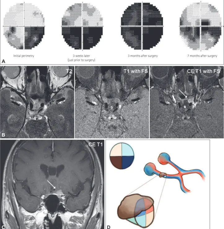
We observed a patient with a rapidly developed inferior altitudinal VFD due to a small meningioma inside the optic canal, which was almost undetectable in standard imaging studies. A 65-year-old man with hypertension and hyperlipidemia complained of blurred vision persisting over several days. He denied having headache, diplopia, or ocular pain. He had a history of medullary infarction and was taking aspirin, antihypertensive medication, and a statin. With the exception of the old infarction, there were no abnormal findings on axi- al T2-weighted MRI scans obtained 10 months previously (Supplementary Fig. 1 in the on- line-only Data Supplement). An ophthalmologic examination revealed decreased visual acu- ity in the left eye (10/20) compared to the right (16/20). Humphrey perimetry revealed an inferior altitudinal VFD in the left eye, which was more severe on the nasal side than on the temporal side (Fig. 1A). There was no evidence of relative afferent pupillary defects, ptosis, proptosis, or disc swelling, or pallor on funduscopy. Ocular movement, intraocular pres- sures, and slit-lamp and optical coherence tomography findings were normal. The acute onset and painless monocular visual loss raised a suspicion of AION, leading to a focus on evaluat- ing the patient’s vascular etiology. However, there were no abnormal findings on MR angiog- raphy (Supplementary Fig. 1 in the online-only Data Supplement). A follow-up visual field test performed 3 weeks later revealed an aggravated altitudinal VFD, especially on the infe- rior temporal side (Fig. 1A). High-spatial-resolution (2-mm slice thickness) MRI with gad- olinium enhancement of the orbit was performed to exclude compressive neuropathy mim- icking AION. The results revealed a 1.2-cm meningioma on the posterior part of the left optic canal causing medial displacement and downward compression of the optic nerve (Fig.
1B and C). The patient underwent a frontotemporal craniotomy and resection of the me- ningioma. The tumor originated from the left side tuberculum sellar and extended into the optic canal. Although he reported a subjective improvement in vision immediately after sur- gery, the VFD was aggravated on the superior side just before surgery (Fig. 1A). Four months after resection of the tumor, his vision had partially recovered (Fig. 1A).
The main mechanism underlying the visual disturbance in compressive optic neuropathy is secondary disruption of the axoplasmic flow and demyelination of the optic nerve.6 There- fore, the onset of VFD usually progresses slowly. Since the optic nerve delivers visual infor- mation received from each of the retinal quadrants to the optic chiasm, the VFD can present in various patterns according to the initial direction of optic nerve compression. In this case the VFD started at the inferonasal area and extended to the inferotemporal area, which can be explained by a medial downward displacement of the optic nerve due to compression from Seung Min Kima,b
Jookyung Leea Soo Geun Joec Jong S. Kima Sun U. Kwona
a Department of Neurology,
Asan Medical Center, University of Ulsan College of Medicine, Seoul, Korea
b Department of Neurology,
Veterans Health Service Medical Center, Seoul, Korea
c Department of Ophthalmology, Gangneung Asan Hospital,
University of Ulsan College of Medicine, Seoul, Korea
pISSN 1738-6586 / eISSN 2005-5013 / J Clin Neurol 2015;11(4):404-406 / http://dx.doi.org/10.3988/jcn.2015.11.4.404
Received July 3, 2015 Revised July 15, 2015 Accepted July 17, 2015 Correspondence Sun U. Kwon, MD, PhD Department of Neurology, Asan Medical Center,
University of Ulsan College of Medicine, 88 Olympic-ro 43-gil, Songpa-gu, Seoul 05505, Korea
Tel +82-2-3010-3960 Fax +82-2-474-4691 E-mail [email protected]
cc This is an Open Access article distributed under the terms of the Creative Commons Attribution Non-Com- mercial License (http://creativecommons.org/licenses/by-nc/3.0) which permits unrestricted non-commercial use, distribution, and reproduction in any medium, provided the original work is properly cited.
JCN
Open Access LETTER TO THE EDITORwww.thejcn.com 405
Kim SM et al.
JCN
the meningioma (Fig. 1D). The VFD was rapid and severe due to the meningioma being located on the optic canal and the diagnosis delay.
Another possible mechanism is ischemia caused by vascu- lar compression. Ischemic symptoms usually present as pos- terior ischemic optic neuropathy (PION) with compression of
the collateral branches from the ophthalmic artery in the op- tic canal.7 However, ischemia is unlikely to be the main mech- anism in this case for two reasons: 1) an altitudinal VFD is relatively rare in PION7 and 2) in this case the VFD worsened according to the direction of nerve compression and improved following surgical resection. Other features associated with
Initial perimetry 3 weeks later
(just prior to surgery) 3 months after surgery 7 months after surgery
Fig. 1. Serial Humphrey perimetries and high-resolution MRI of compressive optic neuropathy with a monocular inferior altitudinal visual field defect (VFD). A: Initial Humphrey perimetry shows an inferior altitudinal VFD in the left eye that is more severe on the nasal side than on the tem- poral side. Follow-up perimetry demonstrates an aggravated VFD and subsequent recovered visual field after surgery. B: Axial contrast-enhanced (CE) T1-weighted MRI with fat suppression (FS) showing an optic nerve meningioma (arrow), which is not clearly defined on T2-weighted and noncontrast T1-weighted images. C: Coronal T1-weighted MRI showing a meningioma (arrow) on the optic canal compressing the optic nerve in a medial downward direction. D: Schematic representation showing the VFD presenting according to the direction of the optic nerve compression.
A
D B
C
406 J Clin Neurol 2015;11(4):404-406
Altitudinal Hemianopsia Caused by Meningioma
JCN
compressive optic neuropathy were absent in this case, which may be due to the smallness of the tumor.
Optic nerve meningioma arising within the optic canal is rare.8 These tumors are usually extremely small despite caus- ing significant visual disturbance and can easily be overlooked on routine MRI. Furthermore, they may not have any orbital signs, and may present with a normal-appearing optic disc.8 Therefore, it is necessary to consider compressive optic neu- ropathy when a patient presents with an altitudinal VFD, even in the absence of other symptoms.
Supplementary Materials
The online-only Data Supplement is available with this arti- cle at http://dx.doi.org/10.3988/jcn.2015.11.4.404.
Conflicts of Interest
The authors have no financial conflicts of interest.
Acknowledgements
This study was supported by a grant from the Korea Healthcare technology R&D Project, Ministry of Health and Welfare, Republic of Korea (HI10C2020).
REFERENCES
1. Hayreh SS, Zimmerman B. Visual field abnormalities in nonarteritic anterior ischemic optic neuropathy: their pattern and prevalence at initial examination. Arch Ophthalmol 2005;123:1554-1562.
2. Traustason OI, Feldon SE, Leemaster JE, Weiner JM. Anterior isch- emic optic neuropathy: classification of field defects by Octopus au- tomated static perimetry. Graefes Arch Clin Exp Ophthalmol 1988;
226:206-212.
3. Balcer LJ. Clinical practice. Optic neuritis. N Engl J Med 2006;354:
1273-1280.
4. Dutton JJ. Optic nerve sheath meningiomas. Surv Ophthalmol 1992;
37:167-183.
5. Cestari DM, Rizzo JF 3rd. The neuroophthalmic manifestations and treatment options of unruptured intracranial aneurysms. Int Oph- thalmol Clin 2004;44:169-187.
6. Vanroose E, Marchau M, Dehaene I, Lammens M. Altitudinal hemi- anopia; a clinical and anatomical entity or a mere coincidence? Case report and review of literature. Acta Neurol Belg 1990;90:254-264.
7. Hayreh SS. Posterior ischaemic optic neuropathy: clinical features, pathogenesis, and management. Eye (Lond) 2004;18:1188-1206.
8. Shapey J, Sabin HI, Danesh-Meyer HV, Kaye AH. Diagnosis and management of optic nerve sheath meningiomas. J Clin Neurosci 2013;20:1045-1056.