pISSN 1229-1137
Original Article
Effects of Sujeom Powder Pharmacopuncture Injected at Jung-wan (CV 12 ) on the Caerulein-induced Acute
Pancreatitis in the Rat
In Soo Kim, Sang Yun Jeon, Tae San Jeong, Sung Sun Kang, Jae Jun Jo and Young Su Lee
*Dept. of Orienal Medicine, Graduate School of Dongshin University
Objectives : This study was designed to investigate Effects of Sujeom powder(SJP) pharmacopuncture Injected at Jung-wan (CV
12) in rats with caerulein-induced acute pancreatitis(AP).
Methods : We examined changes of organ weight, histology, immunohistochemistry and gene expression of cycolooxygenase 2(COX-2) in the pancreas. Twenty adult male Sprague-Dawley rats were divided into four groups as follow: normal(Nor), caerulein-induced(Con), caerulein+SJP pharmacopuncture 0.2mL injected at Jung-wan (CV
12)(SA), and caerulein+SJP pharmacopuncture 0.8 mL injected at Jung-wan (CV
12)(SB) groups.
Pancreatic tissues of rats from all groups were removed for histological observation and light microscopic examination. Interleukin-6(IL-6) levels were determined spectrophotometrically.
Results : The ratio of pancreas/body weights was significantly( p <0.05) increased in the Con, the SA and the SB compared with the Nor, but was slightly decreased in the SA and in the SB groups compared
1)
中脘(CV 12 ) 手拈散약침이
Caerulein으로 유발된 흰쥐의 췌장염에 미치는 영향
김인수ㆍ전상윤ㆍ정태산ㆍ강성순ㆍ조재준ㆍ이영수*
동신대학교 대학원 한의학과
Acceptance : 2012. 11. 2. Adjustment : 2012. 11. 26. Adoption : 2012. 11. 27.
Corresponding author : Young Su Lee, Dongshin University Oriental Medicine Hospital in Affiliation, Internal Medicine of Bi’s system, 141, Wolsan-ro, Nam-gu, Gwang-ju, 503-230, Republic of Korea
Tel : +82-10-2680-4147 E-mail : [email protected]
This is an Open-Access article distributed under the terms of the Creative Commons Attribution Non-Commercial License(http://creativecommons.org/licenses/by-nc/3.0) which permits unrestricted non-commercial use, distribution, and reproduction in any medium, provided the original work is properly cited.
Copylight ⓒ The Journal of Korean Acupuncture & Moxibustion Medicine Society
Abstract
with the Con. Caerulein administration has significantly( p <0.05) increased in the levels of amylase, but the SA, the SB significantly( p <0.05) decreased in the levels of these enzyme. The levels of amylase were increased significantly with caerulein administration, but were inhibited significantly in the SA and in the SB groups. Interleukin-6(IL-6) levels were significantly( p <005) increased in all groups compared with the Nor, especially in the SB. were significantly increased. The levels of Tumor necrosis factor(TNF)-α levels were significantly increased in all groups compared with the Nor. In the conclusion, the datum of IL-6 and TNF-α are suggested that the inflamation was still existed actively at a point of measurement(24 hours later). The COX-2 positive materials are observed in the pancreas from the Con, but these positive materials are decreased in the SJP pharmacopuncture at Jung-wan (CV
12) treatment group.
Conclusion : SJP pharmacopuncture injected at Jung-wan (CV
12) is potentially capable of limiting pancreatic damage during AP by restoring the fine structure of acinar cells and tissues. Therefore we can say that SJP pharmacopuncture Injected at Jung-wan (CV
12) may have beneficial effects in the treatment of caerulein-induded AP. Further studies about the adequate amount of the SJP pharmacopuncture and about more effective route of administration is still required .
Key Words : Sujeom powder(SJP), pharmacopuncture, caerulein, pancreatitis, amylase
Ⅰ. 서 론
췌장염은 단백분해효소 전구물이 췌장 내에서 조 기 활성화됨으로써 일어나는 자가소화의 과정으로 췌 장에 浮腫, 腫脹을 동반하는 염증성 질환이다. 단기간에 치료가 되는 간질성ㆍ부종성 췌장염부터 염증, 탈락, 궤 사가 심해서 전신증상을 유발하는 급성 췌장염
1), 또 한 췌장염이 수개월 혹은 수년 동안 지속되어 췌장을 파괴하는 만성ㆍ재발성 췌장염까지 다양하다
2). 췌장 염의 발생빈도는 나라마다 다르며 우리나라는 근래 서구화된 생활로 그 발생이 증가하고 있으나 정확한 원인과 발생기전은 아직 규명되지 않은 상태이다
3).
한의학에서 췌장염은 주로 心胃痛, 脾心痛, 胃脘痛, 結胸 등의 범주에 속하며 腹痛, 惡心, 嘔吐 및 發熱 黃疸 등의 증상이 나타난다. 치료는 疏肝解鬱, 理氣止 痛, 活血化瘀하는 大柴胡湯加味, 小腹逐瘀湯加味, 膈 下逐瘀湯加味, 蟠蔥散, 手拈散 등이 善用되었다
4).
手拈散은 草果, 玄胡索, 五靈脂, 沒藥으로 구성되어 있으며 朱의 ≪丹溪心法≫에 처음 수록된 이래 心痛, 脾心痛, 九種心痛에 사용된 活血祛瘀止痛劑의 대표적 처방이다
5). 췌장염 모델에 대한 억제효과를 보인 한 의학적 연구로는 金銀花ㆍ蒲公英ㆍ敗醬
6)등의 약물이 보고되었고 복합처방으로는 正傳加味二陳湯
7), 手掂散
8),
加味大黃牧丹皮湯
9)등이 보고되었으나 췌장염과 관련 하여 약침의 효과를 살펴보는 연구는 전무한 실정이다.
약침요법은 질환과 연관된 경혈과 체표 촉진에 의 해 얻은 양성 반응점(壓通點, 阿是穴)에 주사기를 사 용하여 약물을 주입하는 방법으로, 자침과 약물의 효 능을 이용해 생체의 기능을 조정하고 병리상태를 개 선시켜 질병을 치료하는 新鍼療法이다
10).
이에 본 연구에서는 手拈散약침의 췌장염 억제 효 과를 확인하기 위해 caerulein으로 유발된 흰쥐에 투 여량을 달리한 手拈散약침을 中脘에 시술한 후 췌장 중량/체중 비율, 백혈구변화, 호중구ㆍ림프구 함량비 측정, 혈청 amylase, interleukin 6 농도, TNF-α 농도, 광학현미경, COX-2 면역조직화학 등의 실험을 수행 하여 유의한 결과를 얻었기에 보고한다.
Ⅱ. 실험재료 및 방법
1. 재료
1) 실험동물
체중 180±10 g 내외의 7주령 흰쥐(Sprague-Dawley,
male, 20마리)를 샘타고((주), 오산)로부터 구입하였
다. 실험동물은 동신대학교 한의과대학 동물사육실에 서 일정한 조건(온도 : 21± 2 ℃, 습도 : 50-60 %, 12 시간 주기 명/암)하에서 일반 고형사료(샘타코, 흰쥐 용)와 물을 충분히 공급하면서 1주 동안 적응시킨 후 실험에 사용하였다.
2) 手拈散약침의 구성
실험에 사용한 약재는 동신대학교 부속한방병원에서 구입한 후 정선하여 사용하였고, 手拈散약침(Soojeom powder, 이하 SJP)의 구성은 Table 1과 같다.
Herbal name Scientific name Weight(g)
玄胡索 Rhixoma corydalis 40
草果 Fructus amomi Tsaoko 40
五靈脂 Faeces trogopterori 40
沒藥 Myrrha 40
Total amount 160
Table 1. Prescription of Medicinal Herbs
2. 방법
1) 급성 췌장염 유발
정상군을 제외한 모든 실험동물은 약 16시간 전부터 사료 공급을 중단한 다음 caerulein(40 ㎍/㎏, Sigma, USA)을 1시간 간격으로 2회 복강 투여하여 급성 췌 장염을 유발하였다.
2) 약침의 제조
분쇄기를 이용하여 手拈散 약재들을 분쇄하여 삼각 플라스크에 넣고 증류수 2 L를 가하여 약탕기로(대웅약 탕기) 1시간 30분 동안 전탕하여 1 L로 만든다. 전탕 액을 감압여과기(KNF Neuberger Laboport/ 여과지 Adventec, filter paper no. 5A 110 mm 100 circles)로 3회 여과한 후, 감압농축기(BUCHI Rotavapor R-144/
Buchi waterbath B-480)를 이용하여 온도 90 ˚C, 압 력 190 mbar로 감압 농축하였다. 농축액에 95 %에탄 올 30 ml를 가하여 실온에서 교반한 후 방치하여, 침 전물이 생성되게 한 후 다시 여과하였다. 이 여과액을 다시 감압 농축하여 에탄올과 수분을 모두 제거하였을 때 얻은 분말은 34.4 g이었다. 이 분말에 Normal Saline 300 cc를 넣고 완전히 용해될 때까지 magnetic bar를 이용하여 stir한다(Trans sonic T890H(u/s) dissolve, 50 ˚C). 용해시킨 용액은 8,000 rpm으로 20분 동안
Centrifuge(Beckman Coulter Avati J30I)시켜 침전물 은 남기고 상층액만을 취하였다. 이 용액을 10 % NaOH를 이용해 PH 7.4로 맞추고 마지막으로 생체 투여가 가능하도록 세균과 바이러스를 걸러내기 위해 0.45 ㎛와 0.2 ㎛필터(Adventec)를 이용하여 여과하여 약침으로 사용하였다.
3) 실험군 설정 및 약물투여
급성 췌장염을 유발시키지 않은 정상군(이하 Nor 군)과 췌장염을 유발시킨 다음 1시간 후에 각각 생리 식염수 0.2 mL를 투여한 대조군(Con군), 0.2 mL SJP 약침을 투여한 SA군, 0.8 mL SJP 약침액을 투여한 SB군으로 구분하였다. 각 군의 N은 5마리로 하였다.
약침이 시술된 부위는 쥐의 中脘(CV
12)으로서, 白線을 따라 복부 정중선상에서 胸骨劍突尖과 13번째 肋骨端 과 腸骨의 가상 수평선상의 중간점과의 1/2되는 부위 를 취하였다.
3. 관찰방법
1) 체중측정
정상군ㆍ대조군ㆍ실험군 모두 도살하기 직전 체중 을 측정하였다.
2) 췌장중량/체중비율
手拈散약침을 中脘에 시술한 다음 24시간 후, 실험 이 끝난 모든 실험동물은 urethane(700 ㎎/㎏)을 복강 투여하여 마취한 상태에서 채혈을 한 다음 췌장을 절 취한 후 Grady et al
11)의 방법에 의하여 wet pancreas weight/wet body weight 계산식을 통하여 체중에 대한 췌장의 중량을 측정하였다. 측정에는 전자저울(UW420H, Shimadzu, J메무)을 사용하였다.
3) 혈구검사
手拈散약침을 中脘에 시술한 다음 24시간 후, 혈액 은 urethane(700 ㎎/㎏)을 복강 투여하여 마취한 다음 흉곽을 열고 심장 채혈을 하였다. 채혈된 혈액은 EDTA bottle에 넣은 다음 곧바로 Condo et al
12)의 방법에 의 하여 혈구측정기(HEMAVET 950, USA)를 사용하여 백혈구(호중구ㆍ림프구)를 측정하였다.
4) 혈청 amylase 측정
흰쥐에 手拈散약침을 中脘에 시술한 다음 24시간
후에 심장 채혈을 통하여 혈청을 얻은 다음 amylase SL kit(ELITECH, France)를 사용하여 405 nm에서 혈청 amylase의 농도를 측정(PHOTOMETER 5010, Germany)하였다.
5) 혈청 IL-6 농도 측정
手拈散약침을 中脘에 시술한 다음 24시간 후, 혈청 중의 IL-6 농도는 Pezzilli et al
13)의 방법에 의하여 Mouse IL-6 ELISA kit(BD, USA)를 이용하여 SpectraMax M2(Molecular Device, USA)로 450 ㎚에 서 측정하였다. 먼저 각 well에 ELISA dilution 50 μL 와 혈청 50 μL를 넣고 5초 동안 흔들어 준 후, 실온에 서 2시간 동안 incubation하였다. Plate를 wash buffer 로 5회 수세하고, 각 well에 Working detector를 100 μ L 넣고 실온에서 1시간 동안 incubation 하였다.
Wash buffer를 이용하여 수세한 다음 DAB 발색제를 넣고, 다시 30분 동안 incubation 하고 stop solution 을 분주한 다음 측정하였다.
6) 혈청 TNF-α 측정
手拈散약침을 中脘에 시술한 다음 24시간 후, 혈청 중의 TNF-α 농도의 측정은 ELISA (Enzyme-linked immunosorbent assay)를 이용하여 solid phase sandwich ELISA kit (BD OptEIA TNF ELISA kit) 로 측정하였으며, 검출 한계는 13 pg/mL이다.
7) 광학현미경 관찰
手拈散약침을 中脘에 시술한 다음 24시간 후, 흰쥐 의 췌장을 적출하고 10% formalin용액을 사용하여 24 시간 동안 고정시킨 다음(30, 50, 70, 80, 90, 95, 100
Ⅰ, 100Ⅱ) %와 같이 알코올 농도를 상승시켜 탈수한 다음 xylene으로 투명화 과정을 거친 후 파라핀으로 포매하였고, 포매된 조직을 자동조직절편기(MR-3, USA)를 사용하여 6 ㎛두께로 절편을 제작하였다. 절 편한 조직을 slide glass 위에 부착시키고 xylene으로 paraffin을 제거한 다음 100 %, 90 %, 80 % ethanol 과 같이 농도가 낮아지는 순으로 5분씩 담가 함수과 정을 거치게 하였다. Hematoxylin과 Eosin으로 이중 염색을 한 다음 탈수하였다. Canada balsam으로 봉 합한 후 카메라 부착 광학현미경(Olympus BX51, Japan)으로 관찰한 후 사진을 촬영하였다.
8) 면역조직화학
手拈散약침을 中脘에 시술한 다음 24시간 후, 흰쥐
의 췌장을 적출하여 10 % formalin을 사용하여 24시 간 동안 고정시킨 다음, 파라핀으로 포매한 후 자동조 직절편기(MR-3, USA)를 사용하여 조직을 6 ㎛ 두께 로 절편을 제작하였다. 절편한 조직을 slide glass 위 에 부착시킨 다음 이를 xylene에서 파라핀을 제거한 다음 100 %, 90 %, 80 % ethanol과 같이 농도가 낮 아지는 순으로 5분씩 담가 함수과정을 거치게 하였다.
12시간 후 pH 7.2, 0.1 M의 phosphate buffer saline(PBS, 0.9 % NaCl) 용액에 24시간 동안 배양시 킨 다음 15분간 PBS로 세척한 뒤 10 % horse ser- um을 함유한 blocking solution을 사용하여 20분 동 안 배양시키고 다시 PBS용액으로 15분간 세척하였다.
세척한 각각의 조직 위에 polyclonal anti mouse COX-2 항체(Cayman Chemical Co)를 각각 처리하고 습도가 높은 상온의 배양접시에서 2시간 동안 배양시 킨 뒤 15분간 PBS용액으로 세척하였다. 그리고 2차 항체 biotinylated anti-mouse IgG를 처리하여 60분간 배양시킨 후 15분간 PBS용액으로 세척하였다. 이를 다시 3차 항체 avidin-biotinylated enzyme complex (ABC) regent(Vector Lab, CA, USA)를 조직에 처리 30분간 반응시키고 다시 PBS용액에서 15분간 세척하 였다.
DAB(3, 3`-diaminobenzide) 발색시약을 조직에 떨 어뜨려 2분간 발색시키고 난 후 흐르는 물에 과량의 염색시약을 제거하였다. 물기를 제거한 후 여과시킨 hematoxylin에 20초간 대조염색을 한 다음 통상적인 방법에 따라 표본을 제작하여 카메라 부착 광학현미 경(Olympus BX51, Japan)으로 관찰한 후 사진을 촬 영하였다.
9) 통계처리
실험결과는 Mean±SE로 나타냈으며, 대조군과 실 험군 사이의 평균 차이를 검정할 때에는 Student’s t-test로 검정하여 p값이 0.05 미만일 때 통계적으로 유의한 차이가 있는 것으로 판정하였다.
Ⅲ. 결 과
1. 췌장 중량/체중 비율 변화
Con군(0.40±0.0 2%)은 Nor군(0.27±0.01 %)에 비하
여 유의성(
*p<0.05) 있게 증가하였다. 실험군인 SA군
(0.37±0.02 %)과 SB군(0.38±0.02 %)은 Con군에 비해 감소하였으나 Nor군에 비하여 유의성 있게 증가하였 다(Fig. 1).
Fig. 1. The comparison of pancreas/body weight Normal group(Nor) was administrated with saline only.
Control group(Con) was administrated with saline treatment with caerulein. SA and SB groups were administrated with SJP pharmacopuncture 1 hr after treatment with caerulein. All values are mean±SE(n=5).
Significant differences were compared with normal/control at
*p<0.05.
2. 백혈구의 변화
백혈구의 수는 Nor군(3.6±0.31 K/uL)에 비하여 Con군(4.5±0.73 K/uL)과 실험군인 SA군(4.8±0.61 K/uL), SB군(4.2±0.18 K/uL) 모두에서 증가하였으나 유의성 은 없었다(Fig. 2).
Fig. 2. The changes of white blood cells Normal group(Nor) was administrated with saline only.
Control group(Con) was administrated with saline treatment with caerulein. SA and SB groups were administrated with SJP pharmacopuncture 1 hr after treatment with caerulein. All values are mean±SE(n=5).
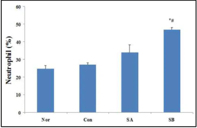
3. 호중구 함량비의 변화
백혈구 중 호중구의 백분율은 Nor군(24.67±1.78 %) 에 비하여 Con군(27.11±1.06 %)에서 다소 증가하였으 나 SA군 (34.03±4.21%)은 약 10 % 증가하였고, SB군 (46.87±1.16 %)근 Nor군과 Con군에 비하여 통계적으 로 유의성(
*#p<0.05) 있게 증가 하였다(Fig. 3).
Fig. 3. The changes of neutrophil content ratio compared with WBC
Normal group(Nor) was administrated with saline only.
Control group(Con) was administrated with saline treatment with caerulein. SA and SB groups were administrated with SJP pharmacopuncture 1 hr after treatment with caerulein. All values are mean±SE(n=5).
Significant differences were compared with normal/control at
#p<0.05.
4. 림프구 함량비의 변화
백혈구 중 림프구의 백분율은 Nor군(69.74±1.45 %)
Fig. 4. The changes of lymphocyte content ratio compared with WBC
Normal group(Nor) was administrated with saline only.
Control group(Con) was administrated with saline treatment with caerulein. SA and SB groups were administrated with SJP pharmacopuncture 1 hr after treatment with caerulein.
All values are mean±SE(n=5). Significant differences were
compared with normal/control at
#p<0.05.
과 Con군(68.99±0.90 %)은 비슷하였으나 SA군(60.65±
4.48 %)에서는 약 9 % 감소하였으며, SB군(48.33±1.15 %) 은 Nor군과 Con군에 비하여 통계적으로 유의성(
*#p<0.05) 있게 감소하였다(Fig. 4).
5. 혈청 Amylase 측정
Amylase의 활성은 Nor군(1098±15.0 U/L)에 비하 여 Con군(2028±106.4 U/L)에서 현저히 높아 통계적 인 유의성(
*p<0.05)을 보여 주었으며, 실험군인 SA군 (1564.8±50.6 U/L)과 SB군(1637.4±62.3 U/L) amylase 의 활성은 Nor군에 비하여 증가(
*p<0.05)하였으나 대 조군에 비하여 유의성(
#p<0.05) 있게 감소하였다(Fig. 5).
Fig. 5. The changes of serum amylase activities Normal group(Nor) was administrated with saline only.
Control group(Con) was administrated with saline treatment with caerulein. SA and SB groups were administrated with SJP pharmacopuncture 1 hr after treatment with caerulein. All values are mean±SE(n=5).
Significant differences were compared with normal/control at
*p<0.05/
#p<0.05.
6. 혈청 IL-6 농도
혈청 내 IL-6의 농도를 측정한 결과, Nor군 (26.204±0.82 pg/mL)에 비하여 Con군(34.72±5.03 pg/mL)과 실험군인 SA군(37.15±0.06 pg/mL)과 SB군 (44.20±5.20 pg/mL) 모두 증가하였으며, 특히 SB군은 통계적으로 유의성(
*p<0.05) 있게 증가하였다(Fig. 6).
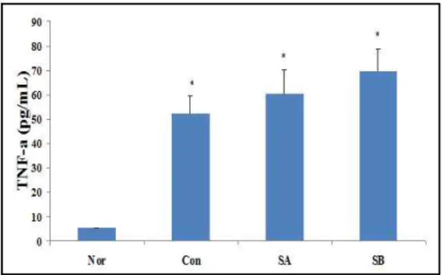
7. 혈청 TNF-α 농도
혈청 내 TNF-α의 농도를 검색한 결과, Nor군 (5.33±0.14 pg/mL)에 비하여 Con군(52.38±7.38 pg/mL)과 실험군인 SA군(60.56±9.90 pg/mL), SB군 (69.63±9.25 pg/mL) 모두 통계적으로 유의성(
*p<0.05)
Fig. 6. The changes of IL-6 concentrations Normal group(Nor) was administrated with saline only.
Control group(Con) was administrated with saline treatment with caerulein. SA and SB groups were administrated with SJP pharmacopuncture 1 hr after treatment with caerulein. All values are mean±SE(n=5). Significant differences were compared with normal/control at
*p<0.05.
Fig. 7. The changes of TNF-α concentrations Normal group(Nor) was administrated with saline only.
Control group(Con) was administrated with saline treatment with caerulein. SA and SB groups were administrated with SJP pharmacopuncture 1 hr after treatment with caerulein. All values are mean±SE(n=5).
Significant differences were compared with normal/control at
*p<0.05.
있게 증가하였다(Fig. 7).
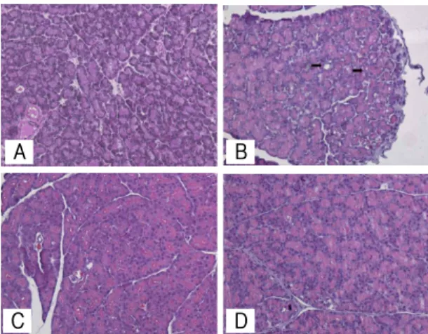
8. 광학현미경 관찰
정상군 샘꽈리세포들은 둥글게 배열되어 있었다.
샘꽈리세포의 핵은 세포의 바닥 쪽에 위치하였고, 핵 주변 세포질은 eosin에 강한 염색성을 보였다(Fig.
8A). 대조군 췌장의 샘꽈리세포에서는 다소의 액포들 이 관찰되었다(Fig. 8B). SA군(Fig. 8C)과 SB군(Fig.
8D) 췌장의 샘꽈리세포에서는 액포가 관찰되지 않았
으며, 세포질의 eosin에 대한 염색성은 보다 높게 관
찰되었다.
Fig. 8. Light micrographs of pancreatitis in 24 hours groups
Morphological changes in caerulein-induced pancreatitis.
Representative hemotoxylin-eosin-stained sections of pancreas were examined by light microscopy in normal rats(A), caerulein treatment control rats(B) shows a number of vacuoles(arrows). SA(C) and SB(D) groups were administrated with SJP pharmacopuncture 1 hr after treatment with caerulein Original magnification set at × 200.
9. COX-2 발현