ISSN 2234-3806 • eISSN 2234-3814
Ann Lab Med 2017;37:313-319
https://doi.org/10.3343/alm.2017.37.4.313
Detection of Serum Antibodies to Hepatitis E Virus Based on HEV Genotype 3 ORF2 Capsid Protein Expressed in Nicotiana benthamiana
Milena Mazalovska, M.S.1, Nikola Varadinov, M.S.1, Tsvetoslav Koynarski, Ph.D.2, Ivan Minkov, Ph.D.1, Pavel Teoharov, Ph.D.3,†, George P. Lomonossoff, Ph.D.4, and Gergana Zahmanova, Ph.D.1
Department of Plant Physiology and Molecular Biology1, University of Plovdiv “Paisii Hilendarski”, Plovdiv, Bulgaria; Department of Animal Genetics2, Faculty of Veterinary Medicine, Trakia University, Stara Zagora, Bulgaria; National Centre of Infectious and Parasitic Diseases3, Sofia, Bulgaria; Department of Biological Chemistry4, John Innes Centre, Norwich Research Park, Norwich, UK
Background: Hepatitis E virus (HEV) causes epidemics in developing countries and is pri- marily transmitted through the fecal-oral route. There have been recent reports on the zoonotic spread of the virus, and several animal species, primarily pigs, have been recog- nized as reservoirs of HEV. Because of its possible spread, there is an urgent need of a method for the cost-effective production of HEV proteins that can be used as diagnostic antigens for the serological detection of anti-HEV antibodies.
Methods: The HEV open reading frame (ORF)2 protein was purified from plant tissue by using immobilized metal-anion chromatography (IMAC). The recombinant protein was used to develop an in-house ELISA for testing anti-HEV antibodies in both human and swine sera. Thirty-six serum samples collected from patients with serologically proven HEV infection with commercial kits were tested for anti-HEV IgG antibodies by using the plant- expressed protein. Forty-five serum samples collected from apparently healthy pigs in Bul- garian farms were also tested.
Results: We confirmed the transient expression and purification of a truncated version of the HEV genotype 3 capsid protein in Nicotiana benthamiana and its usefulness as a di- agnostic antigen. ELISA showed the presence of anti-HEV IgG antibodies in 29 of the 36 human samples. The in-house ELISA showed anti-HEV IgG antibodies in 34 of the 45 pigs.
Conclusions: We describe a method for the production of HEV ORF2 protein in N. ben- thamiana and the usefulness of this protein for the serological detection of anti-HEV anti- bodies in both humans and swine.
Key Words: Hepatitis E virus, Transient expression, ELISA, Nicotiana benthamiana
Received: November 5, 2016 Revision received: December 8, 2016 Accepted: March 8, 2017
Corresponding author: Gergana Zahmanova Department of Plant Physiology and Molecular Biology, University of Plovdiv 24 Tzar Assen str, Plovdiv 4000, Bulgaria Tel: +359-32-261529
Fax: +359-32-261560 E-mail: [email protected]
†Deceased
© Korean Society for Laboratory Medicine This is an Open Access article distributed under the terms of the Creative Commons Attribution Non-Commercial License (http://creativecom- mons.org/licenses/by-nc/4.0) which permits unrestricted non-commercial use, distribution, and reproduction in any medium, provided the original work is properly cited.
INTRODUCTION
Hepatitis E virus (HEV) is the causative agent of epidemic and sporadic viral hepatitis, which has become a major public health concern in both developing and developed regions. Sporadic cases of zoonotic and food-borne infection have been reported in both HEV endemic and non-endemic areas [1]. Several in-
vestigations have shown that domestic pigs in industrialized ar- eas are widely infected with HEV genotype 3, and autochtho- nous cases of hepatitis E have been reported [2, 3]. The mortal- ity rate of HEV infection ranges from 1% to 4%, and can be as high as 25% in pregnant women [4]. HEV is classified in the Hepeviridae family with at least four genotypes (1-4) of the virus infecting humans [5]. Genotypes 1 and 2 are restricted to hu-
2017-03-16 https://crossmark-cdn.crossref.org/widget/v2.0/logos/CROSSMARK_Color_square.svg
mans, while genotypes 3 and 4 are zoonotic and responsible for autochthonous infections in humans [1]. HEV-3 is now consid- ered an emerging pathogen, and is the most common genotype detected in both humans and swine in industrialized nations [2, 6, 7]. Several lines of evidence indicate the occurrence of the zoonotic transmission from pigs, wild boar, and deer to humans [8, 9]. Furthermore, numerous reports have revealed the simi- larities in HEV-3 sequences detected in swine and humans from the same geographic are [10, 11]. In Europe, HEV-3 infections in pig farms are widespread with a prevalence of anti-HEV anti- bodies in pigs reaching 98%, and typically occur at the age of 2-6 months [12]. HEV infection in pig farms is also widespread in the USA [13]. However, to date, there is no information on the circulation of HEV in pigs and other animals in Bulgaria. HEV is not diagnosed routinely in pigs because the infection is often asymptomatic and is not associated with defects in growth [14].
This fact, together with sporadic cases of HEV infection occur- ring after the consumption of undercooked animal meat [15-17], makes HEV a significant zoonotic disease that needs to be prop- erly diagnosed.
HEV is a small, spherical, non-enveloped, single-stranded RNA virus [18]. The HEV genome has three open reading frames (ORFs).
ORF2 encodes the capsid protein, which shows strong immu- nogenicity, and the antibodies generated in response to infec- tion can efficaciously neutralize the virus [19]. Thus, the ORF2 capsid protein is an appropriate candidate for the serological di- agnosis of HEV [20-22].
Definitive diagnosis of HEV infection is based on detection of anti-HEV IgM antibodies or HEV RNA in serum samples. The presence of anti-HEV IgG is typically used to determine the se- roprevalence in a population; however, both IgM and IgG anti- bodies are produced in people with acute HEV infection prior to the manifestation of clinical symptoms, which is potentially diag- nostically relevant [23]. Therefore, patients presenting with in- fection symptoms will likely be positive for both anti-HEV IgM and IgG antibodies, although IgG is less indicative of an acute infection. Although several diagnostic kits for anti-HEV antibod- ies are available, they often provide contradictory results and are quite expensive [24]. Thus, the cost-effective production of re- combinant proteins as diagnostic antigens is needed for the se- rological diagnosis of HEV. Although good expression has been achieved in both mammalian and bacterial cells, the process is quite expensive and cumbersome [25]. Therefore, plants may be a novel source for the cost-effective production of recombi- nant proteins useful for immunological studies. Plants have been explored as platforms for the production of diagnostic and ther-
apeutic recombinant proteins owing to their scalability, safety, and their ability to perform eukaryotic post-translational modification [25, 26].
In this study, we developed a cost-effective and simple method for production of the HEV-3 ORF2 protein in Nicotiana benthami- ana leaves, and demonstrated its usefulness for the serological detection of anti-HEV antibodies in both humans and swine. Tran- sient expression in plant leaves enabled the efficient production of valuable proteins within one week. For this purpose, we used the Cowpea Mosaic Virus (CPMV)-based vector pEAQ-HT [27]
for the expression of the HEV ORF2 protein in plant tissue. The expressed protein was recognized by both human and swine sera and can be used as a diagnostic reagent for the detection of anti-HEV IgG antibodies.
METHODS
1. Synthetic gene and plasmid construction
The nucleotide sequence of the swine genotype 3 HEV capsid protein (GenBank accession number DQ079627.1) lacking the first 109 amino acids (aa) from the N-terminus and 50 aa from the C-terminus (HEV 110-610) was synthesized by Life Tech- nologies (Carlsbad, CA, USA). To maximize the levels of expres- sion, the HEV ORF2 gene was codon-optimized for expression in N. benthamiana.
DNA fragments for insertion of the 6x His-tag at either the N- or C-terminus of HEV 110-610 was amplified by PCR. The primer pair HEV/C-end His-tag/Fw (AAATACCGGTATGGCTACTTCTCCT) and HEV/C-end His-tag/Rv (AGGCCTCGAGCTAATGATGGTGAT- GGTGATGAGCAAGAGCAGAGTGAGGAGCAAG) was used to clone the His-tag at the C-terminus, and the primer pair HEV/N-end His-tag/Fw (AAATACCGGTATGCATCACCATCACCATCATGCTAC- TTCTCCTGCTCCAGATACTGCT) and HEV/N-end His-tag/Rv (GG- CCTCGAGCTAAGCAAGAG) was used for introducing a His-tag at the N-terminus. The PCR fragments were flanked by AgeI and XhoI restriction sites (New England Biolabs, Ipswich, MA, USA), which were used for the cloning of PCR fragments into the pEAQ-HT vector digested with the same enzymes. Finally, the plasmid DNA was purified and confirmed by PCR and se- quencing. The E. coli XL1 blue strain was used for all cloning experiments.
2. Transformation of Agrobacterium tumefaciens
The constructs were transformed into the electrocompetent A.
tumefaciens strain LBA4404. After electroporation at 2.5 kV, Su- per Optimal Broth with catabolite repression (SOC) medium was
immediately added, and the cells were left to recover for 1 hr at 28°C. The cells were then plated on LB agar containing 50 μg/
mL rifampicin and 50 μg/mL kanamycin.
3. Plant growth conditions
N. benthamiana plants were grown in glasshouses maintained at 25°C and watered daily. Supplemental lighting was provided to maintain 16 hr of daylight in the winter months. Plants that were 3–4 weeks old were used for the transient expression ex- periments.
4. Agroinfiltration
Inoculated liquid cultures of A. tumefaciens strain LBA4404 were grown at 28°C in a shaking incubator for 24 hr in Luria-Bertani (LB) medium containing rifampicin and kanamycin. N. bentha- miana leaves were agroinfiltrated via a syringe at an optical den- sity at 600 nm of 0.4.
5. Harvesting
Leaf tissue was harvested at six days post infiltration (dpi). Large- scale sampling was conducted by removing any large veins and non-infiltrated tissue, and recording the leaf sample weight.
6. Protein extraction and sodium dodecyl sulfate-
polyacrylamide gel electrophoresis (SDS-PAGE) analyses
Samples were extracted by adding 3 volumes of the extraction buffer (phosphate buffered saline, PBS, pH 7.4) with complete EDTA-free protease inhibitor cocktail tablets (Roche Diagnostics GmbH, Mannheim, Germany) and homogenized in a blender.Large cell debris was removed by squeezing the homogenate through one layer of Miracloth (Merck KGaA, Darmstadt, Ger- many). NuPAGE Bis-Tris Mini gels of 4–12% or 12% (w/v) acryl- amide (Invitrogen, Carlsbad, CA, USA) and the protein pre-stained standard SeeBluePlus 2 (Invitrogen) or Spectra multicolor broad range protein ladder (TermoFisher scientific, Waltham, MA, USA) were used throughout the experiments.
7. Purification of recombinant HEV 110-610 His-tag protein
The recombinant protein from leaf extracts was purified using immobilized metal-anion chromatography (IMAC) on a Ni-NTA column, according to the manufacturer’s instructions (Qiagen, Hilden, Germany). Briefly, infiltrated leaf tissue was blended with binding buffer containing 10mM imidazole at 3-times the fresh weight of the leaves (FWT). The extract was filtered through two layers of Miracloth, and after centrifugation, the clarified extracts were added onto a Qiagen Ni-NTA column. The column waswashed with 20mM imidazole and was eluted with a total of 4 mL of elution buffer containing 250mM imidazole. Protein con- centrations were determined by densitometry of the stained gels using known amounts of purified bovine serum albumin as a control and by measuring the absorbance at 280 nm. Both pro- teins were purified up to 100 μg/g FWT using IMAC and detected by using SDS-PAGE and western blot.
8. Western blot
The purified proteins were transferred from the SDS-PAGE gel onto a nitrocellulose membrane (Bio-Rad Laboratories Ltd, Hert- fordshire, UK). Membranes were blocked with 5% (w/v) non-fat dried milk in PBS with 0.05% Tween-20 (v/v) (PBST) and incu- bated in the primary monoclonal antibody mouse anti-hepatitis E ORF2 antigen antibody (ab101124; Abcam, Cambridge, UK) diluted 1:3,000 at room temperature for 1 hr and washed with PBST. The bound antibody was detected with secondary anti- mouse antibody-horseradish peroxidase (HRP) (ThermoFisher Scientific) diluted 1:30,000. The emitted luminescence from the ECL detection reagents (GE Healthcare Life Sciences, Buck- inghamshire, UK) was detected with the ImageQuant LAS 500 system (GE Healthcare Life Sciences). For western blot using human and swine serum as the primary antibodies, the mem- brane was incubated in human or swine serum (dilution 1:400), and bound antibodies were detected with secondary anti-hu- man antibody-AP (Sigma-Aldrich, S.Louis, MO, USA) diluted 1:5,000 or secondary anti-pig antibody-AP (KPL, Gaithersburg, MD, USA) diluted 1:10,000, respectively. One-step NBT/BCIP substrate (ThermoFisher Scientific) was used. Western blot is universally accepted approving assay for detection of antibodies and it is considered a gold standard for affirmation of results. In this work, western blot was used to evaluate the quality of our in-house ELISA, by determining the rate of false-positives or false- negatives.
9. Human sera
All human serum samples, with informed consents from the pa- tients, were provided by the National Centre of Infectious and Parasitic Diseases, Sofia, Bulgaria. The serum samples were tested with commercial kits from Dia.Pro (Dia.Pro Diagnostic Bioprobes Srl, Milan, Italy), which detects serum IgG and IgM.
All human sera were positive for both antibodies.
10. Swine sera
Thirty serum samples were collected from apparently healthy pigs (six months old) using blood collected from the ear vein by
veterinarians. Additional 15 serum samples were collected from the intracardiac clot of slaughtered pigs at slaughterhouses post- mortem. Sera were aliquoted and stored at –20°C until use. All swine serum samples were donated by the Department of Ani- mal Genetics, Faculty of Veterinary Medicine, Trakia University, Bulgaria.
11. In-house ELISA based on recombinant HEV 110-610 protein
Microtiter plates (Greiner 96-well flat bottom) were coated with 50 µL/well of serial dilutions of purified protein in duplicate wells (12.5 to 100 ng/well) in PBS (pH 7.4) and incubated overnight at 4°C. After three washes with PBST, plates were incubated with 200 µL/well of blocking solution (PBST–5% [w/v] dry milk) and incubated for 1 hr at room temperature. Serum diluted 1:100 in blocking buffer was then added.
Plates were washed again with PBST before the addition of 50 µL/well of an HRP-conjugated goat anti-human IgG or anti- swine secondary antibody diluted 1:10,000 (KPL) or 1:8,000 (ThermoFisher Scientific), respectively. After incubation with the secondary antibody, plate wells were washed three times before 50 µL/well of the substrate solution (o-phenylenediamine, Sigma- Aldrich) was added. Plates were incubated in the dark at room temperature from 10 to 20 min, and then the reaction was stop- ped by the addition of 50 µL/well of 1M H2SO4, and the plates were read at 492 nm in a plate reader Epoch Microplate Spec- trophotometer (BioTek Instruments Inc., Winooski, VT, USA).
Ten previously characterized negative human serum samples by using commercial kits from Dia.Pro (Dia.Pro Diagnostic Bio- probes Srl) and four negative swine serum samples by using porcine kit PrioCheck HEV Ab (ThermoFisher Scientific) were used as controls to determine the cut-off value for the human
and swine anti-HEV IgG ELISA assay. The cut-off value was cal- culated as absorbance value of the test sample/absorbance value of the negative control or P/N of ≥2.5. Samples with absorbance values above the cut-off (P/N) were considered positive [20].
RESULTS
1. Protein expression and purification of swine genotype 3 HEV 110-610 capsid protein
A synthetic gene of the ORF2 of HEV lacking the first 109 aa at the N-terminus and the last 50 aa at the C-terminus was design ed.
The sequences were codon-optimized for the N. benthamiana genome to increase the yields of expressed protein [28]. The synthetic gene was used as template for insertion of a 6x His- tag at either the N- or C-terminus by the PCR DNA amplification method (see Methods). The resultant PCR products were cloned into pEAQ-HT and, after confirming the sequence of the insert, the constructs were transformed into A. tumefaciens, and N. ben- thamiana leaves were agroinfiltrated.
Plant leaves infiltrated with HEV 110-610 C-end expressed a large amount of a 54.6 kDa protein as revealed by SDS-PAGE followed by staining with Instant Blue (Fig. 1A). Western blot anal- ysis using anti-HEV ORF2 antibody confirmed that the plants suc- cessfully produced HEV 110-610 C-end protein (Fig. 1B). The expressed HEV 110-610 with His-tag at the N-terminus was also analysed by SDS-PAGE (Fig. 1C) and confirmed with western blot (Fig. 1D). The 54.6 kDa band was absent in extracts from tissues infiltrated only with pEAQ-HT (data not shown).
The purified protein was also run on SDS-PAGE gels and de- tected with anti-HEV IgG-positive swine sera (Fig. 2A) and with anti-HEV IgG-positive human sera (Fig. 2B). In addition, the sam- ples were detected with anti-HEV ORF2 antibody as a control
Fig. 1. (A) SDS-PAGE of expressed and purified hepatitis E virus (HEV) 110-610 C-end His-tag protein; (B) Western blot of the gel with an- ti-HEV ORF2 antibody; (C) SDS-PAGE of expressed and purified HEV 110-610 N-end His-tag protein; (D) Western blot of the gel with anti- HEV ORF2 antibody. M, protein marker; 1, crude extract; 2, supernatant; 3, pellet; 4, flow-through; 5, washing step; 6–9, elutes of the 54.6 kDa protein.
Abbreviations: SDS-PAGE, sodium dodecyl sulfate-polyacrylamide gel electrophoresis; ORF2, open reading frame 2.
M 1 2 3 4 5 6 7 8 9 M 1 2 3 4 5 6 7 8 9 M 1 2 3 4 5 6 7 8 9 M 1 2 3 4 5 6 7 8 9
64 kDa 51 kDa
A B C D
(Fig. 2C). Both the human and swine sera recognized a protein band of 54.6 kDa (arrows in Fig. 2) and some aggregates and degradation products.
2. ELISA based on the HEV 110-610 His-tag protein with human sera
The purified HEV 110-610 C-end and N-end His-tag proteins reacted similarly to human sera previously characterized as posi- tive for anti-HEV antibodies. After obtaining the preliminary re- sults with both proteins, we chose to perform all ELISA tests with the HEV 110-610 C-end His-tag protein. Five previously charac- terized negative human sera and five positive human sera were pooled and used for optimization of antigen concentration and serum dilution. Different concentrations of the HEV 110-610 re- combinant protein were used. The optimal serum dilution was determined to be 1/100, and the antigen concentration was 50 ng/well (see Supplemental Data Fig. S1 and Fig. S2).
Thirty-six previously characterized positive human serum sam- ples were tested by ELISA using the HEV 110-610 C-end His-tag protein as the antigen. The in-house ELISA showed anti-HEV IgG antibodies in 29 of the 36 human serum samples (Fig. 3).
Since western blot is more specific and confirmatory than ELI SA, we also performed western blots with all of the human sera. All serum samples showed a positive signal on western blots. The sensitivity of the in-house ELISA assay was 80.5% compared with the commercial kit. From these data, we concluded that the plant-produced HEV 110-610-based ELISA test resulted in seven false-negative results (19.5%) (Fig. 4).
3. Detection of swine HEV antibodies
A total of 45 serum samples collected from healthy pigs in two Bulgarian farms and one slaughterhouse were tested for anti-HEV IgG antibodies. For specific HEV antibody detection in swine, we used both the plant-produced HEV 110-610-based ELISA and western blot. Four previously characterized negative swine sera and four positive swine sera were pooled and used for opti- mization of the antigen concentration and serum dilution. The optimal serum dilution was determined at 1/100, and the anti- gen concentration was 50 ng/well (see Supplemental Data Fig.
S3).
Fig. 2. Western blot of purified hepatitis E virus (HEV) 110-610 His- tag 54.6 kDa protein. Membranes were incubated with swine se- rum (A), human serum (B), and monoclonal antibody (anti-hepatitis ORF2 antibody) (C). M, protein marker; 1, HEV 110-610 N-end His- tag protein; 2, HEV 110-610 C-end His-tag protein (arrows).
Swine serum
98 kDa 62 kDa 49 kDa
Human serum Monoclonal Ab
M 1 2 M 1 2 1 2
A B C
Fig. 3. In–house ELISA of serum samples based on the hepatitis E virus (HEV) 110-610 C-end His-tag protein. The cut-off value (CO) is indicated by a horizontal line. Samples were considered positive (in red), if the absorbance was above the cut-off value (P/N) (see Methods).
19
417
418
Fig. 3. In–house ELISA of serum samples based on the hepatitis E virus (HEV) 110-610 C- 419
end His-tag protein. The cut-off value (CO) is indicated by a horizontal line. Samples were 420
considered positive (in red), if the absorbance was above the cut-off value (P/N) (see Methods).
421 422
423 424
3
2
1
0 Swine Sera Human Sera
Absorbance (492 nm)
CO 0.485
CO 0.142
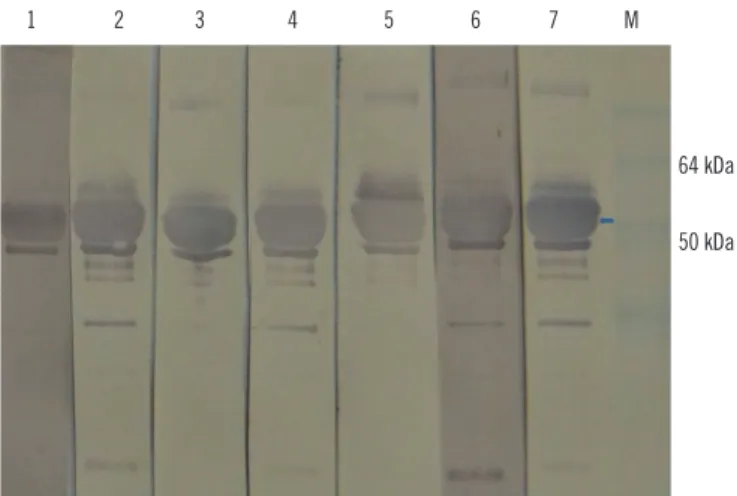
Fig. 4. Western blot probed with hepatitis E virus (HEV) 110-610- based ELISA negative human sera from this study (lanes 1-7). Ar- row indicates the 54.6 kDa protein of interest. There are some ag- gregates and degraded products. Five HEV negative human sera previously characterized by commercial kit were used as control for western blot specificity (data not shown). M, protein marker.
1 2 3 4 5 6 7 M
64 kDa
50 kDa
The in-house ELISA showed antibodies in 34 of 45 pigs, indi- cating that 75.5% were positive for anti-HEV IgG antibodies (Fig. 3).
We then performed western blot with the 11 swine serum sam- ples that scored negative by the ORF2-based ELISA. Four of these 11 samples tested positive by western blot (Fig. 5). This shows that the ELISA test resulted in four false-negative results or a rate of 8%.
DISCUSSION
We provide the first demonstration of the transient expression of truncated HEV 110-610 His-tagged protein in the plant N. ben- thamiana. The specificity of the expressed protein was confirmed by western blot using well-characterized monoclonal antibody, as well as human and pig sera. The optimal concentration of the antigen and serum dilution were assessed for an in-house ELISA for both swine and human sera. We demonstrated that the plant-produced truncated HEV ORF2 protein used as a coat- ing antigen in our in-house ELISA was able to detect anti-HEV IgG antibodies in human sera with similar accuracy compared with that of the commercial kit. Since western blot is considered the gold standard, we used this method to confirm the ELISA- negative results. Anti-HEV IgG was detected in all human serum samples tested by the western blot. It is possible that some lin- ear epitopes have been unmasked compared with the native antigen used in the ELISA. Furthermore, using the same method described herein, the recombinant plant ORF2 can also be used
for the detection of serum IgM, which is considered to be more critical for the diagnosis of acute HEV infection (data not shown).
Moreover, we could confirm the presence of the anti-HEV IgG antibodies in pigs in Bulgaria. Because pigs ordinarily are not tested for HEV infection, additional testing needs to be done to assess the seroprevalence of anti-HEV antibodies in Bulgarian farms.
Plant leaves infiltrated with pEAQ-HT HEV 110-610 with a His-tag at either the N- or C-terminus resulted in the expression of a 54.6 kDa protein at up to 100 μg/g of FWT. For instance, it has been reported that 1 mg of ORF2 protein can be produced per liter of E. coli culture [29]. Using the same protocol, 1 L of culture would produce enough protein to process 10,000 test samples, while 1 kg of plant tissue could produce enough pro- tein to process 1,000,000 test samples at the yield described herein. This implies that one would need 100 L of bacterial cul- ture to process the same number of samples as could be pro- cessed with the protein produced by 1 kg of plant tissue, which is much easier to obtain and much more cost-effective. If the production capacity is scaled up to levels used in the commer- cial production of plant-derived proteins (i.e., hundreds of kilos), then the final cost of the diagnostic reagent can quickly be low- ered, improving its availability to those who need to administer the tests.
In conclusion, the HEV 110-610 recombinant protein can be produced in high yields at a relatively low cost in plant tissue, and could be used as a diagnostic antigen in the ELISA-based detection of anti-HEV antibodies in both human and swine sera.
Authors’ Disclosures of Potential Conflicts of Interest
George P. Lomonossoff declares that he is a named inventor on granted patent WO 29087391 A1, which describes the plant transient expression system used for the work described in this manuscript.
Acknowledgments
The authors’ research on HEV is supported by grants from the Bulgarian Science Fund project DMU03/33, FEBS Collaborative Experimental Scholarship for Central and Eastern Europe, and the UK Biotechnological and Biological Sciences Research Coun- cil (BBSRC) Institute Strategic Programme Grant “Understand- ing and Exploiting Plant and Microbial Secondary Metabolism”
(BB/J004596/1).
Fig. 5. Western blot of the hepatitis E virus (HEV) 110-610 C-end His-tag protein, probed with ELISA negative swine sera from this study (lanes 1–4), and with positive serum (lane 5). M, protein mark- er. Swine sera recognized a protein band of 54.6 kDa (arrow). Four HEV negative swine sera previously characterized by commercial kit were used as control for western blot specificity (data not shown).
1 2 3 4 5 M
72 kDa 52 kDa
REFERENCES
1. Meng XJ. Zoonotic and foodborne transmission of hepatitis E virus. Se- min Liver Dis: 2013:33:041-9.
2. Di Bartolo I, Ponterio E, Castellini L, Ostanello F, Ruggeri FM. Viral and antibody HEV prevalence in swine at slaughterhouse in Italy. Vet Micro- biol 2011;149:330-8.
3. Colson P, Romanet P, Moal V, Borentain P, Purgus R, Benezech A, et al.
Autochthonous infections with hepatitis E virus genotype 4, France. Emerg Infect Dis 2012;18:1361-4.
4. Kumar A, Beniwal M, Kar P, Sharma JB, Murthy NS. Hepatitis E in preg- nancy. Int Journal Gynecol Obstet 2004;85:240-4.
5. Smith DB, Purdy MA, Simmonds P. Genetic variability and the classifi- cation of hepatitis E virus. J Virol 2013;87:4161-9.
6. De Silva AN, Muddu AK, Iredale JP, Sheron N, Khakoo SI, Pelosi E. Un- expectedly high incidence of indigenous acute hepatitis E within South Hampshire: time for routine testing? J Med Virol 2008;80:283-8.
7. Pavio N, Meng XJ, Renou C. Zoonotic hepatitis E: animal reservoirs and emerging risks. Vet Res 2010;41:46.
8. Berto A, Martelli F, Grierson S, Banks M. Hepatitis E virus in pork food chain, United Kingdom, 2009–2010. Emerg Infect Dis 2012;18:1358- 60.
9. Purcell RH and Emerson SU. Hepatitis E: an emerging awareness of an old disease. J Hepatol 2008;48:494-503.
10. van der Poel WH, Verschoor F, van der Heide R, Herrera M-I, Vivo A, Kooreman M, et al. Hepatitis E virus sequences in swine related to se- quences in humans, The Netherlands. Emerg Infect Dis 2001;7:970-6.
11. Takahashi K, Kitajima N, Abe N, Mishiro S. Complete or near-complete nucleotide sequences of hepatitis E virus genome recovered from a wild boar, a deer, and four patients who ate the deer. Virology 2004;330:501-5.
12. Meng XJ. Recent advances in Hepatitis E virus. J Viral Hepat 2010;17:
153-61.
13. Huang FF, Haqshenas G, Guenette DK, Halbur PG, Schommer SK, Pier- son FW, et al. Detection by reverse transcription-PCR and genetic char- acterization of field isolates of swine hepatitis E virus from pigs in differ- ent geographic regions of the United States. J Clin Microbiol 2002;40:
1326-32.
14. Meng XJ, Purcell RH, Halbur PG, Lehman JR, Webb DM, Tsareva TS, et al. A novel virus in swine is closely related to the human hepatitis E virus. Proc Nat Acad Sci USA 1997;94:9860-5.
15. Meng XJ. From barnyard to food table: the omnipresence of hepatitis E virus and risk for zoonotic infection and food safety. Virus Res 2011;161:
23-30.
16. Pavio N, Merbah T, Thébault A. Frequent hepatitis E virus contamina-
tion in food containing raw pork liver, France. Emerg Infect Dis 2014;20:
1925-7.
17. Mizuo H, Yazaki Y, Sugawara K, Tsuda F, Takahashi M, Nishizawa T, et al. Possible risk factors for the transmission of hepatitis E virus and for the severe form of hepatitis E acquired locally in Hokkaido, Japan. J Med Virol 2005;76:341-9.
18. Tam AW, Smith MM, Guerra ME, Huang C-C, Bradley DW, Fry KE, et al.
Hepatitis E virus (HEV): molecular cloning and sequencing of the full- length viral genome. Virology 1991;185:120-31.
19. Meng J, Dai X, Chang JC, Lopareva E, Pillot J, Fields HA, et al. Identifi- cation and characterization of the neutralization epitope(s) of the hepa- titis E virus. Virology 2001;288:203-11.
20. Jiménez de Oya N, Galindo I, Gironés O, Duizer E, Escribano JM, Saiz JC. Serological immunoassay for detection of hepatitis E virus on the basis of genotype 3 open reading frame 2 recombinant proteins pro- duced in Trichoplusia ni larvae. J Clin Microbiol 2009;47:3276-82.
21. Baechlein C, Meemken D, Pezzoni G, Engemann C, Grummer B. Ex- pression of a truncated hepatitis E virus capsid protein in the protozoan organism Leishmania tarentolae and its application in a serological as- say. J Virol Methods 2013;193:238-43.
22. Yamashita T, Mori Y, Miyazaki N, Cheng RH, Yoshimura M, Unno H, et al. Biological and immunological characteristics of hepatitis E virus-like particles based on the crystal structure. Proc Nat Acad Sci USA 2009;
106:12986-91.
23. Hoofnagle JH, Nelson KE, Purcell RH. Hepatitis E. N Engl J Med 2012;
367:1237-44.
24. Bendall R, Ellis V, Ijaz S, Ali R, Dalton H. A comparison of two commer- cially available anti‐HEV IgG kits and a re‐evaluation of anti‐HEV IgG se- roprevalence data in developed countries. J Med Virol 2010;82:799-805.
25. Mett V, Farrance CE, Green BJ, Yusibov V. Plants as biofactories. Bio- logicals 2008;36:354-8.
26. Lomonossoff GP and D’Aoust M-A. Plant-produced biopharmaceuticals:
A case of technical developments driving clinical deployment. Science 2016;353:1237-40.
27. Sainsbury F, Thuenemann EC, Lomonossoff GP. pEAQ: versatile expres- sion vectors for easy and quick transient expression of heterologous pro- teins in plants. Plant Biotechnol J 2009;7:682-93.
28. Love AJ, Chapman SN, Matic S, Noris E, Lomonossoff GP, Taliansky M.
In planta production of a candidate vaccine against bovine papillomavi- rus type 1. Planta 2012;236:1305-13.
29. Taherkhani R, Makvandi M, Farshadpour F. Development of enzyme- linked immunosorbent assays using 2 truncated ORF2 proteins for de- tection of IgG antibodies against hepatitis E virus. Ann Lab Med 2014;
34:118-26.
Supplemental Data Fig. 1. Optimization of antigen concentration.
Previously characterized positive and negative human sera were tested by ELISA against serial dilutions (100 to 12.5 ng/well) of pu- rified hepatitis E virus 110-610 C-end His-tag protein. Data in each panel are shown as the average absorbance of five negative and five positive human sera.
1.5
1.0
0.5
0
100.0 50.0 25.0 12.5
Antigen (ng)
Absorbance (492 nm)
Positive Negative
Supplemental Data Fig. 2. Optimization of human serum dilution.
Different serial dilutions of human sera (1/50 to 1/400) were tested by ELISA against 50 ng of purified hepatitis E virus 110-610 C-end His-tag protein. Data are shown as the average absorbance of five negative and five positive human sera.
2.0
1.5
1.0
0.5
0
50 100 200 400
Serum dilution
Absorbance (492 nm)
Positive Negative
Supplemental Data Fig. 3. Optimization of swine serum dilution.
Different serial dilutions of swine sera (1/50 to 1/400) were tested by an ELISA against 50 ng of purified hepatitis E virus 110-610 C- end His-tag protein. Data are shown as the average absorbance of four negative and four positive swine sera.
2.5
2.0
1.5
1.0
0.5
0
50 100 200 400
Serum dilution
Absorbance (492 nm)
Positive Negative