• 검색 결과가 없습니다.

Surgical Correction of Cor Triatriatum Associated with Pulmonary Artery Thrombosis in an Adult

N/A
N/A
Protected

Academic year: 2021

Share "Surgical Correction of Cor Triatriatum Associated with Pulmonary Artery Thrombosis in an Adult"

Copied!
5
0
0

로드 중.... (전체 텍스트 보기)

전체 글

(1)

*Department of Thoracic and Cardiovascular Surgery, Konkuk University Medical Center, Konkuk University School of Medicine

**Department of Radiology, Konkuk University Medical Center, Konkuk University School of Medicine Received: February 15, 2011, Revised: September 8, 2011, Accepted: September 27, 2011

Corresponding author: Je-Kyoun Shin, Department of Thoracic and Cardiovascular Surgery, Konkuk University Medical Center, Konkuk University School of Medicine, 4-12, Hwayang-dong, Gwangjin-gu, Seoul 143-729, Korea

(Tel) 82-2-2030-7590 (Fax) 82-2-2030-5039 (E-mail) [email protected]
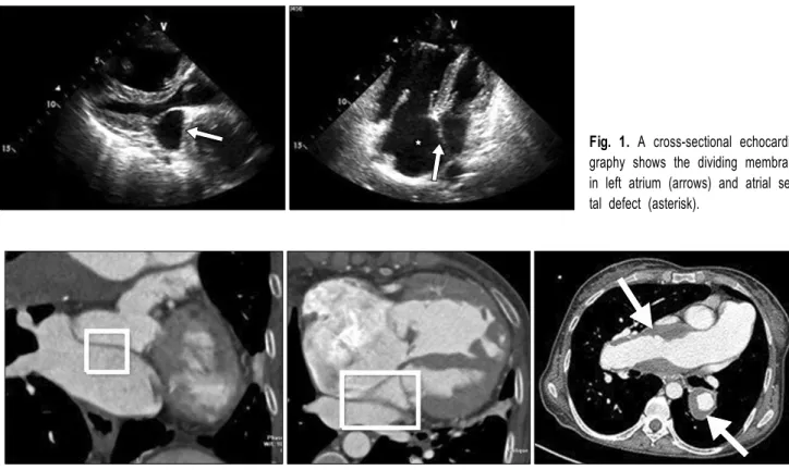
C

The Korean Society for Thoracic and Cardiovascular Surgery. 2011. All right reserved.

CC

This is an open access article distributed under the terms of the Creative Commons Attribution Non-Commercial License (http://creative- commons.org/licenses/by-nc/3.0) which permits unrestricted non-commercial use, distribution, and reproduction in any medium, provided the original work is properly cited.

Jun-Seok Kim, M.D.*, Sung-min Ko, M.D.**, Meong-Gun Song, M.D.*

We herein present a case of a successful correction of cor triatriatum associated with thrombotic pulmonary hyper- tension diagnosed in an adult female patient. We confirmed diagnosis using transthoracic and transesophageal echocardiography in addition to cardiac computed tomography and magnetic resonance imaging. Surgical repair comprised excision of the fibromuscular membranous septum in the left atrium, patch closure of an atrial septal defect, and reconstruction of the pulmonary arteries with a vascular graft. Cor triatriatum complicated pulmonary thrombotic hypertension with atrial septal defect is amenable to surgical correction with satisfactory results.

Key words: 1. Pulmonary arteries 2. Heart septal defects, atrial 3. Hypertension, pulmonary 4. Thrombosis

5. Cor triatriatum

CASE REPORT

A 44-year-old woman was admitted to our hospital for as- sessment of an over 10-year history of fatigue, severe dysp- nea on exertion, functional impairment class III (NYHA), and intermittent hemoptysis. When the patient was admitted to the hospital, she could not walk even 4 or 5 meters and had been misdiagnosed with Eisenmenger syndrome by various clinics. The patient also had clubbing of her fingers and toes.

During her physical examination, she displayed hypotension (91/60 mmHg) with a regular beat of 70 beats/min. Oxygen saturation on room air was approximately 76% and PaO2 41.8 mmHg based on an arterial blood gas analysis (ABGA).

Cardiac auscultation revealed a grade III systolic ejection

murmur at the left upper parasternal border and a slightly ac-

centuated second heart sound. An electrocardiogram showed a

sinus rhythm with a right bundle-branch block, right axis de-

viation, right atrial enlargement, and an S1Q3T3 pattern (an S

wave in lead I, a Q wave, and an inverted T wave in lead

III). Chest radiography displayed significant cardiomegaly, es-

pecially a right–sided heart enlargement and pulmonary ve-

nous congestion. A transthoracic two-dimensional echo-

cardiography (TTE) revealed an abnormal linear echo density

membrane, which divided the proximal superior common pul-

monary venous chamber and the distal true left atrial cavity

(Fig. 1). It also showed a moderate degree of tricuspid in-

sufficiency with severe pulmonary hypertension, in which the

right ventricle systolic pressure (RVSP) was estimated at 122

(2)

Fig. 1. A cross-sectional echocardio- graphy shows the dividing membrane in left atrium (arrows) and atrial sep- tal defect (asterisk).

Fig. 2. Computed tomography shows divided left atrium (square line) and huge thrombus (arrow) in pulmonary artery.

mmHg. This condition was verified by transesophageal echo- cardiography, which revealed cor triatriatum with a large at- rial septal defect (ASD) sized at 35 mm, which was commu- nicating between the right atrial chamber and both proximal and distal chambers of the left side. Prior to surgery, cardiac computed tomography (CT) and magnetic resonance imaging scans were performed. The procedures confirmed the diag- nosis of cor triatriatum, ASD, and severe pulmonary hyper- tension with pulmonary artery aneurysm containing a large intraluminal thrombus, which caused nearly total obstruction of both pulmonary artery passages (Fig. 2). Extremely dilated pulmonary arteries and left-side shifting of interventricular septum were visible, but otherwise the left ventricular func- tion was normal. The ratio of pulmonary blood flow to sys- temic blood flow (QP/QS) was 1.33. No further car- diovascular anomaly was depicted. The patient elected to un- dergo a surgical operation consisting of a median sternotomy.

After a cardiopulmonary bypass (CPB) was initiated, car- dioplegia was then induced under deep hypothermia by infus- ing a cold blood cardioplegic solution into the aortic root. A

large ASD was discovered, and the anomalous membrane in

the left atrium was examined through the ASD. The mem-

brane was successfully resected and the atrial septal defect

was then closed with a bovine pericardial patch. We sub-

sequently continued to perform a thrombectomy in the main

pulmonary artery (MPA) and in the right and left pulmonary

arteries (RPA & LPA), which were nearly obstructed by nu-

merous thrombi located even in the distal branches of both

pulmonary arteries. We also noticed severe dilatation sized at

5 cm in diameter from the distal MPA to both the RPA and

LPA, which were very firm and stiff on palpation. After re-

section of the extremely dilated MPA with both pulmonary

arteries, a complete thrombectomy was performed, even in

the distal branches of both pulmonary arteries by suction. A

24 mm vascular intergard interposition graft was then placed

in an end-to-end fashion between the main pulmonary artery

and the right pulmonary artery. The left pulmonary artery

was anastomosed successively to the 18 mm vascular inter-

position graft in an end-to-side fashion. At the microscopic

level, diffuse formations of fibrinoid thrombi were found in

(3)

Fig. 3. Histological findings of the resected pulmonary artery. Microscopically, diffuse formations of fibrinoid thrombi (asterisk) were found in the arterial wall showing fibrosclerotic degeneration (A, B). There were also infiltration of chronic inflammatory cells, medial fibrosis, and in- timal thickening in the cross-section of arterial walls, suggesting arteriosclerosis due to longstanding pulmonary hypertension (C, D).

Hematoxylin-Eosin stain, A, B: ×200, C, D: ×400.

the arterial wall showing fibrosclerotic degeneration. An in- filtration of chronic inflammatory cells, medial fibrosis, and intimal thickening in the cross-section of the arterial wall were also visible, which suggested the presence of arterio- sclerosis due to longstanding pulmonary hypertension (Fig. 3).

The pulmonary artery thrombectomy and reconstruction were performed in under 35 minutes of total circulatory arrest at 15.0

o

C. The cardiopulmonary bypass time was 290 minutes and aortic cross-clamping time was 202 minutes. Mechanical ventilation was applied postoperatively for 5 days. The pa- tient was transferred from the ICU to the general ward 13 days after the operation.

Pulmonary hypertension rapidly regresses after surgery.

Using a Swan-Ganz catheter to estimate systolic pulmonary

arterial pressure indicated 50∼55 mmHg within 3 days after

surgery, which decreased remarkably when compared to the

122 mmHg revealed by preoperative echocardiography. A

postoperative follow-up echocardiogram and CT revealed sat-

isfactory results by displaying no blood flow disturbance in

the left atrium, successful resection of the obstructing mem-

brane, and no remnants of thrombus in the pulmonary arteries

(Fig. 4). The patient’s oxygen saturation in room air was

98%, PaO

2

90 mmHg, and PH 7.44 checked using ABGA 10

days after surgery. Her exercise tolerance significantly im-

(4)

Fig. 4. Postoperative computed tomography shows successful resection of anom- alous partition (square line) and good circulation of pulmonary artery (arrows).

proved from 20∼30 meters of walking distance to 300 me- ters for 6 minutes by approximately 3 weeks after surgery.

She was prescribed Tracleer

(bosentan) to manage pulmo- nary hypertension and within the 6 months after the operation she was in good condition during periodic visits to an out- patient clinic.

DISCUSSION

Cor triatriatum is a rare congenital anomaly characterized by an abnormal separation of the left atrium into a proximal pulmonary venous chamber and a distal true chamber [1].

Approximately 75% of patients with such malformations die in infancy, because symptoms usually develop early, therefore surgical operation is requisite during the first year of a pa- tient’s life. When older patients present with chronic symp- toms, immediate surgical correction is also necessary [2]. The embryogenesis of cor triatriatum remains controversial and is usually explained by either entrapment theory, as proposed by Van Praagh and Corsini [3], or by a failed incorporation of the common pulmonary veins into the left atrium [4]. Though it is being diagnosed with increased frequency in adulthood, cor triatriatum remains a rare form of congenital heart

disease. In a review by Niwayama of 36 patients diagnosed with the disease, ten were older than 12 years of age [5].

Our 44-year-old patient represents one of the oldest reported

living patients. In this report, we present a 44-year-old wom-

an with cor triatriatum, a large atrial septal defect (ASD)

sized at 35 mm accompanied by chronic pulmonary artery

thrombosis. In about 75% of patients there is an atrial septal

defect between the right atrium and the proximal venous ac-

cessory chamber or the true distal atrium. However, cor tria-

triatum with severe pulmonary hypertension complicated by

chronic pulmonary artery thrombosis is very rare. Pulmonary

hypertension with an atrial septal defect might not have been

influenced by pulmonary venous obstruction because there

was a large atrial septal defect (ASD) of 35 mm, which

looked like a common atrium and communicated between the

right atrial chamber and both the proximal and distal cham-

bers on the left. Proximal pulmonary artery thrombosis has

been reported in patients with ASD and pulmonary artery hy-

pertension (PAH) [6]. In the present case, the thrombi were

considered to have formed in situ as there were no symptoms

suggesting systemic embolization of such large thrombi. The

aggravation of symptoms could have been caused by the in-

creased obstruction of blood flow by the thrombi and minute

(5)

normal coagulation factors, and platelet dysfunction have been determined to be the causes of thrombosis in secondary PAH [7]. The relatively sluggish blood flow in the dilated pulmonary arteries and polycythemia might have contributed to the formation of such large thrombi in this patient. The natural history of large in situ thrombi in the pulmonary ar- teries is unknown, as only a few case reports are available.

In patients with PAH and pulmonary embolism due to such large thrombi, acute worsening of symptoms with right heart failure may lead to death in weeks to months, or occasion- ally, sudden death may occur. In summary cor triatriatum is a rare disease associated with chronic thrombotic pulmonary hypertension that can be treated successfully if the diagnosis is done well and sufficient information is available preopera- tively.

2. Kouchoukos NT, Blackstone EH, Doty DB, Hanley FL, Karp RB. Cor triatriatum. In: Kouchoukos NT, Blackstone EH, Doty DB, Hanley FL, Karp RB. Kirklin/Barratt-Boyes cardiac surgery. 3rd ed. Philadelphia: Churchill Livingstone.

2003;781-9.

3. Van Praagh R, Corsini I. Cor triatriatum: pathologic anat- omy and a consideration of morphogenesis based on 13 postmortem cases and a study of normal development of the pulmonary vein and atrial septum in 83 human embryos.

Am Heart J 1969;78:379-405.

4. Loeffler E. Unusual malformation of the left atrium; pulmo- nary sinus. Arch Pathol (Chic) 1949;48:371-6.

5. Niwayama G. Cor triatriatum. Am Heart J 1960;59:291-317.

6. Schamroth CL, Sareli P, Pocock WA, et al. Pulmonary arte- rial thrombosis in secundum atrial septal defect. Am J Car- diol 1987;60:1152-6.

7. Archer S, Rich S. Primary pulmonary hypertension: a vascu- lar biology and translational research “Work in progress”.

Circulation 2000;102:2781-91.

수치

Fig. 1. A cross-sectional echocardio- echocardio-graphy shows the dividing membrane  in left atrium (arrows) and atrial  sep-tal defect (asterisk).
Fig. 3. Histological findings of the resected pulmonary artery. Microscopically, diffuse formations of fibrinoid thrombi (asterisk) were found in  the arterial wall showing fibrosclerotic degeneration (A, B)
Fig. 4. Postoperative computed tomography shows successful resection of anom- anom-alous partition (square line) and good circulation of pulmonary artery (arrows).

참조

관련 문서

GRIPHON Prostacyclin (PGI2) Receptor Agonist In Pulmonary Arterial Hypertension IPAH idiopathic pulmonary arterial hypertension MCTD mixed connective tissue

Percutaneous polymethylmethacrylate vertebroplasty in the treatment of osteoporotic vertebral body compression fractures: technical aspects. Pulmonary embolism caused

Four circular bone defects with a diameter of 8mm were formed on each of the calvaria of rabbits. Each defect in the calvarium was allocated to 1) control group,.. 2)

A and E, In control group, a small amount of new bone was observed at the margin of bone defect (40×); B and F, In experimental group 1, a large amount of new bone was formed

In 4-week group, the group filled with bone graft with decortication revealed larger new bone formation area than shown in the group that had a defect area

The defect area was filled with new-bone formation around the graft material hydroxy-apatite (open stars) at the periphery and chronic inflammation with

In analyzing those textbooks, 3 main focuses have been observed; an aspect of external appearance, a musical aspect as a practice material, and an

Under this rule, motor vehicle and motor vehicle equipment manufacturers will be required to include in their programs to remedy a safety-related defect or a noncompliance