INTRODUCTION
Mechanical ventilation has been used to support acutely ill patients for several decades. However, the possibility that mechanical ventilation can actually worsen acute lung injury is now widely accepted (1). More recently, what is now known as ventilator-induced lung injury (VILI) has been initially documented experimentally (2) and has received much atten- tion in the clinical fields (3, 4). VILI is mainly viewed as the result of excessive tidal volume, repeated opening and collapse of airways and inflammatory responses that an injurious ven- tilatory pattern may trigger. Broccard and colleagues (5) de- monstrated that animals ventilated with high tidal volume and positive end-expiratory pressure (PEEP) resulted in less extensive histologic change in the prone position than in the supine position. A number of factors could contribute to ben- eficial effects of prone position to alter dorsal lung transpul- monary pressures, including the compressive effects of con- solidated lung, direct transmission of the weight of abdomi- nal content and heart (6).
Lung injury appears to predispose patients to the develop- ment of a systemic inflammatory response that culminates in multiple organ dysfunction syndrome and death (7). Several studies have suggested that inflammatory cells and mediators play an important role in the pathogenesis of VILI (8, 9). Sup-
plementation (or blocking) of various inflammatory cytokines has been found to induce (or abrogate) lung injury (9). Behnia et al. (10) demonstrated that serum aspartate aminotransferase and LDH were significantly higher in the positive pressure ventilation with overdistension of the lungs compared with the control group.
The purpose of this study was 1) to examine serum levels of LDH in the supine and prone position during the VILI, 2) to determine whether the locations of VILI are influenced by body position, and 3) to evaluate whether prone position pre- vents the VILI.
MATERIALS AND METHODS Animals Preparation and Instrumentation
Care of the animals, techniques, and procedures were app- roved by the Animal Care and Use Committee of Chonnam National University Hospital. New Zealand white rabbits weighing 3.21±0.09 kg were anesthetized with an intramus- cular injection of a ketamine hydrochloride (30 mg/kg) and xylazine (0.3 mg/kg). An endotracheal tube (3.5 mm internal diameter) was inserted via a tracheostomy. Mechanical venti- lation was initiated in the pressure-controlled mode (IV-110B,
Sung Chul Lim, Yu Il Kim
Department of Internal Medicine, Chonnam National University Hospital, Gwangju, Korea
Address for correspondence Sung Chul Lim, M.D.
Department of Internal Medicine, Chonnam National University Hospital, 8 Hakdong, Dong-gu, Gwangju 501-757, Korea
Tel : +82.62-220-6574, Fax : +82.62-225-8578 E-mail : [email protected]
*This study was supported by the CUHRI-U-200342.
223
The Role of the Lactate Dehydrogenase and the Effect of Prone Position during Ventilator-induced Lung Injury
To examine the impact of lactate dehydrogenase (LDH) as an early marker of ven- tilator-induced lung injury (VILI) and the effect of prone position during the VILI, we ventilated 28 normal white rabbits (10 supine, 10 prone, 8 controls) for 6 hr or until PaO2/FIO2ratio was <200 mmHg. We applied an identical injurious ventilatory pat- tern (peak inspiratory pressure of 35 cmH2O with a PEEP of 3 cmH2O, I:E ratio of 1:2, and FIO2of 0.40) in the supine and prone group. VILI was assessed by oxygena- tion, gravimetric analysis and histologic grading. Serum levels of LDH progressively increased significantly during the VILI (supine and prone groups) as compared with controls. There was a significant negative correlation between oxygenation and LDH levels (r=-0.619, p<0.001). Wet weight/dry weight ratios (WW/DW) and histologic scores for dependent regions were significantly higher in the supine than the prone group. There were no differences in WW/DW and histologic scores for nondepen- dent regions between the supine and prone group. These findings suggest that serum LDH levels might be an early marker of severity of lung injury. The prone position resulted in a less severe and more homogenous distribution of VILI.
Key Words : Respiratory Distress Syndrome, Adult; Lung Injury, Acute; Ventilator-Induced Lung Injury;
Prone Position; L-Lactate Dehydrogenase
Received : 18 November 2003 Accepted : 12 January 2004
Sechrist infant ventilator, Sechrist industries, Anaheim, CA, U.S.A.), with a peak inspiratory pressure of 15 cmH2O, a PEEP of 3 cmH2O, a frequency of 25 breaths/min, an inspi- ration-to-expiration (I:E) ratio of 1:2, and an inspired oxygen fraction (FIO2) of 0.40. Anesthesia and paralysis were main- tained throughout the experiment by continuous infusions of sodium pentobarbital (2-4 mg/kg/hr) and pancuronium bromide (0.03-0.07 mg/kg/hr).
The internal carotid artery was cannulated with a 20-gauge catheter (Custom Product, Abbott Ireland, Sligo, Republic of Ireland) to monitor systemic arterial pressure and heart rate.
Arterial blood gas samples were analyzed at 37℃(Rapidlab 865, Chiron diagnostics corporation, U.K.) and corrected for body temperature. Rectal temperature was monitored and maintained within the range of 37±1℃using a radiant heat- ing lamp.
Experimental Protocol
After recording baseline hemodynamic and gas exchange, 28 rabbits were randomly assigned to one of three groups (10 supine, 10 prone, 8 control groups). We applied an identical injurious ventilatory pattern (peak inspiratory pressure of 35 cmH2O with a PEEP of 3 cmH2O, I:E ratio of 1:2, and FIO2of 0.40) for 6 hr or until PaO2/FIO2 ratio was <200 mm Hg to compare the extent of VILI occurring in the prone and supine position. Control groups were ventilated with same baseline settings (peak inspiratory pressure of 15 cmH2O, PEEP of 3 cmH2O, I:E ratio of 1:2, and FIO2of 0.40). Carbon dioxide was introduced into the inspiratory limb of the ven- tilator circuit, as necessary to maintain normocapnia (PaCO2, 35 to 45 mmHg) during the induction period. Arterial blood gases were measured at every 1 hr during the experiment.
Blood samples for LDH were obtained every 2 hr and mea- sured by enzyme-linked immunosorbent assay (Olympus, AU 5400, Japan). After completion of the protocol, the heart-lung
block was then removed after exsanguinations of the hep- arinized animals.
Gravimetric analysis
Heparinization and exsanguinations were used to minimize the effect of residual blood on gravimetric indices. Left lung was physically sectioned into dorsal and ventral regions that were weighed separately to obtain wet weights (WW) and then desiccated in a vacuum oven at 60℃to dryness for 4-5 days until a constant dry weight (DW).
Histologic Scoring
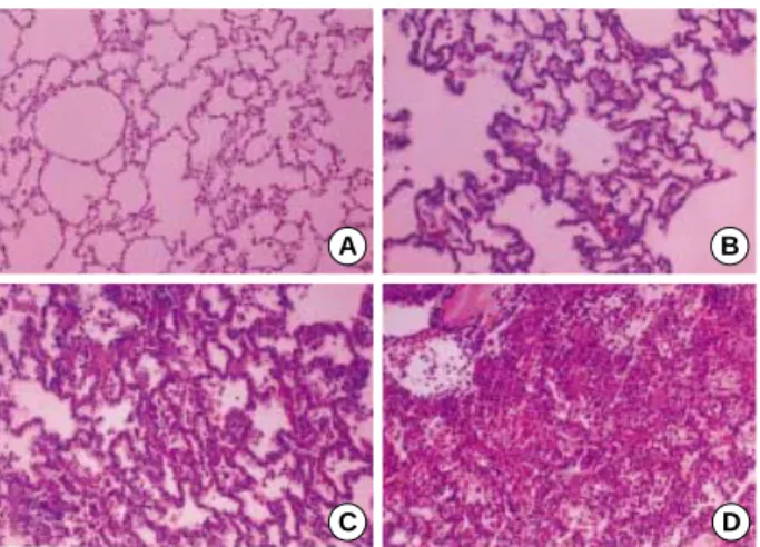
The resected lungs were infused with 4% paraformaldehyde solution from a tracheostomy tube. Subsequently, the trachea was clamped and the lungs were placed in 4% buffered para- formaldehyde solution. The lungs were divided into dorsal and ventral regions along a coronal plane connecting the mid- height caudal and cranial portions of the lungs. Subdivision into caudal and cranial sectors was accomplished by section- ing the lungs (in the transverse plane) along upper and lower lobe fissures. To avoid sampling bias, tissue samples of 3 L were excised (regardless of macroscopic appearance) from the mid-portion of each sector. The samples were sectioned and stained (hematoxylin and eosin). The sections were later char- acterized using a semiquantitative grading system by a pathol- ogist who was blinded as to the experimental protocol and region of sampling. After viewing approximately ten fields per sector under low and high power, each section was assigned a numerical histologic score ranging from 1 to 4, based on the degree of neutrophilic infiltration, hemorrhage, and edema in the interstitial and alveolar spaces as follows (Fig. 1). 1 (normal), normal appearing lung; 2 (mild), mild congestion, interstitial edema, and interstitial neutrophilic infiltrate with occasional red blood cells and neutrophils in the alveolar spaces;
3 (moderate), moderate congestion and interstitial edema with neutrophils partially filling the alveolar spaces but without consolidation; 4 (severe), marked congestion and interstitial edema, with neutrophilic infiltrate nearly filling the alveolar spaces, or with frank lung consolidation. Atelectasis per se was disregarded and not scored as abnormality.
Statistical analysis
All values are reported as mean±SD, unless specified other- wise. Separate one-way analysis of variance (ANOVA) proce- dures were used to test for differences in oxygenation and serum levels of LDH. In each group, changes in measured vari- ables over time were analyzed using a repeated-measures anal- ysis of variance. A statistical package, SPSS 11.0 for Windows (SPSS Inc., Chicago, IL, U.S.A.), was used to analyze the data.
A value of p<0.05 was considered significant.
Fig. 1.Representative examples of the histologic scores (HS). (A) HS 1: normal; (B) HS 2: mild; (C) HS 3: moderate; (D) HS 4: severe.
Hematoxylin and eosin, magnification (×200).
B A
C D
RESULTS
Changes in Hemodynamics and Oxygenation
There were no significant differences in oxygenation and systemic arterial pressure among the groups under baseline conditions. Systemic arterial pressure trended downward dur- ing the VILI in the prone as compared with supine group.
However, there were no significant differences in systemic arterial pressure between supine and prone group. In the supine group, PaO2decreased significantly after 2 hr as compared with baseline values (181.1±26.0 vs. 141.9±46.3 mm Hg;
p<0.05). PaO2decreased significantly after 3 hr as compared with baseline values in the prone group (182.0±19.2 vs.
137.1±47.2 mm Hg; p<0.05). Although PaO2tended to
be lower in the supine than prone group during the VILI, this change did not reach significance between two groups at end of experiment. PaO2remained stable for 6 hr in the control group (Fig. 2).
Relationship between Serum levels of LDH and Oxygenation
Serum levels of LDH elevated progressively during the ex- periment in injurious ventilatory pattern (supine and prone position) when compared with the control groups (Fig. 3).
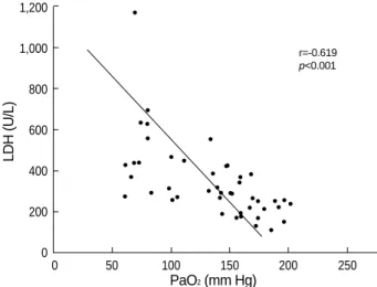
There were no significant differences of serum levels of LDH between supine and prone position at the end experiment (supine: 679±423 U/L; prone: 435±174 U/L; p=0.51). In the control group, the trend toward elevated levels of LDH with time was not statistically significant when compared with baseline values. There was a significant negative corre- lation between oxygenation and LDH levels (r=-0.619, p<
0.001) (Fig. 4).
Gravimetrics
The ratio of wet weight to dry weight (WW/DW) for de- pendent and nondependent regions was significantly greater in injurious ventilatory pattern (supine and prone) than the control group. The WW/DW for dependent regions was significantly higher in the supine than prone group. There were no differences in WW/DW for nondependent regions between supine and prone group. When dependent and non- dependent regions were compared in the same animal, regional WW/DW was significantly greater in dependent in the supine group (dependent: 6.36±0.72; nondependent: 4.78±0.37;
p<0.01). In the prone group, however, no significant differ- ences was found in WW/DW between dependent and nonde-
PaO2(mm Hg)
Time (hr) 200
180 160 140 120 100 80 60 40 20 0
0 1 2 3 4 5 6
Control
Prone
Supine
*
*
*
*
*
* *
*
*
Fig. 2. Comparisons of oxygenation among the groups. PaO2dec- reased significantly after 2 and 3 hr in the supine and prone groups as compared with baseline values, respectively. Data represent the mean±SD. *p<0.05 versus baseline values.
Control Prone Supine
*
*
Fig. 3. Comparisons of lactate dehydrogenase among the groups.
Serum levels of LDH elevated progressively during the VILI (supine and prone position) when compared with control groups. Data represent the mean±SD. *p<0.05 versus baseline values.
LDH (U/L)
Time (hr) 700
600
500
400
300
200
100
00 2 4 6
*
*
Fig. 4. Correlation between lactate dehydrogenase and oxygena- tion. There was a significant negative correlation between oxygena- tion and LDH levels.
LDH (U/L)
PaO2(mm Hg) 1,200
1,000
800
600
400
200
0
0 50 100 150 200 250
r=-0.619 p<0.001
pendent regions (dependent: 4.34±1.20; nondependent:
4.32±1.49; p=0.56) (Fig. 5).
Histology
Gross postmortem inspection of the lungs in the supine group revealed edema and hemorrhage that were most promi- nent in the dependent regions. These changes appeared much less intense in the prone position. Histologic scores for depen- dent and nondependent were significantly higher in injurious ventilatory pattern (supine and prone) as compared with the control group (Fig. 6). In the dependent regions, mean histo- logic scores were significantly higher in the supine than the prone group (supine: 3.06±0.36; prone: 2.20±0.27; p=0.04).
In the nondependent regions, however, mean histologic scores did not differ in both groups (supine: 2.31±0.12; prone:
2.01±0.46; p=0.52). Histologic scores were greater in depen- dent than in nondependent regions for animals ventilated in the supine group (p=0.02). In the prone group, however, no significant differences was found in histologic scores between dependent and nondependent regions (p=0.44).
DISCUSSION
The main findings of this study were that in the VILI mod- els 1) PaO2tended to be lower in the supine than prone group during the VILI. However, this change did not reach signifi- cance between two groups at end of experiment. PaO2remain- ed stable for 6 hr in control group; 2) Serum levels of LDH progressive increased during the induction of VILI in the supine and prone position. There was a significant negative correlation between oxygenation and serum levels of LDH;
3) WW/DW and histologic scores for dependent and nonde-
pendent regions were significantly greater in the injurious ventilatory pattern (supine and prone group) than in the con- trol group. Dependent regions with tissue damage and edema observed in the supine were attenuated by the prone group.
Despite advance in critical care, the mortality rate in patients with acute respiratory distress syndrome (ARDS) remains high at values exceeding 30-40%. Furthermore, most patients who die do so from multiple system organ failure rather than from hypoxia (7). One hypothesis that has recently been ad- vanced to explain this observation is that mechanical ventila- tion per se may be not only exacerbation of underlying lung injury but may also lead to the development of a systemic inflammatory response syndrome (2, 8, 9) and multiple sys- tem organ failure (11, 12).
Two mechanisms are thought to contribute to the develop- ment of VILI: overdistension of the alveoli and shearing forces caused by the stress that occurs where collapsed areas of the lungs are reopened by positive pressure. Exactly how mechani- cal ventilation induces its deleterious effects is as of yet unclear.
Studies in vitro and in vivo have found that both the pattern and the degree of stretch are important in determining cellular response (9, 12). Supplementation (or blocking) of various inflammatory cytokines has been found to induce (or abrogate) lung injury (13). Ranieri et al. (14) demonstrated that the con- centrations of proinflammatory cytokines in both bronchoalve- olar lavage (BAL) and plasma could indeed be decreased in patients ventilated with a lung-protective strategy. A report showed that serum aspartate aminotransferase and LDH were significantly higher in the positive pressure ventilation with overdistension of the lungs compared with the control group (10). These results suggested that serum LDH might be early markers of ventilator-induced lung injury in the rat model.
Our results showed that serum levels of LDH elevated pro- gressively during the experiment in the injurious ventilat-
WW/DW
8
7
6
5
4
3
2
2
0
Dependent Nondependent
*
�
�
Fig. 5. Wet weight to dry weight (WW/DW) ratios among the groups.
Each values represent mean±SD. *p<0.05 versus prone; �p<0.05 versus supine or prone.
Supine Prone Contol
Histologic Score
4
3
2
1
0
Dependent Nondependent
*
�
�
Fig. 6. Histologic scores among the groups. Each values represent mean±SD. *p<0.05 versus prone; �p<0.05 versus supine or prone.
Supine Prone Contol
ory pattern, whereas serum levels of LDH remained stable for 6 hr in the control groups. There was a significant nega- tive correlation between oxygenation and LDH levels.
Patients with ARDS are frequently kept in the supine posi- tion for days to weeks. However, many researchers suggest that the primary horizontal posture should be prone rather than the supine (15). The prone position has been proposed as a relatively simple maneuver to improve oxygenation (16), and an increased end-expiratory lung volume was originally suggested as the main mechanism for oxygenation improve- ment (17). Several studies found that the prone position causes a venous admixture reduction and a more even ventilation distribution, without affecting end-expiratory lung volume (18, 19). More recently, experimental reports demonstrated that the prone position may be reducing the VILI (5, 20).
Broccard and colleagues (20) demonstrated that lung injury induced solely by mechanical forces, the prone position resulted in a less severe and more homogeneous distribution of VILI in the dog models. These observations are consistent with our results. In our VILI model, we observed a dependent predom- inance of tissue edema and histologic change in the supine position. Dependent and nondependent scores were similar in the prone position. However, the exact mechanism for the attenuated lung injury during prone ventilation remains spec- ulative. In the present study we found that PaO2tended to be lower in the supine than prone group during the VILI. How- ever, this change did not reach significance between two groups at the end of experiment. PaO2remained stable for 6 hr in the control group. Based on our results, we suggest that reduction in tidal volume is the more important therapeutic intervention for the significant reduction of VILI. Experi- mentally, use of high levels of PEEP (21), partial liquid ven- tilation (22), and proinflammatory cytokine neutralization (23) might be reducing VILI, but clinical proofs of efficacy are still lacking.
Despite its interesting results, this study was limited in several ways. In particular, our model did not evaluate long- term effects of prone position. Further investigation is need- ed to clarify the effects of mechanical ventilation on the lungs during periods of longer duration. Imai et al. (24) demonstrat- ed that mechanical ventilation can lead to epithelial cell apop- tosis in the kidney and small intestine, accompanied by bio- chemical evidence of organ dysfunction. Further experimen- tal and clinical evidence will be required to explain VILI cor- related with multiple organ dysfunction syndrome and exam- ine other potential strategies for prevention the VILI.
REFERENCES
1. Parker JC, Hernandez LA, Peevy KJ. Mechanisms of ventilator-induc- ed lung injury. Crit Care Med 1993; 21: 131-43.
2. Dreyfuss D, Saumon G. Ventilator-induced lung injury: lessons from experimental studies. Am J Respir Crit Care Med 1998; 157: 294-323.
3. Amato MB, Barbas CS, Medeiros DM, Magaldi RB, Schettino GP, Lorenzi-Filho G, Kairalla RA, Deheinzelin D, Munoz C, Oliveira R, Takagaki TY, Carvalho CR. Effect of a protective-ventilation strategy on mortality in the acute respiratory distress syndrome. N Engl J Med 1998; 338: 347-54.
4. Brochard L, Roudot-Thoraval F, Roupie E, Delclaux C, Chastre J, Fernandez-Mondejar E, Clementi E, Mancebo J, Factor P, Matamis D, Ranieri M, Blanch L, Rodi G, Mentec H, Dreyfuss D, Ferrer M, Brun-Buisson C, Tobin M, Lemaire F. Tidal volume reduction for prevention of ventilator-induced lung injury in acute respiratory dis- tress syndrome. The Multicenter Trial Group on Tidal Volume reduc- tion in ARDS. Am J Respir Crit Care Med 1998; 158: 1831-8.
5. Broccard AF, Shapiro RS, Schmitz LL, Ravenscraft SA, Marini JJ.
Influence of prone position on the extent and distribution of lung injury in a high tidal volume oleic acid model of acute respiratory distress syndrome. Crit Care Med 1997; 25: 16-27.
6. Pelosi P, D’Andrea L, Vitale G, Pesenti A, Gattinoni L. Vertical gra- dient of regional lung inflation in adult respiratory distress syndrome.
Am J Respir Crit Care Med 1994; 149: 8-13.
7. Montgomery AB, Stager MA, Carrico CJ, Hudson LD. Causes of mortality in patients with the adult respiratory distress syndrome. Am Rev Respir Dis 1985; 132: 485-9.
8. Ghosh A, Greenberg ME. Calcium signaling in neurons: molecular mechanisms and cellular consequences. Science 1995; 268: 239-47.
9. Slutsky AS. Lung injury caused by mechanical ventilation. Chest 1999; 116: 9S-15S.
10. Behnia R, Molteni A, Waters CM, Panos RJ, Ward WF, Schnaper HW, TS’Ao CH. Early markers of ventilator-induced lung injury in rats. Ann Clin Lab Sci 1996; 26: 437-50.
11. Slutsky AS, Tremblay LN. Multiple system organ failure. Is mechan- ical ventilation a contributing factor? Am J Respir Crit Care Med 1998; 157: 1721-5.
12. Tremblay LN, Slutsky AS. Ventilator-induced injury: from barotrau- ma to biotrauma. Proc Assoc Am Physicians 1998; 110: 482-8.
13. Chernoff AE, Granowitz EV, Shapiro L, Vannier E, Lonnemann G, Angel JB, Kennedy JS, Rabson AR, Wolff SM, Dinarello CA. A ran- domized, controlled trial of IL-10 in humans. Inhibition of inflamma- tory cytokine production and immune responses. J Immunol 1995;
154: 5492-9.
14. Ranieri VM, Suter PM, Tortorella C, De Tullio R, Dayer JM, Brienza A, Bruno F, Slutsky AS. Effect of mechanical ventilation on inflam- matory mediators in patients with acute respiratory distress syndrome:
a randomized controlled trial. JAMA 1999; 282: 54-61.
15. Messerole E, Peine P, Wittkopp S, Marini JJ, Albert RK. The prag- matics of prone positioning. Am J Respir Crit Care Med 2002; 165:
1359-63.
16. Piehl MA, Brown RS. Use of extreme position changes in acute res- piratory failure. Crit Care Med 1976; 4: 13-4.
17. Douglas WW, Rehder K, Beynen FM, Sessler AD, Marsh HM. Im- proved oxygenation in patients with acute respiratory failure: the prone position. Am Rev Respir Dis 1977; 115: 559-66.
18. Albert RK, Leasa D, Sanderson M, Robertson HT, Hlastala MP. The prone position improves arterial oxygenation and reduces shunt in oleic-acid-induced acute lung injury. Am Rev Respir Dis 1987; 135:
628-33.
19. Lamm WJ, Graham MM, Albert RK. Mechanism by which the prone position improves oxygenation in acute lung injury. Am J Respir Crit Care Med 1994; 150: 184-93.
20. Broccard A, Shapiro RS, Schmitz LL, Adams AB, Nahum A, Marini JJ. Prone positioning attenuates and redistributes ventilator-induced lung injury in dogs. Crit Care Med 2000; 28: 295-303.
21. Dreyfuss D, Soler P, Basset G, Saumon G. High inflation pressure pulmonary edema. Respective effects of high airway pressure, high tidal volume, and positive end-expiratory pressure. Am Rev Respir Dis 1988; 137: 1159-64.
22. Dreyfuss D, Martin-Lefevre L, Saumon G. Hyperinflation-induced lung injury during alveolar flooding in rats: effect of perfluorocarbon instillation. Am J Respir Crit Care Med 1999; 159: 1752-7.
23. Bone RC, Fisher CJ Jr, Clemmer TP, Slotman GJ, Metz CA. Early methylprednisolone treatment for septic syndrome and the adult res- piratory distress syndrome. Chest 1987; 92: 1032-6.
24. Imai Y, Parodo J, Kajikawa O, de Perrot M, Fischer S, Edwards V, Cutz E, Liu M, Keshavjee S, Martin TR, Marshall JC, Ranieri VM, Slutsky AS. Injurious mechanical ventilation and end-organ epithe- lial cell apoptosis and organ dysfunction in an experimental model of acute respiratory distress syndrome. JAMA 2003; 289: 2104-12.