CASE REPORT
J Korean Surg Soc 2011;81:281-286
http://dx.doi.org/10.4174/jkss.2011.81.4.281
JKSS
Journal of the Korean Surgical Society pISSN 2233-7903ㆍeISSN 2093-0488
Received December 2, 2010, Revised January 19, 2011, Accepted February 7, 2011 Correspondence to: Kyung Sik Kim
Department of Surgery, Yonsei University College of Medicine, 250 Seongsan-ro, Seodaemun-gu, Seoul 120-752, Korea Tel: +82-2-2228-2125, Fax: +82-2-313-8289, E-mail: [email protected]
cc Journal of the Korean Surgical Society is an Open Access Journal. All articles are distributed under the terms of the Creative Commons Attribution Non-Commercial License (http://creativecommons.org/licenses/by-nc/3.0/) which permits unrestricted non-commercial use, distribution, and reproduction in any medium, provided the original work is properly cited.
Synchronous double primary cancers associated with a choledochal cyst and anomalous pancreaticobiliary
ductal union
Kang Kook Choi, Sae Byeol Choi
1, Seung Woo Park
2, Hyun Ki Kim
3, Young Nyun Park
3, Kyung Sik Kim
Department of Surgery, Yonsei University College of Medicine, 1Department of Surgery, Korea University College of Medicine, Departments of 2Internal Medicine and 3Pathology, Yonsei University College of Medicine, Seoul, Korea
A 60-year-old female was admitted with epigastric pain lasting a month. Preoperative diagnosis was choledochal cyst with anomalous pancreaticobiliaryductal union (APBDU), C-P type. A papillary mass measuring 2.5 × 1.9 cm was found adjacent to the pancreaticocholedochal junction. Gallbladder (GB) cancer was also observed. Pyloric-preserving pancreatico- duodenectomy (PPPD) was performed. The patient received adjuvant chemotherapy/radiation therapy on the tumor bed.
The gallbladder cancer showed serosal invasion, while the bile duct cancer extended into the pancreas. Although common bile duct (CBD) cancer lesion showed focally positive for p53 and the gallbladder cancer lesion showed negative for p53, the Ki-67 labeling index of the CBD cancer and GB cancer were about 10% and 30%, respectively. Nine months after curative re- section, a stricture on the subhepatic colon developed due to adjuvant radiation therapy. Localized peritoneal seedings were incidentally found during a right hemicolectomy. The patient underwent chemotherapy and had no evidence of tumor re- currence for two years after PPPD.
Key Words: Choledochal cyst, Gallbladder neoplsms, Bile duct neoplasms, Synchronous multiple primary neoplasms
INTRODUCTION
In anomalous pancreaticobiliary ductal union (APBDU), the common channel is abnormally long and the con- nection between the choledochus and pancreatic duct is outside of the duodenal wall [1]. This congenital disorder is frequently associated with choledochal cysts, another congenital condition consisting of localized or diffused di- latation of the biliary tract [1]. Moreover, APBDU are
prone to have benign as well as malignant complications including carcinoma of biliary tract [2].
Although several cases of single carcinoma in the biliary tract associated with APBDU have been reported, syn- chronous double cancer is very rare [3]. We describe here a case of a choledochal cyst combined with APBDU that was first detected in a patient in her sixties as a double pri- mary cancer of the gallbladder and CBD.
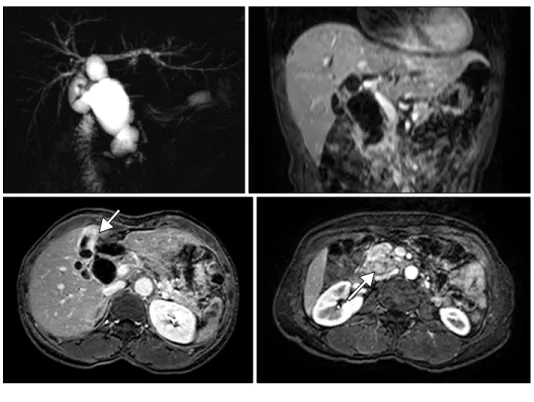
Fig. 1. Magnetic resonance cholan- giopancreatography (MRCP) image including vertical and horizontal sections. MRCP shows fusiform cystic changes involving the entire common bile duct and hilar bile duct compatible with a choledochal cyst (type Ⅰa) associated with the long common channel of the pancrea- ticobiliary junction (C-P type). Irre- gular wall thickening at the fundus and body of the gallbladder was seen. A 2.5 × 1.9 cm mass is also seen around the pancreaticobiliary junc- tion.
Fig. 2. Specimens from patient before and after fixation. The com- mon bile duct (CBD) is markedly dilated, measuring 7 cm in inner circumference and 6.5 cm in length.
Opening along the CBD revealed an ill-defined mass measuring 2 × 1.5 cm. The gallbladder shows an ill-defined thickened wall area in the fundus measuring 3 × 3 cm. GB, gallbladder.
CASE REPORT
A 60-year-old female was admitted with a one month history of epigastric pain. Her medical records indicated she had taken medication for hypertension. Upon admit- tance, right upper quardrant pain were found and the oth- er symptoms including fever and weight loss were absent.
On abdominal physical examination, no palpable mass or tenderness was discovered. Laboratory results revealed a total bilirubin level of 1.2 mg/dL, alanine aminotrans-
ferase of 201 IU/L, aspartate aminotransferase of 318 IU/L, and alkaline phosphatase of 126 IU/L. The levels of tumor markers carcinoembryonic antigen and carbohydrate an- tigen 19-9 were 6.69 ng/mL and 0.1 U/mL, respectively.
Magnetic resonance imaging and magnetic resonance cholangiopancreatico-graphy identified a choledochal cyst with APBDU, choledocho-pancreatic (C-P) type. An irregular-shaped papillary mass measuring 2.5 × 1.9 cm was found adjacent to the pancreaticocholedochal junc- tion with pancreatic invasion and the irregular wall thick-
Fig. 3. Hematoxylin & Eosin stain image of tumor. Tumor shows polypoid growth into the common bile duct (CBD) lumen with pancreatic invasion. (H&E; A, ×40; B, ×100). Adenocarcinoma of the gallbladder invaded fibromuscular connective tissue (H&E; C, ×40; D, ×100).
ening at the fundus and body of gallbladder was also seen.
There was no lymph node enlargement or distant meta- stasis (Fig. 1).
Pylorus preserving pancreaticoduodenectomy (PPPD) was performed without additional hepatic resection. The proximal resection margin was at the level of the common hepatic duct bifurcation. Lymph node dissection was per- formed around the hepatoduodenal ligament, common hepatic artery, celiac axis, and paraaortic area.
On pathologic evaluation, the CBD was markedly di- lated, measuring 7 cm in inner circumference and 6.5 cm in length. Upon opening, we found an ill-defined yellowish tan solid mass abutting on the ampulla of Vater, measur- ing 2 × 1.5 × 1.5 cm. The gallbladder showed an ill-defined thickened wall area in the fundus, measuring 3 × 3 cm (Fig.
2). Microscopic examination revealed moderately-differ- entiated adenocarcinoma of the CBD with extension to the pancreatic head and moderately-differentiated adenocar- cinoma of the gallbladder with extension to the serosa (Fig. 3). Only one regional node was involved by the tumor out of a total of 27 resected lymph nodes. There is no K-ras mutation on codon 12 and 13 in both gallbladder and CBD cancer lesions. In immunohistochemical staining with p53 and Ki-67, although CBD cancer lesion showed focally positive for p53 and the gallbladder (GB) cancer lesion showed negative for p53, the Ki-67 labeling index of the CBD cancer and GB cancer were about 10% and 30%, re- spectively (Fig. 4).
After an uneventful postoperative course, the patient received adjuvant chemotherapy with cisplatin, tega-
Fig. 4. Immunostaining of tumor. p53 and Ki-67 immunostaining (×100; A, B, common bile duct cancer; C, D, gallbladder cancer) and K-ras mutation analysis (E).
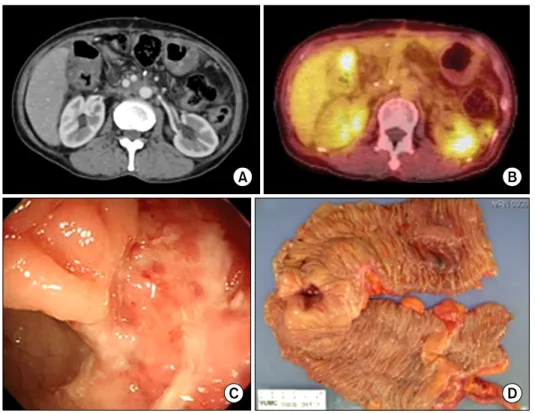
fur-uracil (UFT), and radiation therapy on the tumor bed of the gallbladder with 5,040 cGy. Nine months after cura- tive resection, a stricture developed on the subhepatic co- lon due to adjuvant radiation therapy (Fig. 5). Localized peritoneal seedings were incidentally found during right hemicolectomy. The patient underwent chemotherapy-
with 5FU, Leucovorin and oxaliplatin 6 times. There is no evidence of tumor recurrence for 29 months after Rt. hemi- colectomy with chemotherapy.
Fig. 5. Images of computed tomo- graphy (CT), positron emission tomography (PET), colonoscopy and gross specimen for a com- plication caused by adjuvant radia- tion therapy. A stricture is shown on the subhepatic colon. (A, CT; B, PET;
C, colonoscopy; D, gross specimen).
DISCUSSION
The mode of anomalous union is classified into two types: the pancreatico-choledochal (P-C) type, in which the main pancreatic duct enters the common bile duct, and the C-P type, where the CBD enters the main pancreatic duct [1]. The incidence of P-C type ductal junctions is high- er in patients with GB cancer [4], while the incidence of C-P type junctions is higher in patients with carcinoma in con- genital cystic dilatation [5]. In our experience, seven out of 10 patients with gallbladder cancer associated with APBDU have the P-C type [6].
APBDU is frequently associated with choledochal cysts and the incidence of malignancy in choledochal cysts ranges from 2.5 to 26% [3]. The number of bile duct cancer cases exceeds the number of cases of gallbladder cancer in the dilated bile duct with APBDU. Gallbladder cancer fre- quently occurs, but bile duct cancer also arises, in APBDU not associated with dilatation [7]. According to recent 10-year data from the Japanese Study Group on Pancreati- cobiliary Maljunction, however, the rate of gallbladder cancer is higher than bile duct cancer with biliary dilata- tion [3]. The incidence of gallbladder cancer associated
with APBDU is reported to range from 16.7 to 18.3% [4].
The gallbladder experiences conditions with signifi- cantly greater malignant potential in patients with APBDU, especially in cases not associated with chol- edochal cysts. Neoplastic development in the bile duct of patients with APBDU evolves through a multistep process associated with hyperproliferation and genetic alterations [3]. In our case, the K-ras mutation was not observed at any site of cancer. However, for epithelial cells in both cancer- ous areas with positive Ki-67, it might have caused anoth- er signal regulation apart from K-ras gene.
Early resection of the cyst wall and bile duct with re- construction of the biliary ducts is been recommended. To prevent biliary tract cancers, APBDU with cystic dilata- tions should be excised during childhood. It is usually a surgical disease of infancy or childhood, but 20 to 30% of patients are first diagnosed in adulthood due to incidental findings during medical check-ups or late onset of symp- toms [2]. In the present case, the patient had a choledochal cyst and APBDU without symptoms until her 60s.
Although she was symptom-free, the congenital anoma- lies of this patient eventually developed into biliary tract cancers of the gallbladder and CBD, both compatible with
stage IIIB according to the 7th American Joint Committee on Cancer tumor-node-metastasis classification system.
Radical cholecystectomy, removal of the GB with en-bloc subsegmental resection of the adjacent hepatic paren- chyma of segments IVb and V plus a regional lymphade- nectomy, is suggested for T2 or more advanced GB cancer [8]. Surgeons who prefer aggressive operation often per- form PPPD plus hepatectomy for GB cancer, and the hos- pital mortalities of these cases were higher (10 to 15%) than those (2 to 5%) of PPPD or major hepatectomy [8].
Although the role of radiation therapy in patients with biliary malignancies remains a debate, favorable survivals have been reported in postoperative adjuvant radiation therapy for GB cancer with lymph node invasion [9].
Because simple cholecystectomy with external radio- therapy can be recommended as an alternative treatments, only cholecystectomy with adjuvant chemoradiation ther- apy performed for GB cancer in current report [10].
In conclusion, choledochal cysts and APBDU are closely associated biliary tract anomalies. The malignant poten- tial of these disease entities usually requires early surgical intervention and life-long follow-up.
CONFLICTS OF INTEREST
No potential conflict of interest relevant to this article was reported.
REFERENCES
1. The Japanese Study Group on Pancreaticobiliary Maljunc- tion (JSPBM). The Committee of JSPBM for Diagnostic Criteria. Diagnostic criteria of pancreaticobiliary maljunc- tion. J Hepatobiliary Pancreat Surg 1994;1:219-21.
2. Yamaguchi M. Congenital choledochal cyst. Analysis of 1,433 patients in the Japanese literature. Am J Surg 1980;
140:653-7.
3. Funabiki T, Matsubara T, Miyakawa S, Ishihara S.
Pancreaticobiliary maljunction and carcinogenesis to bili- ary and pancreatic malignancy. Langenbecks Arch Surg 2009;394:159-69.
4. Sandoh N, Shirai Y, Hatakeyama K. Incidence of anom- alous union of the pancreaticobiliary ductal system in bili- ary cancer. Hepatogastroenterology 1997;44:1580-3.
5. Jan YY, Chen HM, Chen MF. Malignancy in choledochal cysts. Hepatogastroenterology 2002;49:100-3.
6. Kang CM, Kim KS, Choi JS, Lee WJ, Kim BR. Gallbladder carcinoma associated with anomalous pancreaticobiliary duct junction. Can J Gastroenterol 2007;21:383-7.
7. Tashiro S, Imaizumi T, Ohkawa H, Okada A, Katoh T, Kawaharada Y, et al. Pancreaticobiliary maljunction: retro- spective and nationwide survey in Japan. J Hepatobiliary Pancreat Surg 2003;10:345-51.
8. Pitt HA. Gallbladder cancer: what is an aggressive ap- proach? Ann Surg 2005;241:395-6.
9. Cho SY, Kim SH, Park SJ, Han SS, Kim YK, Lee KW, et al.
Adjuvant chemoradiation therapy in gallbladder cancer. J Surg Oncol 2010;102:87-93.
10. Mondragón-Sánchez R, González-Geroniz M, Oñate- Ocaña LF, Garduño-López AL, Mondragón-Sánchez A, Bernal-Maldonado R, et al. A retrospective analysis of pa- tients with gallbladder cancer treated with radical re- section versus cholecystectomy plus external radiotherapy.
Hepatogastroenterology 2003;50:1806-10.