bloodresearch.or.kr Blood Res 2015;50:54-65.
Letters to the Editor 55
a total leukocyte count (TLC) of 2.9×10⁹/L. On day 70, he completed his second phase of induction chemotherapy and was started on G-GSF therapy for severe neutropenia (TLC: 0.76×10⁹/L). On day 73, his peripheral blood smear showed marked leukopenia with a predominance of lym- phocytes and 2% blasts without any left shift or increased granulations. This lack of prototypical G-CSF response along with previous MRD positivity and presence of 2% blasts led to a suspicion of relapse, and bone marrow examination was advised. Marrow aspirate showed hypocellular marrow with 14% blasts, and focal areas showed up to 25% blasts (Fig. 1B), along with a lack of a maturing myeloid compo- nent, which suggested relapse. Trephine biopsy, in contrast, showed hypocellularity with only a few scattered CD34 positive blasts. This necessitated flow cytometric evaluation, where the blasts showed negativity for B lymphoblastic markers and expressed myeloid markers, including CD13, CD33, and CD34, suggesting that they were regenerating myeloblasts (Fig. 1C-F). No further therapy was ad- ministered and the patient was kept on close hematological follow-up. The counts recovered after 4 days, with the typi- cal responses of a left shift and increased granulation.
Follow-up marrow examination after complete recovery of blood counts, 10 days after discontinuing G-CSF therapy, revealed cellular marrow elements with 4% blasts (regenerating) and negative MRD (<0.01%).
Post-G-CSF blastocytosis is a problematic and confusing issue that is difficult to differentiate from disease recurrence and hemopoietic recovery. In our case, there were regenerat- ing blasts (>20%) in the bone marrow, with an absence of telltale G-CSF-induced changes, such as presence of im- mature granulocytes, monocytosis, or prominent gran- ulations on peripheral smear. To conclude, the regenerating blast percentage may increase after G-CSF therapy to >20%, without concomitant increases in other mature myeloid precursors.
The lag time before complete response to G-CSF therapy may be as long as 10 days, as seen in our case, posing potential problems in management and a challenge in therapeutic decisions. A careful morphological assessment of the blasts at the time of diagnosis, aided by awareness of these G-CSF-associated aberrant patterns and flow cytometric characterization can help guide management decisions.
Ritesh Sachdev, Tushar Sahni, Smeeta Gajendra, Bhawna Jha, Shalini Goel, Pranav Dorwal
Department of Pathology and Laboratory Medicine, Medanta-The Medicity, Gurgaon, India
Correspondence to: Ritesh Sachdev Department of Pathology and Laboratory Medicine,
Medanta-The Medicity, Sector-38, Gurgaon, Haryana 122 001, India E-mail: [email protected]
Received on Jul. 18, 2014; Revised on Aug. 5, 2014; Accepted on Feb. 12, 2015
http://dx.doi.org/10.5045/br.2015.50.1.54
AuthorsÊ Disclosures of Potential Conflicts of Interest No potential conflicts of interest relevant to this article were reported.
REFERENCES
1. Liu CZ, Persad R, Inghirami G, et al. Transient atypical mono- cytosis mimic acute myelomonocytic leukemia in post-chemo- therapy patients receiving G-CSF: report of two cases. Clin Lab Haematol 2004;26:359-62.
2. Arici M, Hazendaroğlu IC, Erman M, Ozcebe O. Leukoerythro- blastosis following the use of G-CSF. Am J Hematol 1996;52:
123-4.
3. Imataki O, Ohnishi H, Yamaoka G, et al. Marked increase of nor- mal blast morphologically mimicking leukemic clone in acute lymphoblastic leukemia patient following G-CSF therapy. Int J Hematol 2008;88:468-70.
4. Hosokawa T, Tomoda T, Misaki Y, Wakiguchi H, Kurashige T.
Marked increase of peripheral blood myeloblasts following G-CSF therapy in a patient with acute lymphoblastic leukemia.
Acta Paediatr Jpn 1995;37:78-80.
A case of pediatric ALL with t(16;21)(p11.2;q22) and FUS-ERG rearrangement
TO THE EDITOR: t(16;21)(p11.2;q22) is a non-random chro- mosomal translocation that occurs in acute myeloid leuke- mia (AML), myelodysplastic syndrome that evolves to AML, blast crisis of chronic myelogenous leukemia, and rare cases of Ewing’s tumors [1]. AML cases harboring t(16;21)(p11.2;
q22) are associated with poor prognosis and a high relapse rate. This translocation produces a fusion gene between the FUS (fused in sarcoma) gene on chromosome 16 and the ERG (erythroblast transformation-specific related) gene on chromosome 21. The FUS gene is highly related to the EWSR1 (Ewing sarcoma breakpoint region 1) gene, and the FUS protein is an RNA binding protein. On the other hand, the ERG gene encodes an external transcribed spacer (ETS) family transcription factor. In the chimeric protein, the N-terminal FUS transactivation domain fuses to the C-terminal DNA binding ETS domain of ERG. The fusion protein seems to function as a transcriptional activator [2].
However, the occurrence of t(16;21) in acute lympho- blastic leukemia (ALL) is very rare. To our knowledge, it has been reported in only 13 ALL patients, including 10 adults and three children [3-6].
We herein describe the case of a 6-year-old boy who was admitted to our hospital owing to parotid enlargement and cranial nerve VII palsy. Physical examination revealed
Blood Res2015;50:54-65. bloodresearch.or.kr
56 Letters to the Editor
Fig. 2. (A) Partial karyotype showing a reciprocal translocation between 16p11.2 and 21q22; (B) Sequenc- ing analysis of the RT-PCR product identifying a fusion between exon 7 of FUS and exon 6 of ERG.
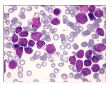
Fig. 1. Morphology of leukemic cells at diagnosis showing L1/L2 blasts.
liver enlargement, and blood evaluation showed a white blood cell count of 6.1×10⁹/L with 13% blasts, hemoglobin level of 10.9 g/dL, and platelet count of 190×109/L. A bone marrow aspirate showed that 81% of the cellularity was replaced by L1/L2 French-American-British morphology lymphoblasts (Fig. 1). Immunophenotyping of the blasts with flow cytometry using a FacSort instrument (Becton Dickinson, San José, CA, USA) indicated a common ALL (B II) according to the European Group for the Immunological Classification of Leukemias. The leukemic cells expressed CD79a, CD22, CD19, CD10, HLA-DR, and were partially positive for TdT, CD34, and CD117 while negative for cytoplasmic -chain.
Conventional cytogenetic analysis of the bone marrow cell culture revealed the following karyotype: 46,XY,t(16;21) (p11.2;q22)[10]/46,XY[10] (Fig. 2A). Subsequently, reverse transcriptase polymerase chain reaction (RT-PCR) and di- rect sequencing were performed to assess the presence of FUS-ERG fusion transcript according to standard procedures [7, 8]. RT-PCR showed a 554-bp specific fragment, and se- quencing analysis confirmed the presence of a chimeric tran- script consisting of FUS exon 7 at the 5’ end fused with ERG exon 6 at the 3’ end (Fig. 2B).
The patient was enrolled in our current protocol for ALL-BFM ALL-IC 2009. His response to the prednisone pre-phase was good. He then underwent induction chemo- therapy consisting of vincristine, daunorubicin, and L-asparaginase. The minimal residual disease (MRD), de- tected by flow cytometry of the bone marrow aspirate, at day 15 of treatment was 0.19%. The patient achieved com- plete remission after the induction phase and was stratified as an intermediate risk patient, with central nervous system involvement owing to facial palsy. However, blast cells were not detected in the cerebrospinal fluid at diagnosis. He re- ceived consolidation chemotherapy with cytarabine, cyclo- phosphamide, and mercaptopurine, followed by high-dose methotrexate. Subsequently, he received late re-induction therapy with dexamethasone, vincristine, doxorubicin, L-as- paraginase, cyclophosphamide, cytarabine, and thiopurine, as well as maintenance therapy with mercaptopurine and methotrexate. He remained free of leukemia 31 months after diagnosis. Flow cytometry analysis of MRD was neg- ative on day 33 (after the induction phase) and on week 12 (prior to the administration of the consolidation phase
bloodresearch.or.kr Blood Res 2015;50:54-65.
Letters to the Editor 57
with high-dose methotrexate). MRD was also confirmed to be negative by quantitative real-time PCR at these time points (day 33 and week 12).
According to the Mitelman database and the literature, about 71 cases of t(16;21) have been reported to date, most of them in AML [9]. The rearrangement of the two chromo- somes involved in such a translocation forms the FUS-ERG fusion gene.
In AML, four types of chimeric transcripts have been described, designated as types A, B, C, and D, which corre- spond to the 255-, 211-, 176-, and 349-bp chimeric products, respectively [2, 8]. These transcripts consist of FUS exons 1 to 6, 1 to 7, or 1 to 8 fused to ERG exons 7 to 17 or 9 to 17. Types A and C are out-of-frame fusion transcripts and likely to have been produced by alternative splicing.
The other two in-frame transcripts, types B and D, have been shown to result from the fusion of FUS and ERG genes at different breakpoint positions in t(16;21). These two types of fusion were predicted to produce the FUS-ERG chimeric protein, which may function as the transcriptional activator responsible for the neoplastic process of several t(16;21)-AML cases. The prognosis of AML patients with this translocation is usually very poor owing to resistance to chemotherapy and a high rate of early relapse.
On the other hand, in ALL, a recent study of 256 adult patients reported that FUS-ERG fusion transcript was found in 14 cases, including 10 with t(16;21) identified by cytoge- netic analysis. Unfortunately, no information regarding the sequencing of the FUS-ERG fusion gene and the follow-up of these patients was provided [3]. Furthermore, three pedia- tric cases of t(16;21) have been reported, but only two of them provide clinical information and results from RT-PCR and sequencing of the FUS-ERG fusion gene [4-6]. The case reported by Kanazawa et al. involved a 1-year-old boy with precursor B cell ALL that did not initially respond to ALL-oriented therapy, but the patient achieved complete remission with an AML treatment protocol. Moreover, Oh et al. described the case of an 8-month-old infant with precursor B cell ALL who achieved complete remission but relapsed 4 months after diagnosis. These patients and the one in our case had the same unusual chimeric transcript that consisted of FUS exon 7 at the 5’ end fused with ERG exon 6 at the 3’ end, which is different from those described in AML [5, 6]. It is worthy of note that despite having the same molecular rearrangement, the three patients had different responses to ALL-oriented treatment. Our patient achieved complete remission with a chemotherapy schedule for ALL, and he remained free of leukemia 31 months after diagnosis. Therefore, it is challenging to establish the prog- nostic value of t(16;21) in ALL patients, mostly owing to the small number of reported cases with this unusual abnor- mality, differences in patient age, treatment heterogeneity, and short follow-up periods.
In conclusion, the present case supports the possible asso- ciation between ALL cases with t(16;21) and a specific type of FUS-ERG fusion transcript first suggested by Oh et al.
However, the role of this transcript in the leukemogenesis of ALL and its prognostic value remain uncertain. Therefore, an analysis of additional ALL cases with t(16;21)(p11.2;q22) and the FUS-ERG fusion chimeric transcript are needed to determine the significance of these findings.
Mariela C. Coccé1, Cristina N. Alonso2, Jorge Rossi3, Maria S. Felice2, Myriam R. Gitter2, Marta S. Gallego1
1Servicio de Genética, 2Servicio de Hemato-Oncología,
3Servicio de Inmunología. Hospital de Pediatría
“Prof. Dr. J. P. Garrahan”, Buenos Aires, Argentina Correspondence to: Mariela C. Coccé Laboratorio de Citogenética, Servicio de Genética, Hospital de Pediatría “Prof. Dr. J. P. Garrahan”, Combate de los Pozos 1881, CP 1245, Buenos Aires, Argentina
E-mail: [email protected]
Received on Jul. 29, 2014; Revised on Sep. 24, 2014; Accepted on Feb. 12, 2015 http://dx.doi.org/10.5045/br.2015.50.1.55
AuthorsÊ Disclosures of Potential Conflicts of Interest No potential conflicts of interest relevant to this article were reported.
REFERENCES
1. Huret JL. t(16;21)(p11;q22). Atlas Genet Cytogenet Oncol Haematol 2005;9:36-8.
2. Ichikawa H, Shimizu K, Hayashi Y, Ohki M. An RNA-binding protein gene, TLS/FUS, is fused to ERG in human myeloid leuke- mia with t(16;21) chromosomal translocation. Cancer Res 1994;54:2865-8.
3. Liu F, Gao L, Jing Y, et al. Detection and clinical significance of gene rearrangements in Chinese patients with adult acute lym- phoblastic leukemia. Leuk Lymphoma 2013;54:1521-6.
4. Heller A, Loncarevic IF, Glaser M, et al. Breakpoint differ- entiation in chromosomal aberrations of hematological malig- nancies: Identification of 33 previously unrecorded breakpoints.
Int J Oncol 2004;24:127-36.
5. Kanazawa T, Ogawa C, Taketani T, Taki T, Hayashi Y, Morikawa A. TLS/FUS-ERG fusion gene in acute lymphoblastic leukemia with t(16;21)(p11;q22) and monitoring of minimal residual disease. Leuk Lymphoma 2005;46:1833-5.
6. Oh SH, Park TS, Choi JR, et al. Two childhood cases of acute leu- kemia with t(16;21)(p11.2;q22): second case report of infantile acute lymphoblastic leukemia with unusual type of FUS-ERG chimeric transcript. Cancer Genet Cytogenet 2010;200:180-3.
7. van Dongen JJ, Macintyre EA, Gabert JA, et al. Standardized RT-PCR analysis of fusion gene transcripts from chromosome aberrations in acute leukemia for detection of minimal residual disease. Report of the BIOMED-1 Concerted Action: inves- tigation of minimal residual disease in acute leukemia. Leukemia 1999;13:1901-28.
8. Kong XT, Ida K, Ichikawa H, et al. Consistent detection of TLS/FUS-ERG chimeric transcripts in acute myeloid leukemia with t(16;21)(p11;q22) and identification of a novel transcript.
Blood Res2015;50:54-65. bloodresearch.or.kr
58 Letters to the Editor
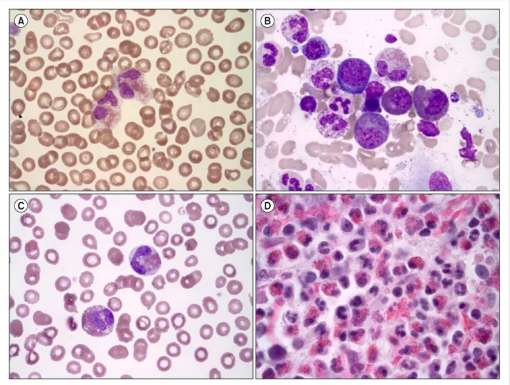
Fig. 1. Peripheral blood (PB) smear and bone marrow (BM) aspirate findings of first case (A, B) and second case (C, D). (A) Eosinophilia (13.0×10⁹/L) was observed in the PB (Wright-Giemsa, ×1,000). (B) Dysplastic eosinophils (sparse and mixed granules) were increased in the BM aspirate (Wright-Giemsa, ×1,000). (C) Eosinophilia (55.316×10⁹/L) was observed in the PB (Wright-Giemsa, ×1,000). (D) The BM section was packed with eosinophils (hematoxylin-eosin, ×400).
Blood 1997;90:1192-9.
9. Mitelman F, Johansson B, Mertens F. Mitelman Database of Chromosome Aberrations and Gene Fusions in Cancer.
Bethesda, MD: National Cancer Institute, 2013. (Assessed May 20, 2014, at http://cgap.nci.nih.gov/Chromosomes/Mitelman)
Chronic eosinophilic leukemia with a FIP1L1-PDGFRA rearrangement:
Two case reports and a review of Korean cases
TO THE EDITOR: According to the 2008 World Health Organization (WHO) guidelines, eosinophilia is associated with hematologic diseases such as chronic eosinophilic leu-
kemia-not otherwise specified (CEL-NOS); idiopathic hy- pereosinophilic syndrome (HES) or idiopathic hyper- eosinophilia; and myeloid and lymphoid neoplasms with eosinophilia, and abnormalities of PDGFRA, PDGFRB or FGFR1 [1]. CEL is diagnosed in cases with increased blasts or cytogenetic/clonal abnormalities. We report 2 cases of myeloid and lymphoid neoplasms (CEL) with the FIP1L1- PDGFRA rearrangement.
CASES
The first case, 27-year-old man was referred to our hospi- tal for a dyspnea workup. The echocardiogram revealed severe tricuspid valve regurgitation and moderate mitral valve regurgitation. A valvuloplasty was performed and diz- ziness developed after the surgery. Brain magnetic resonance imaging revealed microbleeding in the right temporo-occi- pital lobe. He was referred to the hemato-oncology depart- ment for evaluation of eosinophilia. The laboratory findings were as follows: WBC count, 21.1×10⁹/L (eosinophil count,