2 )INTRODUCTI ON
Interferon (IFN) play s a key role in m ediating the
Correspondence : Byung-Kiu Park, Pediatric Oncology Clinic, National Cancer Center, 809 Madu-dong, Ilsan-gu, Koyang, Kyonggi, 4 11-351, Korea Koyang, Korea
Tel: +82-31-920-1242, Fax: +82-31-920-1959 E-mail: [email protected]
antiviral and antiproliferative respon ses as well as in m odulating the immune respon se ( 1,2) . These responses are elicited largely through the tran scriptional activation of the IFN -regulatory genes. These genes possess specific con sen sus sequences w ithin their prom oters and are regulated in part by binding of the interferon regulatory factor s (IRF s), a grow ing family of tran scription factors (3,4), to the consen su s sequences.
= Abs t r ac t =
B ackground: The role of the interferon consensus sequence binding protein (ICSBP), a member of interferon regulatory factor family , in protecting against a vesicular stomatitis virus (V SV) infection has not been firmly elucidated. Thus, it was investigated utilizing the human promyelocytic leukemia HL -60 cells which do not express ICSBP . Methods: HL -60 cells were stably transfected with plasmid containing cDNA for either ICSBP or DNA binding domain (DBD) and tested for their VSV-susceptibilities. The susceptibility of each transfectant group to a VSV infection was determined by a plaque assay at 1 h, 24 h, and 48 h post-infection in the presence (500 IU/ml) or ab sence of interferon α(IFNα). Results: In the ab sence of IFNα, the three groups showed similar sensitivities to a VSV infection. However, when pre-treated with IFN, the viral titers in both the ICSBP and control clones steadily decreased over 48 h of incubation, indicating the existence of IFNα-mediated protection against VSV infection. The IFNα-treated ICSBP clones appeared to be more resistant to infection compared with the control clones, although the difference was not great . On the contrary , the viral titers in the IFNα-treated DBD clones increased at 24 h then decreased by 48 h. Conclu sion : The expression of truncated ICSBP (DBD) does not appear to underlie the impaired protection against a V SV infection in the DBD clones, since even the control clones lacking ICSBP were protected from a VSV infection. This suggests that ICSBP does not play a critical role in the IFNα- mediated anti-VSV response of HL -60 cells, although it appears to confer some resistance to a VSV infection.
K ey W ords: interferon consensus sequence binding protein, DNA binding domain, interferon α, vesicular stomatitis virus infection
In t e r fe r o n c o n s e n s u s s e q u e n c e b i n d i n g p r o t e in : No t e s s e n t i a l fo r i n t e r fe r o n α- m e d i a t e d a n t iv i r a l r e s p o n s e t o v e s i c u l a r s t o m a t i t i s v i r u s i n fe c t i o n i n HL- 6 0 c e l l s
Byung-Kiu Park
D ep artm ent of Pediatrics, Gy eongsang National Univers ity College of Medicine and Gy eongsang Institute of Cancer Research, Chinj u, Korea
The interferon con sen sus sequence binding protein (IC SBP), a m ember of the IRF family , w as originally isolated as the protein that recognized the interferon - stimulated respon se elem ent (ISRE) motif present in the prom oter region of the MH C class I, H -2LD gene (5,6) . Unlike m ost of other IRF protein s, IC SBP exhibits a tissue-restricted pattern of expression and is expressed exclu sively in immune cells, particularly in the m acrophage and lymphoid lineages. IC SBP is induced by IFNα but not by IFNα/β(7 ), and represses the IFNα/β and IFNα/β-inducible prom oter s through the ISRE (8) . Conversely , IC SBP is capable of stimulating the tran scription of certain IFNγ-inducible prom oter s in a gamm a- activated sequence (GA S)-dependent m anner (9) . Studies on the role of IC SBP in the antiviral respon se have been made through several approaches. A recent w ork on IC SBP knockout m ice dem on strated that these m ice are sensitive to particular viral infection s and IC SBP thu s play s a critical role in the establishm ent of an antiviral state ( 10) . How ever, sen sitivity to viral infection s is not a generalized phenomenon and a vesicular stom atitis viru s (V SV) infection was w ell controlled in these m ice . In contrast, another study show ed that U 937 m onocytic cells tran sfected w ith truncated IC SBP cDN A , which retain s the DNA -binding dom ain (DBD) but lack s the regulatory dom ain , did not have antiviral activity again st V SV upon IFN treatm ent ( 11) . This suggests that IC SBP play s an important role in controlling V SV infection s.
A s seen in these two reports, the protective role of IC SBP again st V SV infection has not been firm ly established . Thu s the function of IC SBP in V SV infection was studied in a similar way as reported previou sly ( 11) utilizing the IC SBP and DBD stable clones of HL -60 hum an promyelocytic leukem ia cells.
H owever, in the current study , HL -60 cells, another m odel of the m onocyte-m acrophage lineage, w ere u sed since they w ere reported to express little or no IC SBP (6, 12), while U 937 cells obviou sly did. This character obviously renders HL -60 cells superior to U 937 cells for evaluating the effect of exogenou sly introduced IC SBP . The present study reports that both the IC SBP and
control clones of HL -60 cells are protected from V SV infection s in the presence of IFNα, although the form er clones are m ore resistant . This indicates that IC SBP is not essential for the IFNα-mediated anti-V SV respon se, though it does play a certain role, since even the control clones lacking IC SBP w ere protected from a V SV infection . In addition , the results indicate that the DBD clones of HL -60 cells show an impaired anti-V SV re- spon se to IFNα, which is probably due to the inhibitory effects of DBD on the action of other IRF proteins.
MATERIALS AND METHODS
1. C ell cu lt u r e
HL -60 cells w ere obtained from the Am erican Type Culture Collection (Rockville, MD) . They w ere cultured in a RPMI 1640 medium (Life Technologies, Grand Island, N Y) supplem ented w ith 10% heat-inactivated fetal bovine sera, 2 mM glutam ine, 50 μg/m l gentamicin sulfate, and 50 μM β-mercaptoethanol. The cells were m aintained at 37℃ in a humidified incubator with 5%
CO2. The tran sfected HL -60 cells w ere cultured in the G4 18 (Geneticin , Life Technologies) containing medium at 400 μg/m l.
2. C on s t r u ct ion of I C S B P ex p r e s sion v ect or s
An expression plasm id for the full-length IC SBP w as constructed by subcloning the B amH I fragm ent of a full-length mIC SBP into the X hoI site of pCXN 2 ( 13) . An expression plasm id for DBD was con structed by subcloning the B amHI/H indIII fragm ent of DBD (8) into pCXN 2 .
3. T r an sf ection an d clonin g of st able tr an sf ect an t s
The HL -60 cells ( 1 ×10⁷) were tran sfected w ith 50 μg of control pCXN 2 (w ithout insert) , pCXN 2 containing IC SBP or DBD cDNA by electroporation with a Cell- Porator (Life Technologies) as previou sly described ( 11) . The cells w ere selected with G4 18 at 400 μg/ml for 14 day s then cloned by lim ited dilution at 0 .5 cells/96 m icrotiter well.
4 . Im m u n ob lot a n a ly si s
The cells ( 1× 10⁷) were incubated w ith a ly sis buffer ( 10 mM Tris, pH 7 .5, 10 mM N aCl, 15 mM MgCl2, 1 mM AEB SF , and 0 .05% N onidet P -40), and nuclei were then pelleted ( 14) . The nuclei w ere w ashed and the nuclear extracts were prepared as previou sly described ( 15), then resu spended in a buffer solution containing 20
mM HEPE S, pH 7 .9, 0 .4 M N aCl, 1 mM EDTA , pH 8.0, 1 mM EGTA , 1 mM D TT, and 1 mM AEB SF . Thirty m icrogram s of nuclear protein s w ere resolved on 10% (for IC SBP analy sis) or 15% (for DBD analy sis) SD S-PA GE , electroblotted to Immobilon P nitrocellulose (Millipore, Bedford, MA) , and reacted with the appropriate antibodies according to the ECL protocol provided by the manufacturer (Am er sham , Arlington H eights, IL) . The prim ary antisera w ere either rabbit anti-peptide antibodies that react w ith the C-term inal dom ain of IC SBP (7) or poly clonal rabbit anti-rIC SBP antibodies that react w ith the DBD epitopes. E ach antibody w as diluted either 1:500 (for IC SBP) or 1:300 (for DBD), respectively , in a blocking solution containing PB S, pH 7.1, 5% nonfat dry milk , and 0 .05%
Tween -20 . The target protein s were detected with peroxidase-coupled goat anti-rabbit IgG diluted 1:30,000 .
5 . V ir al in f e ct ion
The cells (3 ×10⁶) w ere incubated w ith the Indiana strain of V SV at 5 plaque-form ing units (PFU) per cell for 1 h at 37 ℃. They w ere sub sequently w ashed and cultured in a complete medium for the indicated tim es, and the viral y ields from the supernatants w ere determ ined by a plaque assay ( 16) . Where indicated, the cells were pretreated w ith recombinant hum an IFNα2A (Lee B iomolecular Laboratories, San Diego) at 500 units/m l overnight and cultured with IFN after infection .
RESULTS
1. Ex pr ession of ICSBP or DB D in HL - 60 tr an sf ect ant s
Clones expressing either IC SBP or DBD (Fig . 1A) w ere screened by immunoblot analy sis. The tw enty clones that were propagated from each of three tran sfection group s w ere screened .
U sing immunoblot analy sis, the IC SBP (Fig . 1B) or DBD peptide (Fig . 1C) w as confirm ed in the respective clones, while none of these peptides w ere expressed in the control clones. Three representative clones from each Fig . 1 . Im m u n o b lot d e te ctio n of IC S BP a n d DBD
p e ptid e s in HL-6 0 s ta b le c lo n e s . (A ) S c h e m a tic illus tra tio n of IC S BP a n d DBD. Th e N-te rmin a l 1 10-a a s e q u e n c e (h a tc h e d) re p re s e nts th e DBD . (B) Nu c le a r e xtra cts (3 0 μg) fro m e a ch tra ns fe cta nt c lo n e w e re re s o lv e d o n 10 % S DS -P AG E a n d re a cte d w ith th e a p p ro p ria te a ntib o d ie s . Ra b b it a ntib o d ie s to th e C-te rm in a l p e ptid e of IC S BP w e re d ilute d 1 :5 0 0 in a b lo c kin g s o lutio n a n d th e IC S BP w a s d e te cte d w ith p e rox id a s e -c o u p le d g o a t a nti-ra b b it Ig G d ilute d 1 :3 0 ,0 0 0 . IC S BP (in d ic a te d by a n a rrow ) is s tro n g ly e x p re s s e d o n ly in th e IC S BP c lo n e s . MW M, m o le c u la r w e ig ht m a rke r; rIC S BP , re co m b in a nt IC S BP p rote in g e n e ra te d in a b a c u lov irus v e cto r2 2 ). (C) DBD w a s s im ila rly d e te cte d a s in (B) , e xc e pt th a t p rote ins w e re re s o lv e d o n 15 % S DS -PAG E a n d th e p rim a ry a ntib o d ie s w e re d ilute d 1 :3 0 0 . DBD (in d ic a te d by a n a rrow ) is d is tin ctly e x p re s s e d in th e DBD clo n e s .
transfection group, shown in Fig. 1, were used for further investigation.
2. S u s cept ib ilit ies of IF Nα- t r eat ed st ab le clon e s t o V SV in f ect ion
To compare the susceptibilities of ICSBP, DBD, and control clones to a VSV infection, three clones from each group were infected with VSV and the viral titers were determined by a plaque assay at various times following infection.
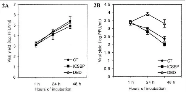
In the absence of IFNα, the ICSBP, DBD, and control clones showed similar sensitivities to a VSV infection over 48 h of incubation (Fig. 2A) . When these clones were pre-treated with IFNαprior to the VSV challenge and cultured with IFN after infection, the viral titers steadily decreased for up to 48 h of incubation in both the ICSBP and control clones. This suggests that IFN can protect the cells from a VSV infection (Fig. 2B).
When the viral titers in the control and ICSBP clones were compared, the titers from the control clones were
higher than those from the ICSBP clones by 1.5-fold (24 h) and 2.2-fold (48 h). This suggests that there is an additional resistance to a VSV infection conferred by ICSBP. Unlike the two other clones, the viral titers in the DBD clones increased at 24 h then decreased by 48 h post-infection, indicating an impaired IFN-mediated resistance to a VSV infection. The amount of viral shedding in the DBD clones was 7.9-fold (24 h) and 9.0-fold (48 h) greater than that from the control clones.
DI SCUSSI ON
It has been reported that ICSBP is necessary for establishing a resistance to various pathogens (10,17-19), despite it being a transcriptional repressor of IFN- regulatory genes. ICSBP-deficient mice succumbed upon challenge with certain viruses such as the vaccinia virus and the lymphocytic choriomeningitis virus as well as other intracellular pathogens. A defective T helper cell type 1 (Th 1) response, a decrease in cytotoxic T
Fig . 2 . IFN α-induce d prote ction a ga inst VSV infe ction in the HL-60 control a nd ICS BP clone s . (A) Ce lls (3 x 10⁶) of the clone s from e a ch group we re infe cte d w it h VSV at 5 P FU pe r ce ll a nd the vira l tite rs in the re s ulting s upe rnata nts we re dete rmined at the indicate d times . Vira l t ite rs from e a ch of thre e tra nsfecta nt groups incre a s e d ste a dily ove r 48 h of incubat ion. Va lue s a re the me a n of vira l t ite rs from thre e clones for e a ch group. Ba rs re pre s e nt t he ra nge of the thre e s e pa rate mea s ure me nts . (B) Whe n incubate d in the pre se nce of IFN α(500 IU/ ml), t he vira l t ite rs in t he ICS BP a nd cont rol clone s de cre a se d ste a dily ove r 48 h of incubation w hile t hos e in the DBD clones increa s e d at 24 h the n de cre a se d by 48 h. Va lue s a nd ba rs a re s a me a s a bove .
lymphocyte activity, as well as the deficient production of IFN γafter stimulation of T cells or macrophages appear to underlie the inability to control these pathogens ( 10,18). Defects in these IFN γ- mediated immune responses that are observed in ICSBP null mice could be explained by a very recent observation that ICSBP acts as a transcriptional activator for IFN γ-inducible promoters (9). In contrast, these mice survived the VSV infection and mounted a normal neutralizing antibody response, which implies that the B and T helper cell compartments as well as the IFN type I system was normal in these mice. In the present study, IFN γ treatment effectively reversed the sensitivities of HL-60 control and ICSBP clones to a VSV infection (Fig. 2). In a certain viral infection, one of the two IFN systems (type I and II) usually dominates (20). Although attempts to examine the effect of IFN γon the anti-VSV response were not made, it can be said that at least the IFN type I system was indispensable for an anti-VSV response.
The HL-60 control clones that did not express ICSBP on the immunoblot analysis (Fig. 1B) could control the VSV infection in the presence of IFN γ, even though the ICSBP clones appeared to do it better (Fig. 2B) . This indicates that ICSBP is not essential for protecting against a VSV infection in response to IFN, though it is helpful to a certain extent. In a previous study (11), both the control and ICSBP clones of U937 cells exhibited a similar resistance to a VSV infection. The presumable reason for this difference is that U937 cells obviously express ICSBP while HL-60 cells do not. Accordingly, exogenously introduced ICSBP might not have produced as much effect on the U937 cells as it did on HL-60 cells. Unlike two other clones, the DBD clones failed to elicit effective antiviral activity against VSV in response to IFN (Fig. 2B) . As the control clones mounted an anti-VSV response effectively, the impaired viral resistance in the DBD clones is not likely to be caused by the expression of truncated ICSBP in these clones.
Similarly, defective ICSBP in U937 DBD clones does not appear to be responsible for the impaired anti-VSV response. In this regard, the results of two previous studies on the role of ICSBP in VSV infections ( 10,11)
might not be discordant.
The IRF family proteins are composed of a conserved DBD in the N-terminal region and a divergent C-terminal region that serves as the regulatory domain (3,4) (Fig.
1A). Since the proteins bind the target genes through the DBD, truncated ICSBP lacking the regulatory domain (DBD) blocks the function of other IRF family proteins by occupying their DNA binding sites, producing a dominant negative effect (21). Considering this, the failure of the DBD clones to mount an antiviral response is probably due to the repression of IFNα-inducible anti- viral genes by DBD. As discussed above, the lack of intact ICSBP might well be excluded as a reason for the impaired antiviral response in the DBD clones. It is of note that although the DBD clones did not elicit an effective antiviral response, their viral titers were lower in the presence of IFNαthan in its absence (Fig. 2). One speculation is that the dominant negative effect by DBD was not sufficient to completely block the binding of other IRF proteins induced by IFNα. Subsequently they might have generated some antiviral activity .
In summary, the present data suggest that ICSBP does not play a crucial role in controlling VSV infections and at least the IFN type I system is important for protecting against a VSV infection.
ACKNOWLEDGEMENTS
I wish to express my special thanks to Dr . Ozato (Laboratory of Molecular Growth Regulation, NICHD, NIH) for providing plasmids, antibodies, and viruses.
REFERENCES
1. Pestka S, Langer JA, Zoon KC, Samuel CE : Interferon and their action. Annu Rev Biochem 56 ; 727-777, 1987
2. Stark GR, Kerr IM, Williams BRG, Silverman RH, Schreiber RD : How cells respond to interferons. Annu Rev Biochem 67 ; 227-264, 1998
3. Nguyen H, Hiscott J, Pitha PM : The growing family of IRF transcription factors. Cyt Growth Fact Rev 8 ;
293-312, 1997
4. Mamane Y, Heylbroeck C, Génin P, Algarté M, Servant MJ, LePage C, DeLuca C, Kwon H, Lin R, Hiscott J : Interferon regulatory factors: the next generation. Gene 237 ; 1-14, 1999
5. Driggers PH, Ennist DL, Gleason SL, Mak W-H, Marks MS, Levi B-Z, Flanagan JR, Appella E, Ozato K : An interferon γ-regulated protein that binds the interferon- inducible enhancer element of maj or histocompatibility complex class I genes. Proc Natl Acad Sci USA 87 ; 3743-3747, 1990
6. Weisz A, Marx P, Sharf R, Appella E, Driggers PH, Ozato K, Levi B-Z : Human interferon consensus sequence binding protein is a negative regulator of enhancer elements common to interferon-inducible genes. J Biol Chem 267 ; 25589-25596, 1992 7. Politis AD, Ozato K, Coligan JE, Vogel SN : Re-
gulation of IFN-γ-induced nuclear expression of IFN consensus sequence binding protein in murine peritoneal macrophages. J Immunol 152 ; 2270-2278, 1994 8. Nelson N, Marks MS, Driggers PH, Ozato K :
Interferon consensus sequence-binding protein, a member of the interferon regulatory factor family, suppresses interferon-induced gene transcription. Mol Cell Biol 13; 588-599, 1993
9. Contursi C, Wang I-M, Gabriele L, Gadina M, O Shea J, Morse HC, III, Ozato K : IFN consensus sequence binding protein potentiates STAT1-dependent activation of IFNγ-responsive promoters in macrophages. Proc Natl Acad Sci USA 97 ; 91-96, 2000
10. Holtschke T, Löhler J, Kanno Y, Fehr T, Giese N, Rosenbauer F, Lou J, Knobeloch K-P, Gabriele L, Waring JF, Bachmann MF, Zingernagel RM, Morse III HC, Ozato K, Horak I : Immunodeficiency and chronic myelogenous leukemia-like syndrome in mice with a targeted mutation of the ICSBP gene. Cell 87 ; 307-317, 1996
11. Thornton AM, Buller RML, DeVico AL, Wang I-M, Ozato K : Inhibition of human immunodeficiency virus type I and vaccinia virus infection by a dominant negative factor of the interferon regulatory factor family expressed in monocytic cells. Proc Natl Acad Sci USA 93 ; 383-387, 1996
12. Schmidt M, Nagel S, Proba J, Thiede C, Ritter M,
Waring JF, Rosenbauer F, Huhn D, Wittig B, Horak I, Neubauer A : Lack of interferon consensus sequence binding protein (ICSBP) transcripts in human myeloid leukemias. Blood 91 ; 22-29, 1998
13. Niwa H, Yamamura K, Miyazuki J : Efficient selection of high expression transfectants with a novel eukaryotic vector. Gene 108 ; 193-199, 1991
14. Antalis TM, Godbolt D : Isolation of intact nuclei from hematopoietic cell types. Nucleic Acids Res 19 ; 4301, 1991
15. Schreiber E, Matthias P, Muller MM, Schaffner W : Rapid detection of octamer binding protein with mini- extracts prepared from a small number of cells. Nucleic Acids Res 17 ; 64 19, 1989
16. Bovolenta C, Lou J, Kanno Y, Park B-K, Thornton AM, Coligan JE, Schubert M, Ozato K : Vesicular stomatitis virus infection induces a nuclear DNA- binding factor specific for the interferon-stimulated response element . J Virol 69 ; 4 173-4 181, 1995
17. Fehr T, Schoedon G, Odermatt B, Holtschke T, Schneemann M, Bachmann MF, Mak TW, Horak I, Zinkernagel RM : Crucial role of interferon consensus sequence binding protein, but neither of interferon regulatory factor 1 nor of nitric oxide synthesis for protection against murine listeriosis. J Exp Med 185 ; 921-931, 1997
18. Giese NA, Gabriele L, Doherty TM, Klinman DM, Tadesse-Heath L, Contursi C, Epstein SL, Morse HC III : Interferon (IFN) consensus sequence-binding protein, a transcription factor of the IFN regulatory factor family, regulates immune responses in vivo through control of interleukin 12 expression. J Exp Med 186 ; 1535-1546, 1997
19. Scharton-Kersten T, Contursi L, Masumi A, Sher A, Ozato K : Interferon consensus sequence binding protein-deficient mice display impaired resistance to intracellular infection due to a primary defect in interleukin 12 p40 induction. J Exp Med 186 ; 1523-1534, 1997
20. Kunzi M, Pitha PM : The Role of Cytokines in Viral Infections. In : Mahy BWJ, Collier L eds. : Topley &
Wilson"s Microbiology and Microbial Infections, p93-2 10, Arnold Publications, London, 1998
2 1. Thornton AM, Ogryzko VV, Dent A, Sharf R, Levi
B-Z, Kanno Y, Staudt LM, Howard BH, Ozato K : A dominant negative mutant of an IFN regulatory factor family protein inhibits both type I and type II IFN- stimulated gene expression and anti-proliferative activity of IFNs. J Immunol 157; 5145-5154, 1996
22. Bovolenta C, Driggers PH, Marks MS, Medin JA,
Politis AD, Vogel SN, Levy DE, Sakaguchi K, Appella E, Coligan JE, Ozato K : Molecular interactions between interferon consensus sequence binding protein and members of the interferon regulatory factor family.
Proc Natl Acad Sci USA 91; 5046-5050, 1994