Received:December 26, 2018, Revised:February 18, 2019, Accepted:March 7, 2019 Corresponding to:Seong Wook Kang http://orcid.org/0000-0002-0076-0822
Division of Rheumatology, Department of Internal Medicine, Chungnam National University Hospital, 282 Munhwa-ro, Jung-gu, Daejeon 35015, Korea. E-mail:kangsw@cnuh.co.kr
Copyright ⓒ 2019 by The Korean College of Rheumatology. All rights reserved.
This is an Open Access article, which permits unrestricted non-commerical use, distribution, and reproduction in any medium, provided the original work is properly cited.
Neuromyositis: A Rare Extramuscular Manifestation of Dermatomyositis
Chan Keol Park, Su-Jin Yoo, In Seol Yoo, Jinhyun Kim, Seung Cheol Shim, Seong Wook Kang
Division of Rheumatology, Department of Internal Medicine, Chungnam National University Hospital, Daejeon, Korea
Dermatomyositis (DM) and polymyositis (PM) are representative idiopathic inflammatory myopathies characterized by sym- metric and progressive proximal muscle weakness. Especially, DM is identified by characteristic skin lesions and has many ex- tramuscular manifestations including various cardiac abnormalities, interstitial lung disease, and malignancy. However, in- volvement of peripheral nervous system in DM/PM is very rare and less known. The term “Neuromyositis” was introduced by Senator in 1893 to describe the concomitant involvement of the peripheral nervous system in DM/PM. Since then, a very few cases of neuromyositis have been reported mainly in the United States and Europe. Therefore, the pathogenetic mechanism and disease progression are unclear. In recent years, a few more cases were reported in Asia, specifically, China and Japan; how- ever, none in Korea. Here, we describe a case of DM-associated neuromyositis in a 42-year-old man in Korea and review pre- vious publications through literature research. (J Rheum Dis 2019;26:211-218)
Key Words. Dermatomyositis, Peripheral nervous system diseases, Nerve conduction, Electromyography
INTRODUCTION
Dermatomyositis (DM) and polymyositis (PM) are two major groups of idiopathic inflammatory myopathies (IIM) that are characterized by chronic inflammation in the skeletal muscle, resulting in muscle weakness [1,2].
Especially, DM is identified by characteristic cutaneous manifestations such as heliotrope rash, Gottron’s pap- ules, cuticular changes, a photodistributed erythema or poikiloderma, a scaly alopecia and hyperkeratosis of the palms and fingers, known as “mechanic’s hands” [3]. IIM have many extramuscular manifestations which include dysphagia, various cardiac abnormalities, interstitial lung disease (ILD) and malignancy. Especially, the frequency of cancer development is definitely increased in DM and slightly increased in PM [2]. However, peripheral neuro- pathy is very rarely reported during the course of DM/PM and less known. Concomitant involvement of a periph- eral nervous system in DM/PM has been known as neuro-
myositis which was first introduced by Senator in 1893 [4]. A few cases of a peripheral neuropathy in DM/PM have been reported; however, even a single study was not conducted yet to examine peripheral nerve in a large pop- ulation diagnosed with DM/PM. Consequently, the clin- ical presentation and pathogenetic mechanism of neuro- myositis are unclear. In addition, well-defined diagnostic criteria of this entity are still controversial. After the clas- sical etiologies of peripheral neuropathies are ruled out, the diagnosis of neuromyositis can be accepted.
Previously, Onder et al. [5] reported the neuropathic in- volvement in 8 patients diagnosed with DM/PM.
Although the electrophysiologic studies showed all 8 pa- tients with neuropathic involvement, only 1 patient diag- nosed with neuromyositis because the others had co- morbidities that could cause neuropathies, such as malig- nancies (e.g., breast cancer and lung cancer), other rheu- matic diseases (e.g., Sjögren’s syndrome, scleroderma, and familial Mediterranean fever), and diabetes mellitus.
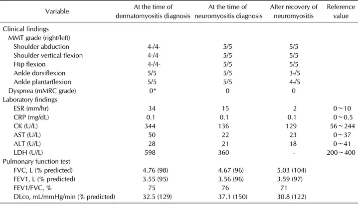
Table 1. Summary of clinical and laboratory findings of the present case
Variable At the time of
dermatomyositis diagnosis
At the time of neuromyositis diagnosis
After recovery of neuromyositis
Reference value Clinical findings
MMT grade (right/left)
Shoulder abduction 4-/4- 5/5 5/5
Shoulder vertical flexion 4-/4- 5/5 5/5
Hip flexion 4-/4- 5/5 5/5
Ankle dorsiflexion 5/5 5/5 3-/5
Ankle plantarflexion 5/5 5/5 4-/5
Dyspnea (mMRC grade) 0* 0 0
Laboratory findings
ESR (mm/hr) 34 15 2 0∼10
CRP (mg/dL) 0.1 0.1 0.1 0∼0.5
CK (U/L) 344 136 129 56∼244
AST (U/L) 50 22 23 0∼37
ALT (U/L) 28 21 18 0∼41
LDH (U/L) 598 360 - 200∼400
Pulmonary function test
FVC, L (% predicted) 4.76 (98) 4.67 (96) 5.03 (104)
FEV1, L (% predicted) 3.55 (95) 3.56 (96) 3.59 (97)
FEV1/FVC, % 75 76 71
DLco, mL/mmHg/min (% predicted) 32.5 (129) 37.1 (150) 30.8 (122)
MMT: manual muscle test, mMRC: modified Medical Research Council dyspnea scale, ESR: erythrocyte sedimentation rate, CRP:
C-reactive protein, CK: creatine kinase, AST: aspartate aminotransferase, ALT: alanine aminotransferase, LDH: lactate dehydrogenase, FVC: forced vital capacity, FEV1: forced expiratory volume in 1 second, DLco: diffuse lung capacity for carbon monoxide. *Not troubled by breathlessness except on strenuous exercise.
Here, we report a case of DM-associated neuromyositis in a 42-year-old man and review the previous publications through literature research.
CASE REPORT
A 42-year-old man visited our clinic with limb weakness and skin lesions on the face and hands. His symptoms oc- curred 3 weeks before visiting our clinic. His upper and lower extremities revealed grade 4- proximal muscle weakness symmetrically on manual muscle test (MMT) (MMT grade 4- indicates full range of motion against gravity, with less than moderate but more than minimum resistance) (Table 1). He also exhibited DM-associated skin lesions such as erythematous rashes on the nose and forehead, Gottron’s papules on the extensor surfaces over the metacarpophalangeal joints and proximal inter- phalangeal joints of both hands, and roughening and cracking on the skin of the tips and sides of the fingers (mechanic’s hands).
To identify the cause of his symptoms, we performed various tests. Laboratory tests showed a white blood cell
count of 4,480/μL, hemoglobin level of 14.7 g/dL, plate- let count of 171,000/μL, erythrocyte sedimentation rate of 34 mm/hr, and C-reactive protein level of 0.1 mg/dL.
The levels of aspartate aminotransferase, alanine amino- transferase, and lactate dehydrogenase were 50, 28, and 598 U/L, respectively (0∼37/0∼41/200∼400 U/L).
Creatine kinase (CK) levels were elevated to 344 U/L (56∼
244 U/L). Viral serologic test results for hepatitis B, hep- atitis C, and human immunodeficiency virus were negative. The patient also tested negative for tumor mark- ers, such as carbohydrate antigen 19-9, carcinoembryonic antigen, and alpha-fetoprotein. Thyroid function tests, hemoglobin A1c levels, electrolytes, blood urea nitrogen levels, and serum creatinine levels were also normal.
Antinuclear antibody (Ab) tests indicated weak positivity with a speckled pattern. Rheumatoid factor, anti-CCP Ab, anti-RNP Ab, and anti-Jo1 Ab were not detected, but an- ti-Ro52 Ab and anti-MDA5 Ab were positive among my- ositis specific antibodies.
Unusually, although there was definite muscle weak- ness on physical examination, electromyography (EMG) of the proximal muscles of the upper and lower ex-
Table 2. Results of nerve conduction study
Nerve 1st study 2nd study 3rd study
Lat Amp CV Lat Amp CV Lat Amp CV
Motor nerve
Left peroneal 3.0 ms 5.3 mV 47.7 m/s 3.92 ms 3.2 mV 52.4 m/s 4.04 ms 7.2 mV 53.4 m/s Right peroneal 3.42 ms 2.8 mV 47.2 m/s 4.13 ms 1.14 mV 48.8 m/s 4.90 ms 3.3 mV 50.0 m/s Sensory nerve
Left peroneal superficial 2.4 ms 5.7 μV 58.3 m/s 2.98 ms 5.7 μV 47.0 m/s 2.69 ms 13.2 μV 52.0 m/s Right peroneal superficial 0.0 ms 0.0 μV 0.0 m/s 3.09 ms 1.46 μV 45.3 m/s 2.51 ms 7.3 μV 55.8 m/s The 1st study means a study performed at the time of dermatomyositis diagnosis. The 2nd study means a study performed at the time of neuromyositis diagnosis. The 3rd study means a study performed after recovery of neuromyositis. Lat: latency, Amp:
amplitude, CV: conduction velocity.
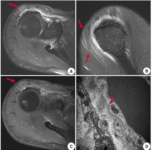
Figure 1. T2-weighted magnetic resonance images (A, B) and en- hanced T1-weighted magnetic resonance image (C) showed high signal intensity in right su- praspinatus and anterior deltoid muscles (arrows). (D) Electron microscopy of right deltoid muscle biopsy demonstrated focal infiltration of fat globules, focal areas of myofibrillar loss, and several tubuloreticular structures (arrow) in the cyto- plasm of endothelial cells, which were consistent with dermato- myositis (×30,000).
tremities showed normal findings. In contrast, a nerve conduction study (NCS) showed some abnormal findings. Peripheral motor NCS showed low-amplitude compound muscle action potential (CMAP) on the left abductor pollicis brevis and right extensor digitorum bre- vis (EDB). Furthermore, the peripheral sensory NCS showed a low-amplitude sensory nerve action potential (SNAP) and a slow conduction velocity in the left median
nerve and absent SNAP on the right superficial peroneal nerve. These NCS findings suggested partial nerve injury on the left median nerve and right peroneal nerve (Table 2). However, an additional EMG study was not performed on the affected sites because there were no clinical symp- toms or signs of peripheral neuropathy. Because the EMG study on the proximal muscles was normal, we conducted a muscle biopsy to diagnose DM after confirming my-
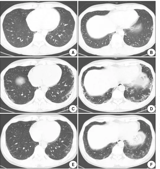
Figure 2. Initial chest computed tomography (CT) images showed multifocal patchy ground glass opacities (GGOs) in subpleural portion of both lower lungs (A, B). Follow-up chest CT images obtained 1 month after the ini- tiation of therapy showed ag- gravation to reticulation with GGOs (C, D). Follow-up chest CT images obtained at the time of neuromyositis diagnosis showed improving state of pre- vious noted reticulation and GGOs (E, F).
ositis with magnetic resonance imaging on the right up- per extremity where symptoms were most severe (Figure 1). A muscle biopsy from the right deltoid muscle did not showed typical findings of DM on light microscopic examination. However, electron microscopic examina- tion showed focal infiltration of fat globules, focal areas of myofibrillar loss, and several tubuloreticular structures in the cytoplasm of endothelial cells (Figure 1), which were consistent with DM [6].
Another important finding was that a chest computed tomography (CT) showed multifocal patchy ground glass opacity in subpleural portion of both lower lungs. These finding suggested non-specific interstitial pneumonia or cryptogenic organizing pneumonia (Figure 2). However, the patient did not complain of dyspnea and pulmonary function tests were also normal (Table 1). There were no other causes to provoke his problems such as infections, comorbidities, other rheumatic diseases, or history of drug use. Moreover, there was no evidence of malignancy
in the screening tests including as tumor markers, ab- dominal CT, chest CT, esophagogastroduodenoscopy, or colonoscopy.
In conclusion, He was diagnosed with DM based on fol- lowing findings that meet the criteria of Bohan and Peter [7]: (1) weakness in the proximal muscles of the limbs;
(2) elevation of skeletal muscle enzyme levels; (3) histo- logic examination findings of the biopsy of the right del- toid muscle that indicates DM; and (4) skin lesions on the face and hands such as heliotrope rash, Gottron’s pap- ules, and mechanic’s hands. Table 1 summarizes the im- portant clinical and laboratory findings of the present case.
After the diagnosis of DM-ILD was established, the pa- tient was initially treated with high-dose steroids (prednisolone at a daily dose of 1 mg/kg). After steroid therapy, his skin rash and muscle strength improved and his elevated muscle enzyme levels returned to the normal range. However, even though there were no respiratory
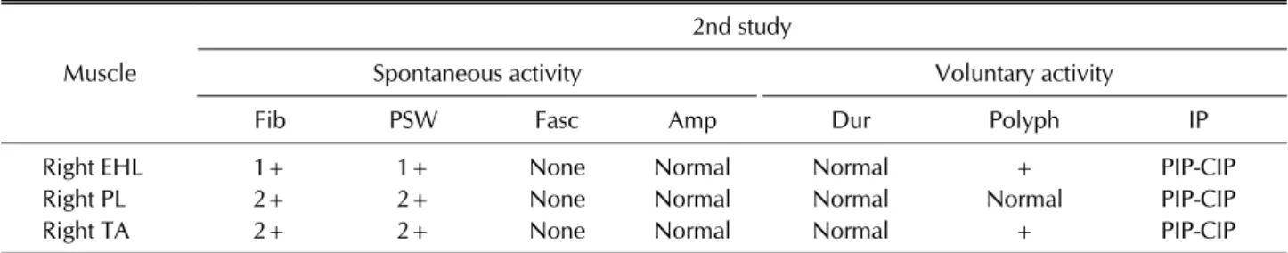
Table 3. Results of electromyography study
Muscle
2nd study
Spontaneous activity Voluntary activity
Fib PSW Fasc Amp Dur Polyph IP
Right EHL 1+ 1+ None Normal Normal + PIP-CIP
Right PL 2+ 2+ None Normal Normal Normal PIP-CIP
Right TA 2+ 2+ None Normal Normal + PIP-CIP
The 2nd study means a study performed at the time of neuromyositis diagnosis. EHL: extensor hallucis longus, PL: peroneus longus, TA: tibialis anterior, Fib: fibrillation potentials, PSW: positive sharp waves, Fasc: fasciculation, Amp: amplitude, Dur: duration, Polyph: polyphasic, IP: interference pattern, PIP: partial interference pattern, CIP: complete interference pattern.
symptoms, such as dyspnea, follow-up chest CT per- formed 1 month after the initiation of steroid therapy showed worsening of ILD (Figure 2). Therefore, azathio- prine (1.5 mg/kg/day) was added to the medication.
Four months after diagnosis with DM-ILD, he com- plained of right ankle weakness that developed gradually for 5 days. On MMT, his right ankle showed grade 3- dor- siflexion and grade 4- plantarflexion (MMT grade 3- in- dicates less than full range of motion against gravity but more than 50%) (Table 1). Other physical examinations on the right lower leg revealed normoactive deep tendon reflex and decreased sensation in the lateral side. We per- formed electrodiagnostic test again to determine the ex- act state. The peripheral motor NCS on both lower ex- tremities showed low-amplitude CMAP on the right EDB. The peripheral sensory NCS on both lower ex- tremities showed delayed latency and low-amplitude SNAP on the right superficial peroneal nerve (Table 2).
The EMG study on the right lower extremity showed ab- normal spontaneous activities in the right tibialis anterior (TA), peroneus longus, and extensor hallucis longus (EHL) and polyphasic motor unit action potential in the right EHL and TA (Table 3). These findings suggested axonal neuropathy on the right common peroneal nerve.
At that time, there was no clinical evidence to suggest that DM had worsened. The strength of the proximal muscles of the upper and lower extremities still im- proved, and laboratory findings such as CK level were all normal (Table 1). In addition, the steroid dose was gradu- ally reduced to 70% of the initial dose, and ILD was also improved after addition of azathioprine (Figure 2).
Moreover, other causes such as trauma and infection, which may cause right ankle weakness, could not be found. Therefore, ultimately, we diagnosed the patient with neuromyositis, a peripheral nerve involvement in
DM. Because of neuromyositis, we decided to slowly tap- er the steroid and increase the dose of azathioprine (1.5 mg/kg/day→2.0 mg/kg/day).
Since then, right ankle weakness improved gradually and almost completely recovered after 6 months without any other further treatment. The recovery of neuro- myositis was confirmed in the follow-up electro- diagnostic test. EMG and peripheral motor NCS on both lower extremities showed normal results. Only the pe- ripheral sensory NCS on both lower extremities showed low-amplitude SNAP on the right superficial peroneal nerve (Table 2). However, this result was also sig- nificantly improved compared to that in the first test at the time of DM diagnosis as well as that in the previous test at the time of neuromyositis diagnosis. Thereafter, he has gradually reduced steroid and is now taking low-dose steroid and azathioprine. There was no recurrence of neu- romyositis as well as exacerbation of DM, and other neu- rologic problems did not occur in any region for about 10 months after neuromyositis on his right ankle improved.
DISCUSSION
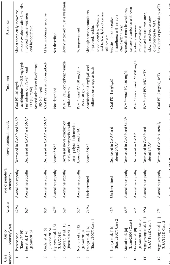
We have herein shown the extremely rare case of con- comitant peripheral neuropathy and DM. In addition, we conducted literature search and compared our case with previous publications. A literature search of the PubMed database was carried out using search terms “dermato- myositis,” “polymyositis,” “neuromyositis,” “peripheral neuropathy” and “peripheral nervous system involve- ment” in various combinations. Table 4 summarizes the clinical features of past cases and the present case.
Involvement of the peripheral nervous system is fre- quently observed in other autoimmune diseases. The prevalence rates were reported to be 40%∼50% in rheu-
Table 4. Previously reported cases of neuromyositis, including the present case Case numberAuthor (country/year)Age/sexType of peripheral neuropathyNerve conduction studyTreatmentResponse 1Present case (Korea/2019)42/MAxonal neuropathyDecreased in CMAP and SNAPOral PD (40 mg/d)+ azathioprine (2∼2.5 mg/kg/d)Almost completely recovered muscle weakness after 6 months 2Irie et al. [14] (Japan/2016)69/FAxonal neuropathyDecreased in CMAP and SNAPFirst admission: IVMP→oral PD (15 mg/d) Second admission: IVMP→oral PD (40 mg/d)
Improved muscle weakness and hypoesthesia 3Onder et al. [5] (Turkey/2015)80/FAxonal neuropathyDecreased in CMAP and SNAPOral PD (dose is not described)At least moderate response 4Nguyen et al. [13] (USA/2014)67/FAxonal neuropathyAbsent SNAPNot describedNot described 5Cojocaru et al. [15] (Romania/2011)59/FAxonal neuropathyDecreased nerve conduction study and compatible with acute polyradiculoneuritis
IVMP, IVIG, cyclophosphamide pulse therapySlowly improved muscle weakness 6Nomura et al. [12] (Japan/2010)52/FAxonal neuropathyAbsent CMAP and SNAPIVMP→oral PD (50 mg/d)→ IVIG 30 g×5 dNo improvement 7França et al. [16] (Brazil/2007) Case 171/MUndeterminedAbsent SNAPAzathioprine (3 mg/kg/d) and followed on a regular basisAlthough sensory complaints improved, residual proprioceptive, vibratory, and tactile dysfunction are still present 8França et al. [16] (Brazil/2007) Case 241/FUndeterminedDecreased in CMAP and absent SNAPOral PD (1 mg/kg/d)Persistent proprioceptive hypesthesia with sensory ataxia after 1 year 9Matsui et al. [8] (Japan/2003) Case 168/FAxonal neuropathyDecreased in CMAP and SNAPIVMP→oral PD (30 mg/d)Normalized muscle weakness, sensory disturbance unknown 10Matsui et al. [8] (Japan/2003) Case 248/FAxonal neuropathyDecreased in CMAP IVMP, twice→oral PD (50 mg/d)Gradually improved muscle weakness 11Vogelgesang et al. [11] (USA/ 1995) Case 19/MAxonal neuropathyDecreased in CMAP and absent SNAPIVMP, oral PD, IVIG, MTXImproved muscle weakness, slowly resolved sensory disturbances by MTX 12Vogelgesang et al. [11] (USA/1995) Case 27/FAxonal neuropathyDecreased CMAP bilaterallyOral PD (2 mg/kg), MTXResolution of paresthesia by MTX M: male, F: female, CMAP: compound muscle action potential, SNAP: sensory nerve action potential, PD: prednisolone, IVMP: intravenous methylprednisolone pulse, IVIG: intravenous immunoglobulin, MTX: methotrexate.
matoid arthritis, 5%∼27% in systemic lupus eryth- ematosus, 10%∼22% in Sjögren’s syndrome, and 5%∼
67% in systemic sclerosis [8,9]. However, the involve- ment of the peripheral nervous system in IIM is less known and the examination of peripheral nerves in a large population with DM/PM has not been conducted yet.
Even Bohan et al. [10] who created the most commonly applied classification criteria for DM/PM, did not inves- tigate neuropathy in 153 patients with DM/PM. Up to now, only a few neuromyositis cases have been reported.
Even though neuromyositis is defined as concomitant myopathy and neuropathy, they are generally considered to be different. However, clinical features alone may be in- sufficient to distinguish myopathy from neuropathy.
Because of such an uncertain association, definite diag- nostic criteria of this entity have not yet been established and are still controversial. In particular, the heterogeneity of associated nerve pathology and lack of unifying patho- genetic mechanism of prior case reports hinder the devel- opment of consistent diagnostic criteria for neuromyositis.
Furthermore, it is difficult to diagnose neuromyositis due to the necessity of excluding other etiologic conditions causing neuropathy. In fact, Wang et al. [9] analyzed NCS in 186 patients with DM/PM in 2010 and found only 4 pa- tients exhibited peripheral neuropathy in the absence of predisposing factors of neuropathy. Consequently, it is crucial to conduct a comprehensive evaluation in order to exclude other causes of neuropathy which can lead to the accurate diagnosis of neuromyositis. Electrodiagnostic tests, such as EMG and NCS, are useful tools in identify- ing neuromyositis. In addition to electrodiagnostic tests, muscle and nerve biopsies are often used to confirm the diagnosis. When the patient had a problem on his right ankle, we also performed extensive examinations to ex- clude the other causes and none was found. We planned to conduct nerve biopsy of the affected region if the symp- toms persisted. However, it was no longer needed as the symptom showed sufficient improvement.
The pathogenetic mechanism is not clear between DM/PM and peripheral neuropathy. The histologic stud- ies of previous publication suggest potential mechanism of neuromyositis. Matsui et al. [8] proposed a vasculitic process induced by overproduction of vascular endothe- lial growth factor (VEGF) in patients with DM compli- cated with peripheral neuropathy. Vogelgesang et al. [11]
described capillary endothelial ischemia as similar to muscle pathology on nerve biopsy. On the contrary, Nomura et al. [12] demonstrated no expression of VEGF
on immunohistochemical study of biopsied specimens in 2010. It is anticipated that factors other than VEGF-asso- ciated vasculitis or capillary endothelial lesions may play an important role in the development of neuromyositis.
Some researchers presumed that membrane attack com- plex formation led to nerve involvement in patients with DM and eventually to nerve injury [13]. As such, previous histologic studies have not clearly demonstrated homo- genous features; therefore, it is imperative to carry out further studies of large case series which include patho- logic specimen (i.e., both muscle and nerve biopsies) in order to clarify the argument.
In our literature survey, the studies of electrodiagnostic and nerve biopsy in patients with neuromyositis ex- hibited that the pathologic type was predominantly axo- nal neuropathy, which is consistent with our patient (Table 4). These data are compatible with findings of Wang et al. [9]. In their study, distal axonal neuropathy is a predominant damage in the patients with concomitant myopathy and neuropathy. In 2016, Irie et al. [14] pub- lished a case of DM complicated with axonal neuropathy and analyzed 9 previous cases of DM with peripheral neuropathy. They also confirmed that most cases were as- sociated with axonal neuropathy [8,11-15]. These results imply that neuromyositis can be anticipated when axonal neuropathy in patients with DM/PM is detected.
To date, no treatment guidelines have been proposed for neuromyositis. In previous reports, immunotherapies, such as intravenous methylprednisolone pulse (IVMP), are the most commonly used treatment for neuro- myositis, based on clinical backgrounds associated with inflammatory myositis. However, the effects of im- munotherapy have varied (Table 4). Some patients had a partial response to oral prednisolone and additional im- munosuppressant [16], while the patient with neuro- myositis in the study of Onder et al. showed a relatively good response [5]. Other studies stated that IVMP fol- lowed by oral prednisolone therapy was effective [8,14].
Conversely, IVMP and intravenous immunoglobulin therapy had no effect in another study [12]. In some cas- es, patients required a secondary immunosuppressant, such as cyclophosphamide and methotrexate in addition to IVMP [11,15]. In a present case, the neurologic symp- tom of the patient was improved just by maintaining a moderate dose of steroid and increasing a dose of azathio- prine used for DM-ILD. However, because the existing treatments were not effective among some patients, it is essential to develop a well-established treatment strategy
supported by sufficient clinical evidences.
We have summarized and analyzed previously pub- lished cases in addition to our case, except for the study of Wang et al. [9], where patients’ details were not described (Table 4). Since 1893, neuromyositis has been primarily reported in the United States and Europe. In Asia, a few more cases were recently reported in China and Japan.
There were no clear differences in treatments and prog- nosis of the neuromyositis between Western and Asian countries including our case. As mentioned, diagnostic criteria and treatment strategies for neuromyositis are not concrete at the moment. Therefore, if a large-scale study of neuromyositis including epidemiology are car- ried out, a more effective treatment will be available for race and geography. In that sense, our case can serve as a basis for such a research. We also emphasize that neuro- myositis is one of the most important extramuscular manifestations that should be considered when treating patients with DM.
SUMMARY
Herein, we reported the case of neuromyositis asso- ciated with DM in a 42-year-old male. To the best of our knowledge, it is the first case of DM-associated neuro- myositis published in Korea. Because of the lack of a well-designed study for neuromyositis, the association between IIM and peripheral neuropathy is unclear and controversial. Still, peripheral neuropathy can be one of the important extramuscular manifestations in patients with DM/PM. Therefore, further studies are much need- ed to elucidate the relationship between DM/PM and pe- ripheral neuropathy.
CONFLICT OF INTEREST
No potential conflict of interest relevant to this article was reported.
REFERENCES
1. Lundberg IE, de Visser M, Werth VP. Classification of myositis. Nat Rev Rheumatol 2018;14:269-78.
2. Dalakas MC, Hohlfeld R. Polymyositis and dermatomyositis.
Lancet 2003;362:971-82.
3. Callen JP. Dermatomyositis. Lancet 2000;355:53-7.
4. Senator H. Ueber acute polymyositis und neuromyositis.
Deutsche Med Wochenschrift 1893;19:933-6.
5. Onder H, GökcemYıldız F, Temucin ÇM. Neuromyositis:
clinical and electrophysiological study of 8 cases. Global Adv Res J Med Med Sci 2015;4:347-50.
6. De Visser M, Emslie-Smith AM, Engel AG. Early ultra- structural alterations in adult dermatomyositis. Capillary abnormalities precede other structural changes in muscle. J Neurol Sci 1989;94:181-92.
7. Bohan A, Peter JB. Polymyositis and dermatomyositis (first of two parts). N Engl J Med 1975;292:344-7.
8. Matsui N, Mitsui T, Endo I, Oshima Y, Kunishige M, Matsumoto T. Dermatomyositis with peripheral nervous system involvement: activation of vascular endothelial growth factor (VEGF) and VEGF receptor (VEGFR) in vas- culitic lesions. Intern Med 2003;42:1233-9.
9. Wang Y, Cui LY, Chen L, Liu MS, Qi X, Li BH, et al. Nerve conduction studies in patients with dermatomyositis or polymyositis. Chin Med J (Engl) 2010;123:523-6.
10. Bohan A, Peter JB, Bowman RL, Pearson CM. Computer-as- sisted analysis of 153 patients with polymyositis and dermatomyositis. Medicine (Baltimore) 1977;56:255-86.
11. Vogelgesang SA, Gutierrez J, Klipple GL, Katona IM.
Polyneuropathy in juvenile dermatomyositis. J Rheumatol 1995;22:1369-72.
12. Nomura M, Watanabe T, Mikami H, Ishikawa H, Yasui K, Yamazaki T, et al. Adult dermatomyositis with severe poly- neuropathy: does neuromyositis exist? Neurol Sci 2010;
31:373-6.
13. Nguyen TP, Bangert C, Biliciler S, Athar P, Sheikh K.
Dermatomyositis-associated sensory neuropathy: a unify- ing pathogenic hypothesis. J Clin Neuromuscul Dis 2014;16:7-11.
14. Irie T, Shigeto H, Koge J, Yamaguchi H, Murai H, Kira JI.
Dermatomyositis complicated with asymmetric peripheral neuritis on exacerbation: a case report and literature review.
Clin Exp Neuroimmunol 2016;7:373-80.
15. Cojocaru IM, Socoliuc G, Sapira V, Bastian A, Alexianu M, Moldovan M. Dermatomyositis and polyradiculoneuritis, a rare association. Rom J Intern Med 2011;49:217-21.
16. França MC Jr, Faria AV, Queiroz LS, Nucci A. Myositis with sensory neuronopathy. Muscle Nerve 2007;36:721-5.