- 34 -
서 론
측경부에 낭성 병변이 존재하는 경우 감별해야 할 질환으로 는 양성 종물로 새열 낭종, 기형종, 유피낭, 혈관종, 림프관종 등이 있으며, 이 가운데 제2형 새열낭종이 가장 흔하다.1) 그러 나 악성 종양에 의한 경부 림프절 전이도 측경부에 낭성 종괴 로 나타 날 수 있으며, 원발 부위로는 편도선, 설기저부 등의 구 인두와 비인두, 갑상선, 타액선 암종 등이 있다.2)
66세 남자 환자가 우측 측경부의 낭성 종괴로 내원하여 제2 형 새열 낭종으로 추정되었으나, 조직검사상 갑상선 유두상 암
종의 전이로 진단되었다. 이 후 근치 수술한 증례로 측경부 낭종 에서 양성 병변과 전이성 암종의 감별 진단 방법 및 치료 과정 등에 관하여 문헌고찰과 함께 보고하는 바이다.
증 례
66세 남자 환자가 5년 전부터 발견된 좌측 상측경부의 무통 성 종괴를 주소로 내원하였다. 신체 검사상 좌측 level II에 4×
2.5cm 크기의 비교적 부드럽고 가동성이 있는 종물이었으며, 최근 5개월간 성장하는 양상이었다. 후두내시경 소견상 성대 마비 등의 신경학적 이상 소견은 없었다. 갑상선 기능 검사를 포함한 일상적인 혈액 및 뇨 검사도 정상 범위였다. 경부 전산 화 단층 촬영에서 3.0×2.2cm 크기의 내부에 조영 증강되는 부분이 있는 저음영의 종물 소견이었으며, 우측 갑상선에 1.2×
1.0cm 정도의 저음영 결절 소견이 관찰되었다(Figs. 1A-C).
초음파 소견은 좌측 갑상선에 5mm 크기의 저에코성 결절과
Received : April 26, 2012 / Revised : May 8, 2012
Accepted : May 8, 2012
교신저자 : 김승우, 134-791 서울 강동구 둔촌동 6-2 중앙보훈병원 이비인후과
전화 : (02) 2225-1384 ・ 전송 : (02) 2225-1385 E-mail : [email protected]
대한 두경부 종양 학회지 제 28 권 제 1 호 2012
제2형 새열 낭종으로 오인된 전이성 갑상선 유두상 암종 1예
중앙보훈병원 이비인후과
김승우·김정민·김춘동
=
Abstract
=Metastatic Papillary Thyroid Carcinoma Masquerading as Type II Branchial Cleft Cyst : A Case Report
Seung Woo Kim, MD, Jung Min Kim, MD, Choon Dong Kim, MD Department of Otolaryngology-Head and Neck Surgery, VHS Medical Center, Seoul, Korea
In case of lateral cervical cystic lesions, the differential diagnoses include branchial cleft cyst(BCC), terato- ma, dermoid, hemangioma and lymphangioma etc. But sometimes metastatic cystic lymph nodes may exist in lateral neck. In such circumstance, the primary lesions are known to stem from oropharynx, nasopharynx, sali- vary and thyroid gland etc. A-66-year-old-male came to our clinic, due to the lateral cervical mass for 5 years.
We performed the neck CT, sonography and sono-guided FNAC. He was initially diagnosed with the benign cyst such as BCC. We performed the excisional biopsy on left level II, but the pathologic report was revealed as metastatic papillary thyroid carcinoma(PTC). And then he received the total thyroidectomy with neck dis- section. The final diagnosis was cystic metastasis from PTC. We learn a valuable lesson form this case in the following. Even if the simple cervical cyst is presumed with radiology and clinical pattern, more careful con- siderations on the basis of history and radiologic findings are mandatory.
KEY WORDS
:Metastatic papillary thyroid carcinomaㆍType II branchial cleft cyst.online©MLComm
- 35 -
1cm 정도의 균등 에코성 결절이었으며, 내부 석회화 등은 없 었다(Fig. 1D). 초음파 유도하 미세 세포검사에서 경부 종괴에 선 갈색의 장액성 액체가 흡인되었고, 병리 결과는 조직구가 관 찰되는 양성 낭종 소견이었으며, 갑상선 결절의 세포검사에서 는 결절성 증식증으로 보고되었다. 이상의 소견을 종합하여 새열낭종 등의 양성 질환을 의심하고 수술을 계획하였다. 낭 종을 파열 없이 절제 하였으며, 주변 조직과의 심한 유착이나, 비정상적 길 등은 발견되지 않았다. 최종 조직검사 결과상 1.8×1.4×1.4cm 크기의 유두상 전이성 암종으로 진단되었다. 술 후 시행한 양전자 단층 촬영 검사상 좌측 갑상선에 표준 흡수값 이 18.9인 과대사 병변이었고, 그 외 전신 전이의 소견은 없었 다. 이차 수술로 갑상선 전적출술, 중심 림프절 제거술과 좌측 선택적 경부 청소술(level II-IV)을 시행하였다. 최종 조직검사 상 좌측 갑상선의 두 결절 모두 유두상 암종으로 진단되었으 며, 경부 청소술 표본에서는 추가 경부 전이소견은 없었다(Fig.
2). 술 후 150mCi 방사능 요오드 동위원소 치료 시행하였고, 18개월 동안 재발 없이 추적 관찰 중이다.
고 찰
측경부에 생기는 낭성 종물 중 가장 흔한 것은 새열 낭종
이다.1) 그러나 간혹 구인두, 비인두, 갑상선, 타액선 등의 암종의 전이에 의한 림프절도 존재 할 수 있다.2) 이런 경우 가장 흔한 갑 상선 암의 세포 형태는 유두상 암종이지만, 수질암, 미분화 암 의 경우도 보고되 있다.2) 그 외 갑상선에 종양 없이 발생학적 이 상으로 측경부에 잔존해 있던 갑상선 조직에서의 악성 낭성종 괴, 새열낭종성 암(branchiogenic carcinoma), 기존 양성 새열 낭종 주변에 발생한 갑상선 암종 등이 발생 할 수 있다.3,4) 이 런 낭성 변환은 갑상선과 전이된 림프절 모두에서 일어 날 수 있다.5) 갑상선 암의 림프절 전이가 낭성 변화를 하는 기전에는 전이된 림프절에 괴사가 일어나서 형성된 가성 낭종이라는 설 과 림프조직으로 이루어진 낭성 림프절을 암종 성분이 둘러싸 는 진성 낭종이라는 두가지 가설이 있다.6)
일반적으로 새열 낭종은 30~40대에 호발하므로, 40대 이 상, 과도한 알코올 섭취, 흡연력, 이전 암의 병력이 있는 경우에 는 전이성 암종의 가능성이 높아진다.2) Gourin and Johnson 등 에 의하면 121예의 측경부 낭종 가운데 9.9%에서 전이성 림프 절이었으며, 40대 이상에서는 23.5%로 그 빈도가 크게 증가 하였다.7) 그러나 40대 이전에서도 37명의 측경부 낭종 환자 에서 4명의 환자가 전이성 갑상선 암종으로 11%의 빈도를 보고한 경우도 있다.8) 낭종성 경부 종괴의 세침흡인검사에의 정확도는 보고자에 따라서 위음성이 50~67% 정도로 높게 보
A B C D
Fig. 1. Preoperative enhanced neck CT and ultrasonographic images. A, B : Enhanced axial CT scans show about 3.0×2.2cm sized mass with thickened cystic wall(A, arrow head), internal septation(A, arrow) and solid component(B, arrow) in left level II. C : En- hanced coronal image shows 1.2×1.0cm oval-shaped hypodensity in the left thyroid(arrow). D : The images shows 1.3×1.1cm sized isoechoic nodule in the lower pole of left thyroid.
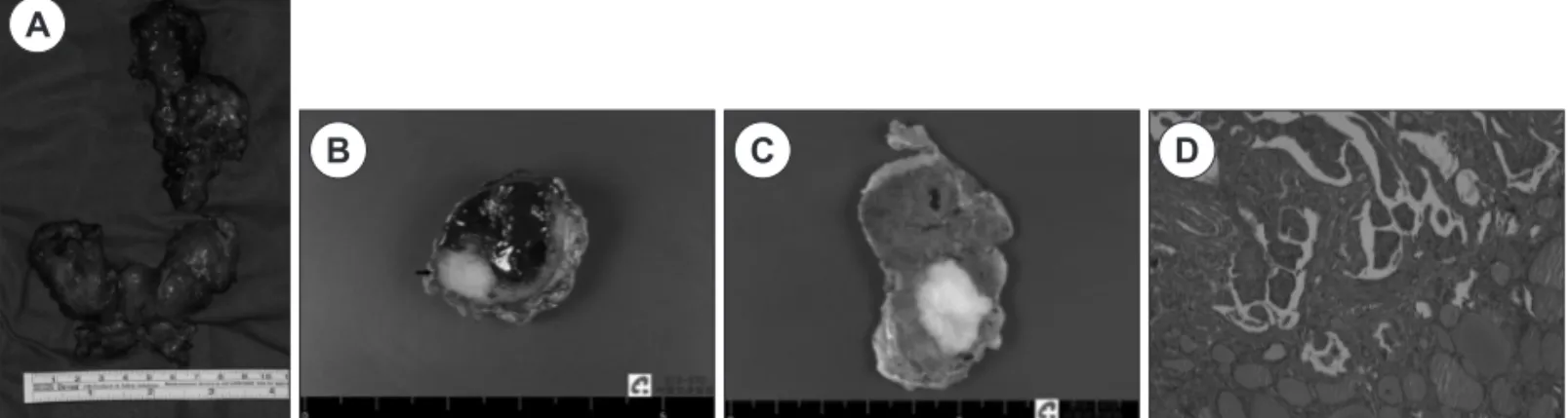
Fig. 2. This photograph shows gross and microscopic findings. A : The surgical specimen after the total thyroidectomy with neck dis- section. B : The gross specimen of metastatic cystic lymph node. It is measured 1.8×1.4×1.4cm with extranodal extension. The cut surface shows well-demarcated solid lesion(arrow). C : The cut surface of left thyroid shows well-margined whitish solid portion. D : It shows papillary growth pattern with colloidal structure(H&E,×100).
A
B C D
- 36 -
고되 있다.9) 또한 이런 낭성 전이성 림프절은 조직학적으로 유 두암의 특징적인 소견을 보이지 않는 경우가 흔하기 때문에 본 증례와 같이 세침흡인검사로 진단하기 어려운 경우가 많다.5) 그 러나 점액성의 초콜렛 색깔의 액체가 흡인되고 세침 흡인물의 갑상샘글로불린의 농도가 증가되면 의심 할 수 있다.10)영상학적으로도 전형적인 양성 낭종과 전이성 낭성 림프절 은 다른 소견을 보인다. 악성 림프절을 시사하는 초음파 소견 으로는 미세석회화를 동반한 비균질성 에코, 종괴의 가로와 세 로축비(transverse-longitudinal dimension ratio)의 상승, 낭 종 내부에 고에코성 요소의 존재, 두꺼운 낭종 벽, 내부의 격막 이 존재하는 경우다.8,11) 악성 림프절에서 전형적인 완전한 낭 종 소견은 드문 것으로 알려져 있다. 이에 상응하는 전산화 단 층 촬영소견으로는 Som 등이 제시한 기준인 중심성 괴사, 종 괴 주변의 조영 증강이 근육보다 큰 경우 그리고 석회화 요소 등이 있으며,8,11) 여기에 낭종내 조영증강 물질의 존재를 강조한 보고도 있다.12)
유두상 갑상선 암종의 낭성 전이의 경우 갑상선의 다발성 침 범, 기관주위 림프절, 흉선 등의 침범이 흔하므로 치료는 갑상 성 전절제술, 선택적 경부 청소술, 그리고 고농도 요오드 동위 원소 치료가 표준이다. 이소성 갑상선암과 새열 낭종에서 발생 한 암종의 경우 갑상선에 병변이 없지만 갑상선 전적출술을 시 행하는것이 권장된다.13) 임상적으로 갑상선에 병변이 없거나, 잠복성이어서 술 전 진단이 불가능한 경우는 경부 낭성 림프절 절제시 동결절편 검사를 이용하여, 그 결과에 따라서 경부 청 소술을 준비해야 한다.14) 그러나 일반적으로는 낭성 전이성 종괴 가 비낭성의 경우 보다 예후는 좋은 것으로 알려져 있다.13)
후향적으로 생각해 보면 이런 증례의 경험 부족으로 고령, 흡 연력, 영상 소견에서 낭종 내부에 고형물질과 격막이 존재 하 는 등의 악성시사 소견을 간과했던 것 같다. 또한 다른 보고된 유사 증례와 달리 낭종의 위치가 전형적인 2형 낭종의 위치 였 으며, 본 병원의 특성상 고령의 환자가 많으며, 갑상선 세포검사 결과 결절성 증식증으로의 보고 등도 진단의 혼선에 영향을 미 친것 같다.
결 론
측경부에 낭성 종괴가 존재하는 경우 전이성 암종에 의한 경우가 있다. 양성 낭성 종괴와의 감별 진단은 올바른 병력 청 취와 신체 검사, 세심한 영상 소견 관찰 그리고 흡인물의 갑상 샘글로블린 수치 등으로 가능하다.
중심 단어 : 전이성 갑상선 유듀상 암종 ・새열 낭종.