Diffuse Sclerosing Variant of Papillary
Thyroid Carcinoma
Sonography and Specimen Radiography
mong various imaging modalities, sonography is generally considered the most accurate imaging modality for the eval-uation and characterization of thyroid nodules1and preop-erative or intraoppreop-erative staging.2Although sonography is widely used in the management of thyroid nodules, this proven efficient modality has its limitations in detecting microcalcifications.3,4
The diffuse sclerosing variant of papillary thyroid carcinoma is a rare variant of papillary thyroid carcinoma, which is mostly char-acterized by the presence of diffusely scattered microcalcifications with or without the presence of an associated mass, diffuse hetero-geneous hypoechogenicity of the background thyroid parenchyma on sonography, and a higher incidence of cervical lymph node metastases.5–7In cases of the diffuse sclerosing variant, which man-ifests as microcalcifications only, sonography may have its limita-tions in detecting microcalcificalimita-tions and assessing the disease extent. Several recent articles reported that specimen radiography could show microcalcifications more easily and the additional can-cer foci that contained microcalcifications, which were not detected on sonography in patients with thyroid carcinoma.4,8
Hyun Kyung Jung, MD, Soon Won Hong, MD, Eun-Kyung Kim, MD, Jung Hyun Yoon, MD, Jin Young Kwak, MD
Received June 21, 2012, from the Departments of Diagnostic Radiology (H.K.J., E-.Y.K., J.Y.K.) and Pathology (S.W.H.), Research Institute of Radio-logical Science, Yonsei University College of Medicine, Seoul, Korea; Department of Diagnos-tic Radiology, Inje University College of Medicine, Haeundae Paik Hospital, Busan, Korea (H.K.J.); and Department of Diagnostic Radiology, Bun-dang CHA Medical Center, CHA University, School of Medicine, Seongnam, Korea (J.H.Y.). Revision requested July 16, 2012. Revised manu-script accepted for publication August 20, 2012.
Address correspondence to Corresponding author: Jin Young Kwak, MD, Department of Radiology, Yonsei University College of Medicine, 50 Yonsei-ro, Seodaemun-gu, Seoul 120-752, Korea.
E-mail: [email protected]
A
The purpose of this pictorial essay is to show the limitations of sonography and com-plementary usefulness of specimen radiography in detecting microcalcifications of the diffuse sclerosing variant of papillary thyroid carcinoma, which mostly manifests as dif-fusely scattered microcalcifications in the thyroid gland.
Key Words—diffuse sclerosing variant of papillary thyroid carcinoma; sonography; specimen radiography; thyroid gland
Sonographic and Specimen Radiographic
Technique
Sonography was performed with a 7–15-MHz linear array transducer (HDI 3000 or 5000; Philips Healthcare, Bothell, WA) or a 5–12-MHz linear array transducer (iU22; Philips Healthcare). When using both machines, compound imaging was performed in all cases. Preopera-tive real-time sonography was performed by 1 of 2 board-certified radiologists with 7 or 11 years of experience in thyroid imaging. After surgery, specimen radiographs were obtained with a Selenia full-field digital mammography system (Lorad/Hologic, Danbury, CT), which is a dedi-cated mammography unit. The system, based on a detec-tor with amorphous selenium, uses a direct capture 70-μm pixel device and yields a 2560 × 3328 matrix image with an 18 × 24-cm paddle. The system was set to allocate 16-bit images and store them at 12 bits. Routine views of thyroid specimens were obtained (focal spot size, 0.3 mm). These images were displayed on a pair of high-resolution 5-megapixel liquid crystal display monitors (MFGD 5621HD; Barco NV, Kortrijk, Belgium) that were part of the review workstation (Selenia Softcopy; Lorad/Hologic) with soft copy reading software (MeVis BreastCare; MeVis Medical Solutions, Bremen, Germany).
Clinical Features and Importance
Previous studies reported the prevalence of the diffuse sclerosing variant of papillary thyroid carcinoma to range from 0.3% to 5.3%.6,9It occurs more frequently in young patients (mean age, 19.5–34.7 years) and in women.10 Serum thyroid antibodies were more frequently increased in patients with a diagnosis of the diffuse sclerosing variant.9 In addition, it has been known to have a high incidence of cervical lymph node metastases, which frequently are bilat-eral, and lung metastasis and, therefore, a more unfavorable 5,7,11
with or without an associated suspicious mass, and the presence of cervical lymph nodes that are suspicious for metastases (Figures 1–5).13Although the prominent sono-graphic “snowstorm appearance” has been well known for the diffuse sclerosing variant, cases presenting as localized microcalcifications were also reported.14 Histopathologi-cally, these microcalcifications are known to correlate with psammoma bodies, extensive fibrosis, and lymphocytic infiltrations.13The diffuse sclerosing variant has unique pathologic features, which are characterized by diffuse involvement of one or both thyroid lobes, dense fibrosis, extensive squamous metaplasia, patchy lymphocytic infil-tration with germinal centers, and numerous psammoma bodies with characteristic nuclear features of papillary thy-roid carcinoma (Figures 1–5).6The diffuse sclerosing vari-ant tends to invade lymphatic vessels in the early stages of the disease and disseminate within the thyroid gland with-out making mass lesions. The extensive lymphocytic infil-trations correlate with a high incidence of lymph node metastasis and post-therapy disease persistence.7
Because of its appearance of diffuse enlargement of the thyroid gland, heterogeneous hypoechogenicity on sonog-raphy, and increased serum thyroid antibodies, the diffuse sclerosing variant of papillary thyroid carcinoma can be con-fused with chronic thyroiditis, and correct diagnosis is often delayed.15However, the abundant psammoma bodies on histopathologic examination are typical for the diffuse scle-rosing variant only, not in patients with chronic thyroiditis. Specimen radiography can show microcalcifications more easily than sonography (Figures 1–5), along with additional cancer foci that contain microcalcifications in patients with a diagnosis of thyroid carcinoma.4,8A recent article reported that specimen radiography is useful for accurate diagnosis and predicting the extent of thyroid car-cinoma.4This article showed that among a total of 122 patients, the microcalcifications within the thyroid were only detected by specimen radiography in 27 patients
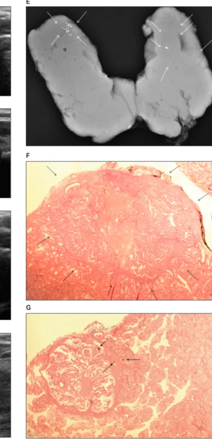
Figure 1. Diffuse sclerosing variant of papillary thyroid carcinoma in a 39-year-old woman. A and B, Transverse and longitudinal sonograms of
the right thyroid gland showing a poorly defined, markedly hypoechoic mass (white arrows) containing microcalcifications (black arrows). C and
D, Transverse and longitudinal sonograms of the left thyroid gland showing no abnormal lesion. E, Specimen radiograph showing scattered faint
amorphous microcalcifications in both lobes of the thyroid gland (white arrows). Microcalcifications located in the left thyroid gland were proven as additional cancer foci in this patient, and the diagnosis was confirmed as the diffuse sclerosing variant. F and G, Pathologic specimens show-ing a main mass (F, black arrows) in the right thyroid gland and additional cancer foci with psammoma bodies (G, black arrows) in the left thyroid gland (hematoxylin-eosin, original magnification ×100).
A C B D E G F
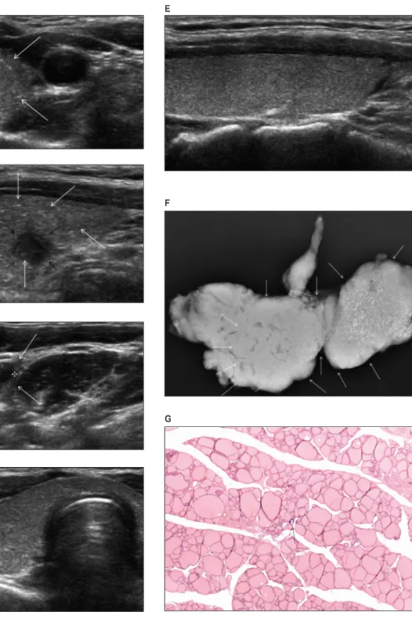
Figure 2. Diffuse sclerosing variant of papillary thyroid carcinoma in a 29-year-old woman. A and B, Transverse and longitudinal sonograms of
the left thyroid gland showing a poorly defined hypoechoic mass (black arrows) and microcalcifications (white arrows) both within and outside the mass. C, Transverse sonogram of the left neck at level IV showing a 1-cm lymph node containing echogenic microcalcifications (white arrows).
D and E, Transverse and longitudinal sonograms of the right thyroid gland showing no abnormal findings. F, Specimen radiograph showing
dif-fusely scattered microcalcifications (white arrows) in both thyroid glands. Microcalcifications of the right thyroid gland were detected only on specimen radiography, which showed additional cancer foci in this patient. G, Pathologic specimen of the right lobe showing psammoma bodies (black arrows), which correlated with the findings on specimen radiography (hematoxylin-eosin, original magnification ×100).
A C B E G F
shows that specimen radiography can potentially con-tribute in 3 ways: (1) by showing that the diffuse scleros-ing variant of papillary thyroid carcinoma should be considered when sonography shows microcalcifications that are not associated with a nodule; (2) by encouraging ultrasound system manufacturers to make changes to their
equipment to improve microcalcification detection rates; and (3) by encouraging pathologists to use specimen radi-ography, assuming that any improvement in sensitivity can be shown to be beneficial in some way, such as by detect-ing microcalcifications that might not be found with rou-tine specimen processing.
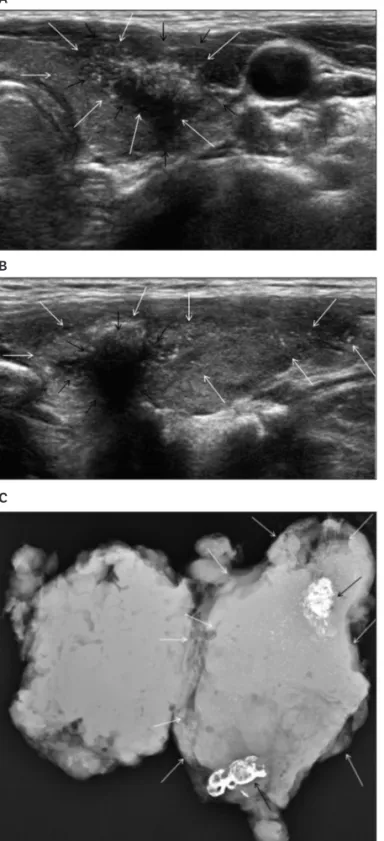
Figure 3. Diffuse sclerosing variant of papillary thyroid carcinoma in
a 33-year-old woman. A and B, Transverse and longitudinal sono-grams of the left thyroid gland showing a poorly defined hypoechoic mass (black arrows) and microcalcifications (white arrows) both within and outside the mass. C, Specimen radiograph showing diffusely scat-tered microcalcifications (white arrows) and dystrophic calcifications (black arrows) in the left thyroid gland. D, Pathologic specimen show-ing a main mass with conglomerated cancer foci (black arrows) and abundant psammoma bodies (blue arrows; hematoxylin-eosin, origi-nal magnification ×100). E, Pathologic specimen showing psammoma bodies (black arrows) also located outside the main mass. The final diagnosis was confirmed as the diffuse sclerosing variant (hema-toxylin-eosin, original magnification ×100).
A
C
B D
Figure 4. Diffuse sclerosing variant of papillary thyroid carcinoma in a 46-year-old woman. A and B, Transverse and longitudinal sonograms of the right
thyroid gland showing a poorly defined hypoechoic mass (black arrows) with internal microcalcifications (white arrows). C and D, Transverse and lon-gitudinal sonograms of the right central neck showing a suspicious lymph node containing internal microcalcifications (white arrows). E, Specimen radiograph showing amorphous microcalcifications in the right thyroid gland (white arrow) and the right central neck (black arrow). F, Pathologic spec-imen showing a mass within the thyroid gland (black arrows; hematoxylin-eosin, original magnification ×100). G, Pathologic specspec-imen showing psam-moma bodies (black arrows) in the right central neck lymph node, suggesting metastasis (hematoxylin-eosin, original magnification ×100).
A
C B
E
Conclusions
Although sonography is an essential modality in the eval-uation of thyroid nodules, specimen radiography is more sensitive than sonography for detection of microcalcifica-tions of thyroid carcinoma, especially in the diagnosis of the diffuse sclerosing variant of papillary thyroid carci-noma. However, new diagnostic techniques that can detect microcalcifications in the thyroid along with the disease extent and that can be performed preoperatively have not yet been established. This new technique is expected to be of further use in the future, thus helping management for patients with thyroid carcinoma.
References
1. Pacini F, Schlumberger M, Dralle H, Elisei R, Smit JW, Wiersinga W; European Thyroid Cancer Taskforce. European consensus for the man-agement of patients with differentiated thyroid carcinoma of the follicu-lar epithelium.Eur J Endocrinol 2006; 154:787–803.
2. Lew JI, Solorzano CC. Use of ultrasound in the management of thyroid cancer. Oncologist 2010; 15:253–258.
3. Kim EK, Park CS, Chung WY, et al. New sonographic criteria for rec-ommending fine-needle aspiration biopsy of nonpalpable solid nodules of the thyroid. AJR Am J Roentgenol 2002; 178:687–691.
4. Kwak JY, Kim EK, Hong SW, Kim MJ, Moon HJ, Park CS. Value of spec-imen radiographs in diagnosing multifocality of thyroid cancer. Br J Surg
2010; 97:517–524.
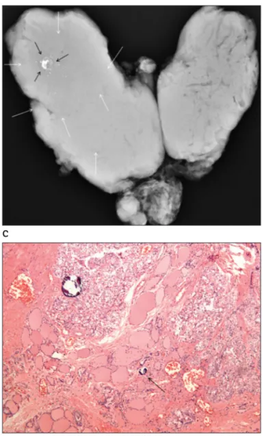
Figure 5. Diffuse sclerosing variant of papillary thyroid carcinoma in a
35-year-old woman. A and B, Transverse and longitudinal sonograms of the right thyroid gland showing a poorly defined hypoechoic mass (black arrows) with internal microcalcifications (white arrows). C, Specimen radiograph showing dystrophic calcifications (black arrows) and addi-tional areas of scattered microcalcifications (white arrows) consisting of a broader extent of the right thyroid gland compared to the sonograms.
D, Pathologic specimen showing psammoma bodies (black arrow),
which are located outside the main mass (hematoxylin-eosin, original magnification ×100).
A
C
B
5. Zhang Y, Xia D, Lin P, Gao L, Li G, Zhang W. Sonographic findings of the diffuse sclerosing variant of papillary carcinoma of the thyroid.
J Ultrasound Med 2010; 29:1223–1226.
6. Kwak JY, Kim EK, Hong SW, et al. Diffuse sclerosing variant of papillary carcinoma of the thyroid: ultrasound features with histopathological cor-relation. Clin Radiol 2007; 62:382–386.
7. Lee JY, Shin JH, Han BK, et al. Diffuse sclerosing variant of papillary car-cinoma of the thyroid: imaging and cytologic findings.Thyroid 2007;
17:567-573.
8. Kwak JY, Kim EK, Hong SW, et al. Diffuse slcerosing variant of papillary carcinoma of the thyroid gland: specimen radiographic features with histopathological correlation. J Clin Endocrinol Metab 2009; 94:1491–
1492.
9. Chow SM, Chan JK, Law SC, et al. Diffuse sclerosing variant of papillary thyroid carcinoma: clinical features and outcome. Eur J Surg Oncol 2003;
29:446–449.
10. Lam AK, Lo CY. Diffuse sclerosing variant of papillary carcinoma of the thyroid: a 35-year comparative study at a single institution. Ann Surg Oncol
2006; 12:176–181.
11. Caplan RH, Wester S, Kisken AW. Diffuse sclerosing variant of papillary thyroid carcinoma: case report and review of the literature. Endocr Pract
1997; 3:287–292.
12. Fujimoto Y, Obara T, Ito Y, Kodama T, Aiba M, Yamaguchi K. Diffuse sclerosing variant of papillary carcinoma of the thyroid: clinical impor-tance, surgical treatment and follow-up study. Cancer 1990; 66:2306–
2312.
13. Kobayashi K, Fukata S, Amino N, Miyauchi A. A case with diffuse scle-rosing variant of papillary carcinoma of the thyroid: characteristic features on ultrasonography. J Med Ultrasonics 2006; 33:159–161.
14. Kwak JY, Kim EK, Son EJ, et al. Papillary thyroid carcinoma manifested solely as microcalcifications on sonography. AJR Am J Roentgenol 2007;
189:227-–231.
15. Martin-Pérez E, Larrañaga E, Serrano P. Diffuse sclerosing variant of pap-illary carcinoma of the thyroid. Eur J Surg 1998; 164:713–715.