Copyright © 2017. The Korean Society for Radiation Oncology
www.e-roj.org
This is an Open Access article distributed under the terms of the Creative Commons Attribution Non-Commercial License (http://creativecommons.org/
licenses/by-nc/4.0/) which permits unrestricted non-commercial use, distribution, and reproduction in any medium, provided the original work is properly cited.
227
Radiat Oncol J 2017;35(3):227-232 https://doi.org/10.3857/roj.2017.00101 pISSN 2234-1900 · eISSN 2234-3156
Purpose: The purpose of this study was to evaluate the prognostic value of the lymph node ratio (LNR), which was defined as the proportion of involved nodes of all dissected nodes, in pN1 breast cancer.
Materials and Methods: We retrospectively analyzed the clinical data of patients with pN1 breast cancer (n = 144) treated at Keimyung University Dongsan Medical Center, Daegu, Korea between 2001 and 2010. The median age was 46 years (range, 27 to 66 years). The LNR was 0.01–0.15 (low LNR) in 130 patients and >0.15 (high LNR) in 14 patients. Sixty-five patients (45.1%) had T1 tumors, 74 (51.4%) had T2 tumors, and 5 (3.5%) had T3 tumors. Eighty-eight patients (61.1%) underwent total mastectomy and 56 (38.9%) underwent partial mastectomy. Fifty-nine patients (41.0%) underwent radiotherapy and 12 (8.3%) underwent regional radiotherapy. The median follow-up period was 65 months.
Results: The 5- and 10-year disease-free survival (DFS) rates were 92.7% and 82.4%, respectively. Univariate analyses revealed that high LNR (p = 0.004), total mastectomy (p = 0.006), no local radiotherapy (p = 0.036), and stage T2 or T3 (p = 0.010) were associated with worse DFS. In multivariable analysis, only high LNR (p = 0.015) was associated with worse DFS.
Conclusion: High LNR is an independent prognostic factor in pN1 breast cancer and could be an indication for adjuvant radiotherapy in these patients.
Keywords: Breast neoplasm, N1, Lymph node ratio
Clinical significance of the lymph node ratio in N1 breast cancer
Jaeho Kim, MD
1, Jin Hee Kim, MD, PhD
1, Ok Bae Kim, MD, PhD
1, Young Kee Oh, PhD
1, Seung Gyu Park, MD
21
Department of Radiation Oncology, Keimyung University School of Medicine, Daegu;
2
Department of Radiation Oncology, Dogae Health Subcenter, Gumi, Korea
Received 04 January 2017, Revised 08 February 2017, Accepted 27 March 2017.
Correspondence: Jin Hee Kim, MD, PhD, Departments of Radiation Oncology, Keimyung University Dongsan Medical Center, Keimyung University School of Medicine, 56 Dalseong-ro, Jung-gu, Daegu 41931, Korea. Tel: +82-53-250-7665, Fax: + 82-53- 250-7984, E-mail: [email protected]
Introduction
Many randomized trials and a meta-analysis have shown that adequate postoperative locoregional radiotherapy for node-positive breast cancer reduces locoregional recurrence and improves the survival rate [1,2]. Therefore, the National Comprehensive Cancer Network guidelines state that locoregional radiation therapy should be considered for node- positive patients after axillary lymph-node dissection [3]. There
is also general consensus that locoregional radiotherapy should
be considered for patients with four or more involved axillary
nodes [4]. However, although the American Joint Committee
on Cancer (AJCC) staging system reflects the disease state
by emphasizing the prognostic importance of the absolute
number of positive lymph nodes, there may be a discrepancy
between the absolute number of positive nodes and the
substantive extent of axillary-node metastasis, especially
in patients with up to three positive nodes. Moreover, the
decision to administer regional radiotherapy based on the patient’s pN1 node status differs between physicians [5-8].
In this context, the lymph node ratio (LNR), which is defined as the proportion of positive axillary lymph nodes of the total number of axillary lymph nodes removed, may resolve this problem [9-11]. Veronesi et al. [12] suggested that the LNR may reduce the discrepancy between clinical evaluation and the actual status of the lymph nodes that is due to the differing practices among physicians.
Therefore, recent studies have focused on the LNR in patients with pN1 breast cancer [13,14]. In this retrospective study, we assessed the prognostic value of the LNR and its potential use as an indication for locoregional radiotherapy after mastectomy in patients with pN1 breast cancer.
Materials and Methods
1. Patients
We performed a retrospective analysis of the clinical data of patients with pN1 invasive breast cancer who were treated at Keimyung University Dongsan Medical Center, Daegu, Korea between 2001 and 2010. The Institutional Review Board approval from Keimyung University Dongsan Medical Center on October 25, 2016. Eligible criteria were as follows: patients with unilateral breast cancer who underwent total mastectomy or partial mastectomy with whole breast radiotherapy, one to three positive lymph nodes, and no distant metastasis at the time of diagnosis. Patients who underwent neoadjuvant chemotherapy before surgery were excluded. In patients with a positive resection margin, re-excision was performed to achieve a margin-free status. In total, 149 patients were initially screened for this study, of which 5 patients underwent sentinel lymph node biopsy and were excluded from the study. Therefore, 144 patients were included in this study.
All patients received postoperative adjuvant chemotherapy, with an adriamycin/cyclophosphamide–paclitaxel regimen in most patients (139, 93.3%). All of the patients underwent either total mastectomy or partial mastectomy with axillary lymph node dissection. Lymph node dissection was usually performed up to level II. None of the patients had any serious comorbidity.
The local radiotherapy field consisted of the whole breast in partial mastectomy patients or the chest wall in total mastectomy patients. All of the partial mastectomy patients underwent local radiotherapy. Of the total mastectomy patients, only three underwent local radiotherapy and all of them underwent regional radiotherapy. The breast or chest
wall was irradiated with 6 MV photons; the median dose of local radiotherapy was 5,040 cGy (range, 4,500 to 5,400 cGy), with 180 cGy per fraction. An electron boost was applied to the tumor bed in all partial mastectomy patients after local radiotherapy (median, 1,000 cGy/5 fractions). The regional radiotherapy field included the supraclavicular axillary lymph nodes. The internal mammary lymph nodes (IMN) were also included in patients with poor prognostic factors such as primary tumor located in an inner quadrant, large tumor, or lymphovascular invasion. Among patients who underwent regional radiotherapy, four patients received radiotherapy with an IMN field. The dose of regional radiation therapy was 5,000 cGy, with 200 cGy given per fraction. The median duration of radiotherapy was 44 days.
2. Clinical and pathological factors and lymph node status
Age, tumor size, operation type, local radiotherapy, regional lymph node radiotherapy, adjuvant hormone therapy, pathology, resection margin, lymphovascular invasion, extracapsular extension, molecular subtype, histological grade, and LNR were used to assess the risk of recurrence.
Staging was based on the 7th edition of the AJCC Cancer Staging Manual. A close margin was defined as the presence of invasive carcinoma within 2 mm of the surgical resection margin. Luminal type A was defined as hormone receptor- positive, HER2-negative and Ki67 <14%. Luminal type B was defined as hormone receptor-positive plus either Ki67 ≥14%
or HER2-positive. HER2-enriched was defined as hormone receptor-negative and HER2-positive. Triple negative was defined as hormone receptor-negative and HER2-negative.
The LNR was arbitrarily set to 0.01 units and the log- rank test was performed with disease-free survival (DFS) to determine the appropriate LNR cutoff value. Based on the results of these analyses, we used a cutoff value of 0.15, which yielded the most significant result and divided patients into two groups according to the LNR, low LNR (≤0.15) and high LNR (>0.15).
3. Follow-up and endpoints
DFS was defined as the time from the date of diagnosis to locoregional recurrence, distant metastasis, or contralateral invasive breast cancer. The patients were followed-up every 3–6 months after surgery with history and physical examinations.
Mammography was performed every 12 months. Additional
imaging studies were performed in patients with suspicious
clinical signs or symptoms.
www.e-roj.org 229
https://doi.org/10.3857/roj.2017.00101
4. Statistical analysis
Data were analyzed with SPSS ver. 18.0 for Windows (SPSS Inc., Chicago, IL, USA). The Kaplan–Meier method was used to analyze DFS and statistical significance was determined using log-rank tests. Cox stepwise regression analysis was used for multivariable analysis. Statistically significant variables in the univariate analysis (p < 0.05) were included in the Cox regression model. Statistical significance was set at p < 0.05.
Results
The characteristics of the 144 patients with pN1 breast cancer are summarized in Table 1. Of 144 evaluable patients, 65 (45.1%), 74 (51.4%), and 5 (3.5%) had T1, T2, and T3 lesions, respectively. More than 20 lymph nodes were dissected in 59 (41.0%) patients. Based on the LNR cutoff value of 0.15, the low LNR group comprised 130 (90.3%) patients and the high LNR group comprised 14 (9.7%) patients. The LNR was statistically independent of other prognostic factors such as age, operation type, T stage, resection margin, lymphovascular invasion, molecular subtype, and histologic grade.
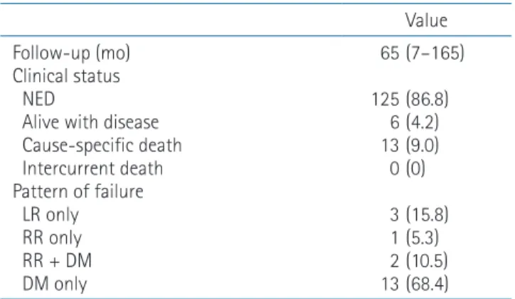
The median follow-up time was 65 months (range, 7 to 165 months). The 5- and 10-year DFS rates were 92.7% and 82.4%, respectively (Fig. 1). The patterns of failure are listed in Table 2. When all 19 recurrences were classified according to regional radiotherapy, recurrence was noted in 18 (13.6%) patients who did not undergo regional radiotherapy while only 1 (8.3%) patient in the regional radiotherapy group suffered distant metastasis. None of the patients in the regional radiotherapy group experienced locoregional recurrence. There were no cases of contralateral breast cancer recurrence. None Table 1. Patient characteristics
Characteristic Value
Age (yr) <40 ≥40 Tumor size T1 T2
T3 Number of positive lymph nodes 1 2
3 Number of dissected lymph nodes ≤20
>20 LNR Low (≤0.15) High (>0.15) Operation type Total mastectomy Partial mastectomy Local radiotherapy No Yes
Regional radiotherapy No Yes
Pathology IDC Other
Histologic grade I & II
III Unknown LVI No Yes Unknown ECE No Yes Unknown
Resection margin (mm) Close (<2)
Clear (≥2) Unknown Molecular subtype Luminal A
a)Luminal B
b)HER2-enriched
c)Triple negative
d)Unknown
Adjuvant hormone therapy No Yes
46 (27–66) 8 (5.6) 136 (94.4)
65 (45.1) 74 (51.4) 5 (3.5) 80 (55.6) 46 (31.9) 18 (12.5) 17 (5–43) 85 (59.0) 59 (41.0) 0.09 (0.02–0.23) 130 (90.3)
14 (9.7) 88 (61.1) 56 (38.9) 85 (59.0) 59 (41.0) 132 (91.7) 12 (8.3) 139 (96.5)
5 (3.5) 62 (43.1) 72 (50.0) 10 (6.9) 57 (39.6) 74 (51.4) 13 (9.0) 100 (69.4)
21 (14.6) 23 (16.0) 21 (14.6) 122 (84.7) 1 (0.7) 63 (43.8) 41 (28.5) 7 (4.9) 22 (15.3)
11 (7.6) 31 (21.5) 113 (78.5) Values are presented as median (range) or number (%).
LNR, lymph node ratio; IDC, invasive ductal carcinoma; LVI, lym- phovascular invasion; ECE, extracapsular extension.
a)
Hormone receptor-positive, HER2-negative and Ki67 <14%.
b)
Hormone receptor-positive & Ki67 ≥14% or HER2-positive.
c)
Hormone receptor-negative and HER2-positive.
d)
Hormone receptor-negative and HER2-negative.
Table 2. Clinical status and patterns of failure
Value Follow-up (mo)
Clinical status NED
Alive with disease Cause-specific death Intercurrent death Pattern of failure LR only RR only RR + DM DM only
65 (7–165) 125 (86.8)
6 (4.2) 13 (9.0) 0 (0) 3 (15.8) 1 (5.3) 2 (10.5) 13 (68.4) Values are presented as median (range) or number (%).
NED, no evidence of disease; LR, local recurrence; RR, regional
recurrence; DM, distant metastasis.
230 www.e-roj.org
https://doi.org/10.3857/roj.2017.00101of the patients experienced adverse effects associated with radiotherapy.
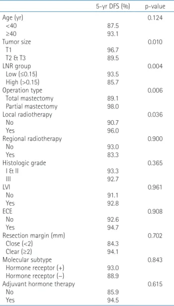
In univariate analyses, large tumor, high LNR, absence of local radiotherapy, and operation type (total mastectomy) were associated with poor DFS. In the subsequent multivariable analysis, only high LNR was significantly associated with poor DFS (Tables 3, 4).
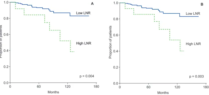
To further evaluate the prognostic role of LNR, we
analyzed DFS in 132 patients who did not undergo regional radiotherapy, of which 13 (9.8%) were in the high LNR group.
DFS was significantly worse in the high LNR group than in the low LNR group (Fig. 2).
Discussion and Conclusion
It is widely accepted that postoperative locoregional radiation therapy reduces locoregional recurrence and mortality in patients with lymph-node-positive breast cancer [1]. A meta- analysis conducted by the Early Breast Cancer Trialists' Collaborative Group (EBCTCG) and a report of the MA.20 study indicate that regional radiotherapy significantly reduced the overall recurrence rate in patients with lymph-node-positive breast cancer, even when adjuvant systemic chemotherapy was administered [15,16]. The EBCTCG study also showed that the absolute benefit of radiotherapy for N1 breast cancer patients is small, but more effective radiotherapeutic regimens introduced since that study should provide better outcomes.
Regional radiotherapy may have considerable benefits in selected patients with N1 breast cancer. However, the prognostic factors associated with reduced risk of locoregional recurrence after regional radiotherapy have not been reliably defined when dividing patients into subgroups. Therefore, there are currently no prognostic factors that provide clear support for locoregional radiotherapy in patients with pN1 breast cancer.
1.0
0.8
0.6
0.4
0.2
0.00 60 120 180
5-year DFS : 92.7%
10-year DFS : 82.4%
Proportion of patients
Months
Fig. 1. Kaplan–Meier estimates of disease-free survival (DFS).
1.0
0.8
0.6
0.4
0.2
0.00 60 120 180
p = 0.003
Proportion of patients
Months
High LNR Low LNR
p = 0.004 High LNR Low LNR 1.0
0.8
0.6
0.4
0.2
0.00 60 120 180
Proportion of patients
Months
High LNR Low LNR
B A
1.0
0.8
0.6
0.4
0.2
0.0
p = 0.003
Proportion of patients
High LNR Low LNR
p = 0.004 High LNR Low LNR 1.0
0.8
0.6
0.4
0.2
0.00 60 120 180
Proportion of patients
Months
High LNR Low LNR
B A
Fig. 2. Comparison of disease-free survival between patients with a low lymph node ratio (low LNR, solid line) and patients with a high
lymph node ratio (high LNR, dashed line). (A) All patients. (B) Patients who did not undergo regional radiotherapy.www.e-roj.org 231
https://doi.org/10.3857/roj.2017.00101