142
서 론
유방암 환자에서 액와 림프절 곽청술은 정확한 병기를 결정하며 전신요법의 필요 유무를 판단 및 예후를 예측할 수 있게 한다. 그 외에도 액와 부위의 국소 재발을 방지하 는 치료 효과를 기대한다. 그러나 림프절 전이가 없는 환 자에서는 그 치료 효과가 의문시되며 보조 화학요법의 시 행 여부도 종양의 크기 및 여러 생물학적 표지자를 이용 하여 결정하게 된다. 그러나 액와부 곽청술 전에 림프절 전이를 미리 정확하게 예측할 수 있는 방법은 아직 밝혀 내지 못하고 있다. 다만 종양의 크기가 매우 유의성이 높 은 인자로 인정되며 그 외 핵분화 등급 및 림프관 침습 유 무 등이 공통적으로 유의성이 있는 인자로 인정되고 있 다.(1,2) 액와 림프절 곽청술은 정도의 차이는 있지만 50%
정도에서 감각이상, 통증, 부종, 팔의 운동장애 그리고 감 염 등의 합병증(3)이 나타난다. 따라서 이러한 합병증을 줄이기 위하여 크기가 작은 종양이나, 고령 그리고 예후 가 좋을 것으로 예상되는 일부 유방암에서는 선택적으로 액와 림프절 곽청술을 생략하거나 최근 호응을 받는 감시 림프절(sentinel node) 검사로 대체하는 등의 시도가 이루 어지고 있다. 특히 5 mm 이하의 종양 등에서는 5% 미만 의 림프절 전이가 있으므로 생략하는 것이 효과적일 수 있다는 주장도 있다.(4)
저자는 이들 여러 조건 중에서 종양의 크기에 따른 림 프절 전이율을 조사하여 종양의 크기에 따른 액와 림프절 곽청술 생략이 가능한 가에 대하여 조사하여 보았다.
방 법
1985년 1월부터 1998년 12월까지 미국 뉴욕 코넬의과대 학의 Strang Cancer Prevention Center에 등록된 유방암 환 자 중 종양의 크기가 2 cm 이하인 경우를 후향적 조사를 하였다. 이 중 종야의 크기에 대한 명확한 수치가 확인되
T1 유방암 환자의 액와 림프절 전이율
건국대학교 의과대학 외과학교실 함 희 원
Incidence of Axillary Lymph Node Metastases in T1 Breast Cancer
Hee-Won Ham, M.D.
Department of Surgery, KonKuk University College of Medicine, Seoul, Korea
Purpose: Tumor size is the strongest predictor of axillary
node metastases. Some authors have reported that axillary dissection in T1a breast cancer is not required because the rate of incidence is less than 5%. However I have doubts concerning the omission of axillary dissection in small breast cancers. Therefore, I investigated the incidence of axillary node metastases in T1 breast cancer according to size for the purpose of using this data as a reference for determining whether or not to dissect axillary lymph nodes.Methods: Data of patients registered at the Strang Cancer
Prevention Center affiliated with the New York Hospital- Cornell Medical Center, from January 1988 to December 1998 were reviewed. After review of charts and pathologic reports for tumor size, age at operation and lymph node status, 592 patients were proven to have primary breast tumor 2 cm in size or smaller. The size of the tumor was determined as the largest diameter of the invasive lesion when possible.Results: Lymph node metastases were seen in 7 of 68
cases in the 0.1∼0.5 cm T1a (10.3%), 29 of 182 in 0.6∼1.0 cm T1b (15.9%), 50 of 206 in 1.1∼1.5 cm (24.3%) and 55 of 136 in 1.6∼2.0 cm tumor size range (40.4%).
Conclusion: Although positive node occurrence was lower
in small size tumors, significant number of patients with T1a invasive tumors have a positive node. Therefore, a small size of tumor alone is not an indicator for the omission of axillary dissection. (Journal of Korean Breast CancerSociety 2002;5:142-146)
ꠏꠏꠏꠏꠏꠏꠏꠏꠏꠏꠏꠏꠏꠏꠏꠏꠏꠏꠏꠏꠏꠏꠏꠏꠏꠏꠏꠏꠏꠏꠏꠏꠏꠏꠏꠏꠏꠏꠏꠏꠏꠏꠏꠏꠏ
Key Words: Breast cancer, Lymph-node metastases, T1
중심 단어: 유방암, 림프절 전이, T1책임저자:함희원, 서울시 광진구 화양동 1번지 ꂕ 143-130, 민중병원 외과
Tel: 02-450-9682, Fax: 02-458-1134 E-mail: [email protected]
접수일:2001년 3월 7일, 게재승인일:2001년 3월 9일 본 논문은 건국대학교 재단 법인 지원으로 이루어짐.
ꠏꠏꠏꠏꠏꠏꠏꠏꠏꠏꠏꠏꠏꠏꠏꠏꠏꠏꠏꠏꠏꠏꠏꠏꠏꠏꠏꠏꠏꠏꠏꠏꠏꠏꠏꠏꠏꠏꠏꠏꠏꠏꠏꠏꠏꠏꠏꠏꠏꠏꠏꠏꠏꠏꠏꠏꠏꠏꠏꠏꠏꠏꠏꠏꠏꠏꠏꠏꠏꠏꠏꠏꠏꠏꠏꠏꠏꠏꠏꠏꠏꠏꠏꠏꠏꠏꠏꠏꠏꠏꠏꠏꠏꠏꠏꠏꠏꠏ 지 않거나 수술 전 화학요법 치료를 받은 경우, 양측성 유
방암 그리고 액와 림프절 곽청술이 생략된 경우를 제외한 592명을 대상으로 의무 기록지와 병리 검사보고서를 바탕 으로 종양의 크기, 수술시 나이 그리고 전이된 림프절 수 를 확인하였다. 종양의 크기는 침윤성 부위만의 수치를 이용하였으며 따로 언급이 없는 경우에는 병리 보고서에 기술되어 있는 크기를 이용하여 각 5 mm 단위로 구분하 였다. 전이 림프절에 대한 조사에서는 액와 림프절 곽청 술을 시행한 경우에는 범위에 관계없이 다 포함 시켰으며 4개 이하의 감시 림프절만 제거한 종양 55예도 포함 시켰 다. 이 대상 환자의 자료를 이용하여 연령, 종양 크기의 분포도 및 각 크기에 따른 림프절 전이율 그리고 전이된 림프절의 수를 조사하였다. 크기에 따른 림프절 전이율의 상관관계 여부는 SPSS 프로그램의 Pearson correlation을 이용하여 확인하였다.
결 과
1) 조사대상의 연령별 분포도 및 크기별 분포도 592명의 나이별 분포도는 40대가 30.2%로 가장 많았으 며 다음은 50대로 25.3%였으며 평균 54.5세였다(Table 1).
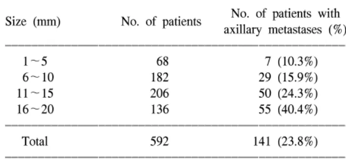
종양 크기에 따른 분포도는 5 mm 이하가 11.5%, 6∼10 mm가 30.7%, 11∼15 mm는 34.8% 그리고 16∼20 mm가 23.0%로 11∼15 mm 크기가 가장 많았다.
2) 종양의 크기에 따른 림프절 전이율
592명의 환자에서 평균 17.2개의 림프절들을 절제하였 으며 354개의 전이된 림프절을 발견하였다. 종양의 크기 에 따른 림프절 전이율은 Table 2와 같다. 림프절 전이율 은 종양의 크기에 따라 정비례하여 증가하는 유의한 상관 관계가 있었다(significant Pearson correlation at the 0.01 level).
5 mm 이하 종양 68예 중에는 1 mm의 미세 침윤 소견 을 가진 종양이 18예가 있었으며 이 중 1예에서 림프절 전이가 있었다. 그 1예는 multicentric focus를 가진 상피내 암 중 일부에서 미세 전이가 발견된 경우였다.
3) 전이된 림프절 숫자에 따른 분포도
각 군의 전이된 림프절 개수가 1개, 2개 이하 혹은 3개 이하인 경우를 조사한 결과는 Table 3과 같다. 종양의 크 기가 5 mm 이하인 경우는 전부 2개 이하의 전이만 있었 으며 전체의 84%에서 3개 이하만의 전이만 있었다.
고 찰
유방암 환자에서 액와 림프절 곽청술의 목적은 림프절 전이 유무를 확인하여 정확한 병기를 알아 그 예후를 예 측하게 하며 전신요법의 치료여부를 결정하게 한다. 그리 고 전이된 림프절에 대한 국소 재발을 줄이는 치료 효과 를 기대하지만, Fisher등(5)에 의하면 액와 림프절 전이가
Table 1. Age distribution
ꠚꠚꠚꠚꠚꠚꠚꠚꠚꠚꠚꠚꠚꠚꠚꠚꠚꠚꠚꠚꠚꠚꠚꠚꠚꠚꠚꠚꠚꠚꠚꠚꠚꠚꠚꠚꠚꠚꠚꠚꠚꠚꠚꠚꠚꠚꠚꠚꠚꠚꠚ Age group No. of patients Distribution (%) ꠏꠏꠏꠏꠏꠏꠏꠏꠏꠏꠏꠏꠏꠏꠏꠏꠏꠏꠏꠏꠏꠏꠏꠏꠏꠏꠏꠏꠏꠏꠏꠏꠏꠏꠏꠏꠏꠏꠏꠏꠏꠏꠏꠏꠏꠏꠏꠏꠏꠏꠏ
21∼30 11 1.9
31∼40 61 10.3
41∼50 179 30.2
51∼60 150 25.3
61∼70 127 21.4
71∼80 56 9.5
81∼ 8 1.4
ꠏꠏꠏꠏꠏꠏꠏꠏꠏꠏꠏꠏꠏꠏꠏꠏꠏꠏꠏꠏꠏꠏꠏꠏꠏꠏꠏꠏꠏꠏꠏꠏꠏꠏꠏꠏꠏꠏꠏꠏꠏꠏꠏꠏꠏꠏꠏꠏꠏꠏꠏ
592 100
ꠏꠏꠏꠏꠏꠏꠏꠏꠏꠏꠏꠏꠏꠏꠏꠏꠏꠏꠏꠏꠏꠏꠏꠏꠏꠏꠏꠏꠏꠏꠏꠏꠏꠏꠏꠏꠏꠏꠏꠏꠏꠏꠏꠏꠏꠏꠏꠏꠏꠏꠏ
Table 3. Percentage of patients with each one, below 2 and below 3 positive node
ꠚꠚꠚꠚꠚꠚꠚꠚꠚꠚꠚꠚꠚꠚꠚꠚꠚꠚꠚꠚꠚꠚꠚꠚꠚꠚꠚꠚꠚꠚꠚꠚꠚꠚꠚꠚꠚꠚꠚꠚꠚꠚꠚꠚꠚꠚꠚꠚꠚꠚꠚꠚꠚꠚꠚꠚꠚꠚꠚꠚꠚꠚꠚꠚꠚꠚꠚꠚꠚꠚꠚꠚꠚꠚꠚꠚꠚꠚꠚꠚꠚꠚꠚꠚꠚꠚꠚꠚꠚꠚꠚꠚꠚꠚꠚꠚꠚꠚꠚꠚꠚꠚꠚꠚꠚꠚꠚꠚ
No. of patients Only 1 Below 2 Below 3
Size (mm)
with positive node positive node positive node positive node
ꠏꠏꠏꠏꠏꠏꠏꠏꠏꠏꠏꠏꠏꠏꠏꠏꠏꠏꠏꠏꠏꠏꠏꠏꠏꠏꠏꠏꠏꠏꠏꠏꠏꠏꠏꠏꠏꠏꠏꠏꠏꠏꠏꠏꠏꠏꠏꠏꠏꠏꠏꠏꠏꠏꠏꠏꠏꠏꠏꠏꠏꠏꠏꠏꠏꠏꠏꠏꠏꠏꠏꠏꠏꠏꠏꠏꠏꠏꠏꠏꠏꠏꠏꠏꠏꠏꠏꠏꠏꠏꠏꠏꠏꠏꠏꠏꠏꠏꠏꠏꠏꠏꠏꠏꠏꠏꠏꠏ
1∼5 7 4 (57.1%) 7 (100%) -
6∼10 29 17 (58.6%) 25 (86.2%) 26 (89.7%)
11∼15 50 26 (52.0%) 35 (70%) 41 (82.0%)
16∼20 55 24 (43.6%) 36 (65.5%) 43 (78.2%)
ꠏꠏꠏꠏꠏꠏꠏꠏꠏꠏꠏꠏꠏꠏꠏꠏꠏꠏꠏꠏꠏꠏꠏꠏꠏꠏꠏꠏꠏꠏꠏꠏꠏꠏꠏꠏꠏꠏꠏꠏꠏꠏꠏꠏꠏꠏꠏꠏꠏꠏꠏꠏꠏꠏꠏꠏꠏꠏꠏꠏꠏꠏꠏꠏꠏꠏꠏꠏꠏꠏꠏꠏꠏꠏꠏꠏꠏꠏꠏꠏꠏꠏꠏꠏꠏꠏꠏꠏꠏꠏꠏꠏꠏꠏꠏꠏꠏꠏꠏꠏꠏꠏꠏꠏꠏꠏꠏꠏ Table 2. Incidence of axillary metastases according to tumor size ꠚꠚꠚꠚꠚꠚꠚꠚꠚꠚꠚꠚꠚꠚꠚꠚꠚꠚꠚꠚꠚꠚꠚꠚꠚꠚꠚꠚꠚꠚꠚꠚꠚꠚꠚꠚꠚꠚꠚꠚꠚꠚꠚꠚꠚꠚꠚꠚꠚꠚꠚ No. of patients with Size (mm) No. of patients
axillary metastases (%) ꠏꠏꠏꠏꠏꠏꠏꠏꠏꠏꠏꠏꠏꠏꠏꠏꠏꠏꠏꠏꠏꠏꠏꠏꠏꠏꠏꠏꠏꠏꠏꠏꠏꠏꠏꠏꠏꠏꠏꠏꠏꠏꠏꠏꠏꠏꠏꠏꠏꠏꠏ
1∼5 68 7 (10.3%)
6∼10 182 29 (15.9%)
11∼15 206 50 (24.3%)
16∼20 136 55 (40.4%)
ꠏꠏꠏꠏꠏꠏꠏꠏꠏꠏꠏꠏꠏꠏꠏꠏꠏꠏꠏꠏꠏꠏꠏꠏꠏꠏꠏꠏꠏꠏꠏꠏꠏꠏꠏꠏꠏꠏꠏꠏꠏꠏꠏꠏꠏꠏꠏꠏꠏꠏꠏ
Total 592 141 (23.8%)
ꠏꠏꠏꠏꠏꠏꠏꠏꠏꠏꠏꠏꠏꠏꠏꠏꠏꠏꠏꠏꠏꠏꠏꠏꠏꠏꠏꠏꠏꠏꠏꠏꠏꠏꠏꠏꠏꠏꠏꠏꠏꠏꠏꠏꠏꠏꠏꠏꠏꠏꠏ
ꠏꠏꠏꠏꠏꠏꠏꠏꠏꠏꠏꠏꠏꠏꠏꠏꠏꠏꠏꠏꠏꠏꠏꠏꠏꠏꠏꠏꠏꠏꠏꠏꠏꠏꠏꠏꠏꠏꠏꠏꠏꠏꠏꠏꠏꠏꠏꠏꠏꠏꠏꠏꠏꠏꠏꠏꠏꠏꠏꠏꠏꠏꠏꠏꠏꠏꠏꠏꠏꠏꠏꠏꠏꠏꠏꠏꠏꠏꠏꠏꠏꠏꠏꠏꠏꠏꠏꠏꠏꠏꠏꠏꠏꠏꠏꠏꠏꠏꠏꠏꠏꠏꠏꠏꠏꠏꠏꠏꠏꠏꠏꠏꠏꠏꠏ Table 4. Axillary lymph node metastases in T1a breast cancer comparison with others
ꠚꠚꠚꠚꠚꠚꠚꠚꠚꠚꠚꠚꠚꠚꠚꠚꠚꠚꠚꠚꠚꠚꠚꠚꠚꠚꠚꠚꠚꠚꠚꠚꠚꠚꠚꠚꠚꠚꠚꠚꠚꠚꠚꠚꠚꠚꠚꠚꠚꠚꠚꠚꠚꠚꠚꠚꠚꠚꠚꠚꠚꠚꠚꠚꠚꠚꠚꠚꠚꠚꠚꠚꠚꠚꠚꠚꠚꠚꠚꠚꠚꠚꠚꠚꠚꠚꠚꠚꠚꠚꠚꠚꠚꠚꠚꠚꠚꠚꠚꠚꠚꠚꠚꠚꠚꠚꠚꠚ
Period of No. of Positive
Author
data collection patients node (%)
ꠏꠏꠏꠏꠏꠏꠏꠏꠏꠏꠏꠏꠏꠏꠏꠏꠏꠏꠏꠏꠏꠏꠏꠏꠏꠏꠏꠏꠏꠏꠏꠏꠏꠏꠏꠏꠏꠏꠏꠏꠏꠏꠏꠏꠏꠏꠏꠏꠏꠏꠏꠏꠏꠏꠏꠏꠏꠏꠏꠏꠏꠏꠏꠏꠏꠏꠏꠏꠏꠏꠏꠏꠏꠏꠏꠏꠏꠏꠏꠏꠏꠏꠏꠏꠏꠏꠏꠏꠏꠏꠏꠏꠏꠏꠏꠏꠏꠏꠏꠏꠏꠏꠏꠏꠏꠏꠏꠏ
1. Pandelidis S(8) 1980∼85 75 2.7 mammographic detected T1a
2. Silverstein M(6) 1979∼94 51 4 nonpalpable lesion
3. Whitten T(9) 1987∼94 82 4 pathologic slide review
4. Chontos A(10) 1987∼94 66 4.5
5. Visser T(11) 1983∼95 141 5.7
6. Port E(12) 1989∼91 81 7.4 slide review
7. Shetty M(13) 1980∼95 51 7.8 exclude multifocal lesion
8. Fein D(4) 1970∼95 68 8.8
9. White R(14) 1983∼92 123 9.8
10. Sinn H(15) 1985∼89 60 10 slide revew
11. Olivotto I(16) 1993∼96 187 10.2 BCOD data*
12. Current study 1988∼98 68 10.3
13. Ries L(17) 1983∼87 1491 11 files of SEER
14. Mustafa I(18) 1984∼95 230 11.3 tumor registry data
15. Smart CR(19) 1975 - 17.2 files of SEER
16. Carter CL(20) 1977∼82 339 20.6 files of SEER
ꠏꠏꠏꠏꠏꠏꠏꠏꠏꠏꠏꠏꠏꠏꠏꠏꠏꠏꠏꠏꠏꠏꠏꠏꠏꠏꠏꠏꠏꠏꠏꠏꠏꠏꠏꠏꠏꠏꠏꠏꠏꠏꠏꠏꠏꠏꠏꠏꠏꠏꠏꠏꠏꠏꠏꠏꠏꠏꠏꠏꠏꠏꠏꠏꠏꠏꠏꠏꠏꠏꠏꠏꠏꠏꠏꠏꠏꠏꠏꠏꠏꠏꠏꠏꠏꠏꠏꠏꠏꠏꠏꠏꠏꠏꠏꠏꠏꠏꠏꠏꠏꠏꠏꠏꠏꠏꠏꠏ
*BCOD = breast cancer outcomes database maintained by the British Columbia cancer agency (BCCD) in Vancouver, British Colombia, Canada.
Table 5. Axillary lymph node metastases in T1b and T1c breast cancer comparison with others
ꠚꠚꠚꠚꠚꠚꠚꠚꠚꠚꠚꠚꠚꠚꠚꠚꠚꠚꠚꠚꠚꠚꠚꠚꠚꠚꠚꠚꠚꠚꠚꠚꠚꠚꠚꠚꠚꠚꠚꠚꠚꠚꠚꠚꠚꠚꠚꠚꠚꠚꠚꠚꠚꠚꠚꠚꠚꠚꠚꠚꠚꠚꠚꠚꠚꠚꠚꠚꠚꠚꠚꠚꠚꠚꠚꠚꠚꠚꠚꠚꠚꠚꠚꠚꠚꠚꠚꠚꠚꠚꠚꠚꠚꠚꠚꠚꠚꠚꠚꠚꠚꠚꠚꠚꠚꠚꠚꠚ
Period of Patients Positive node Patients Positive node
Author
data collection with T1b of T1b (%) with T1c of T1c (%)
ꠏꠏꠏꠏꠏꠏꠏꠏꠏꠏꠏꠏꠏꠏꠏꠏꠏꠏꠏꠏꠏꠏꠏꠏꠏꠏꠏꠏꠏꠏꠏꠏꠏꠏꠏꠏꠏꠏꠏꠏꠏꠏꠏꠏꠏꠏꠏꠏꠏꠏꠏꠏꠏꠏꠏꠏꠏꠏꠏꠏꠏꠏꠏꠏꠏꠏꠏꠏꠏꠏꠏꠏꠏꠏꠏꠏꠏꠏꠏꠏꠏꠏꠏꠏꠏꠏꠏꠏꠏꠏꠏꠏꠏꠏꠏꠏꠏꠏꠏꠏꠏꠏꠏꠏꠏꠏꠏꠏ
1. Ciatto S(21) 1970∼92 123* 7.3* 157* 14.0*
2. Reger V(22) 1977∼87 144 10 356 27.2*
3. Pandelidis S(8) 1980∼95 188 11.7 185 20
4. Fein D(4) 1970∼95 306 13.1 627 25.8
5. Giuliano A(23) 1988∼94 68 13 171 30
6. Porter E(12) 1989∼91 166 14.5 - -
7. Ries L(17) 1983∼87 5306 15.8* 15864* 30.0*
8. Current study 1988∼98 182 15.9 342 30.7
9. Shetty M(13) 1980∼95 208 16.3 499 28.9*
10. Visser T(11) 1983∼95 580 16.4* - -
11. Olivotto I(16) 1993∼96 832 16.6 1957 28.7*
12. Ptaszynski A(24) 1989∼92 149 16.8 488 20.1
13. Bartha A(3) 1979∼95 245 17 - -
14. Mustafa I(18) 1984∼95 1411 17.3 - -
15. White R(14) 1983∼92 808 19.4 - -
16. Chontos A(10) 1987∼94 358 19.6 591 31.5
17. Ravdin P(25) - 712 19.7 2567 33.2
18. Smart C(19) 1975 - 20.2 - 29.2
19. Carter C(20) 1977∼82 996 20.6 6984 33.2
ꠏꠏꠏꠏꠏꠏꠏꠏꠏꠏꠏꠏꠏꠏꠏꠏꠏꠏꠏꠏꠏꠏꠏꠏꠏꠏꠏꠏꠏꠏꠏꠏꠏꠏꠏꠏꠏꠏꠏꠏꠏꠏꠏꠏꠏꠏꠏꠏꠏꠏꠏꠏꠏꠏꠏꠏꠏꠏꠏꠏꠏꠏꠏꠏꠏꠏꠏꠏꠏꠏꠏꠏꠏꠏꠏꠏꠏꠏꠏꠏꠏꠏꠏꠏꠏꠏꠏꠏꠏꠏꠏꠏꠏꠏꠏꠏꠏꠏꠏꠏꠏꠏꠏꠏꠏꠏꠏꠏ
* = Recalculated number from original data to compare with others.
ꠏꠏꠏꠏꠏꠏꠏꠏꠏꠏꠏꠏꠏꠏꠏꠏꠏꠏꠏꠏꠏꠏꠏꠏꠏꠏꠏꠏꠏꠏꠏꠏꠏꠏꠏꠏꠏꠏꠏꠏꠏꠏꠏꠏꠏꠏꠏꠏꠏꠏꠏꠏꠏꠏꠏꠏꠏꠏꠏꠏꠏꠏꠏꠏꠏꠏꠏꠏꠏꠏꠏꠏꠏꠏꠏꠏꠏꠏꠏꠏꠏꠏꠏꠏꠏꠏꠏꠏꠏꠏꠏꠏꠏꠏꠏꠏꠏꠏ 없는 환자에서의 액와 림프절 곽청술은 그 환자의 생존율
에 영향이 없다고 하였다. 따라서 유방암 환자들의 액와 림프절 전이 유무를 미리 알 수 있다면 전이가 없는 환자 에서는 림프부종, 감각이상 등의 여러 합병증을 유발하는 액와부 곽청술을 피할 수 있을 것이다. 그러나 여러 학자 들이 이러한 예측인자들에 대하여 조사하였지만 액와부 곽청술을 시행하기 전에 이러한 전이를 미리 알 수 있는 절대적 방법은 발견되지 않았다. 대부분 인정하듯이 Bartha 등(1)과 Fein등(2)은 종양의 크기에 따라 전이가 정비례하 며 림프관 침습이 있거나 조직 및 핵분화 등급이 공통적 으로 유의한 차이가 있다고 하였으며 Silverstein등(6)은 촉 지 유무가 또한 중요한 예측인자라고 하였다.
림프 전이율에 대한 이번 조사의 결과를 받아들이기 위 하여서는 몇 가지 변수에 대한 이해가 요구된다. 첫째로, 본 조사는 단지 병리 조직학적 보고서에 의존하여 자료를 수집하였기에 종양의 크기 측정이 수술 시기에 따라 다소 차이가 있을 수 있다. 1989년부터 American Joint Commit- tee on Cancer (AJCC)의 병리학적 종양 크기에 대한 규정 이 비침윤성과 침윤성 부위가 같이 있는 경우에는 침윤성 의 크기만으로 측정한다고 바뀌었다.(7) 따라서 본 조사에 서의 종양 크기도 침윤성과 비침윤성 크기를 같이 기술되 어 있는 경우에는 침윤성 부위의 크기를 자료로 이용하였 지만 1989년 이전의 보고서에는 그러한 구분을 하지 않았 을 경우를 생각해 볼 수 있다. 그러나 본 조사의 자료에서 는 이전 시기의 크기가 작은 종양의 수가 상대적으로 적 어 그 영향이 그리 크지는 않으리라 생각된다. 둘째로는 종양 크기가 작으면서 고령이거나 조직학적으로 예후가 좋다고 생각되어지는 일부 유방암에서는 림프절 절제가 생략되었었다. 본 조사에서도 T1a 12예, T1b 12예 그리고 T1c 19예로 모두 43예에서 림프절 곽청술이 생략되었지만 이들이 본 조사의 결과에 어떠한 영향을 미치는가에 대하 여서는 예측할 수 없어 무시하였다.
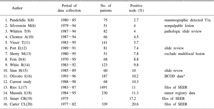
액와 림프절 곽청술을 생략할 수 있는지 관심이 집중되 는 5 mm 이하의 종양에서 림프절 전이에 대해선 적어도 50예 이상 조사한 보고에 의하면 2.7%에서 20.6%까지의 전이가 있다고 발표되었었다(Table 4).(2,6,8-20) 이들 조사 결과 중에서 5% 미만의 전이를 보고한 Pandelidis등(8)과 Silverstein등(6)은 촉지되지 않고 유방 촬영으로 발견된 유 방암에 대한 결과이며 17% 이상 전이가 있다는 Smart등 (19)과 Carter등(20)의 발표는 SEER (Surveillance, Epide- miology and End Results)의 자료를 조사한 것으로 이들의 자료들이 수집 등록된 시기가 1982년 이전이며 여러 기관 에서 보고된 서류에 의존한 자료이므로 정확한 조직학적 조사에 의한 결과인지에 의문을 가질 수 있다. 그러나 그 외 대다수의 조사에서는 7∼12% 정도의 림프절 전이가 있었다고 발표되었으며 본 조사에서의 10.3%와 유사하였 다. 100예 이상씩 조사 보고된 5 mm보다 크고 1 cm 이하
인 T1b 종양에서의 림프절 전이는 7.3%에서 20.6%까지 보고되었지만 역시 촉지되지 않는 종양만 조사한 경우 및 조사 대상 기간이 오래된 경우를 제외한다면 대부분 13∼
19% 정도로 보고되었다. 그리고 1 cm보다 크고 2 cm 이 하인 T1c 종양에서의 림프절 전이는 대부분 30% 정도로 보고되었다(Table 5).(1,2,8,10-14,16-25) 본 조사에서도 이 들 결과와 유사하게 T1b는 15.9%, 그리고 T1c는 30.7%이 었었다. 그리고 본 조사의 10∼15 mm 크기와 16∼20 mm 크기에서의 전이율은 24.3%와 40.4%로 Shetty등(13)의 23.0%와 35.8% 그리고 Olivotto등(16)의 25.0%와 33.6%처 럼 유사한 차이가 있음을 확인할 수 있었다.
결 론
액와 림프절 전이율은 유방암 종양의 크기에 정비례한 다. 그리고 진단 기기의 발달 및 사람들의 유방암 검진 참 여도가 높아짐에 따라 유방암은 점점 더 작은 시기에 발 견되어지고 따라서 이들의 액와 림프절 곽청술 시행 유무 에도 많은 관심 및 논란이 있다. 앞에서 비교해 본 바와 같이 T1b, T1c 종양에서의 림프절 전이율은 여러 조사의 결과가 대부분 유사하였지만 T1a 에서는 그 조사 대상의 조건에 따라 많은 차이를 나타내는 것을 볼 수 있다. 그러 나 종양 크기만으로 조사한 경우에는 10% 이상의 전이가 있다고 발표된 보고들이 많았으며 이번 조사에서도 10.3%
의 전이가 있음을 확인하였다. 그러므로 종양의 크기만으 로 액와 림프절 곽청술의 생략 여부를 결정하기에는 미흡 하다고 사료된다. 그러나 5 mm 이하의 종양에서 나타난 전이는 2개 이하의 림프절에만 국한되어 있었다. 따라서 5 mm 이하의 작은 종양이라도 액와 림프절 곽청술을 생 략하기보다는 최근 많은 연구가 있는 감시 림프절 절제술 을 이용하여 선별하여 시행하는 것이 바람직하다고 사료 된다.
감사의 글
본 논문을 위하여 도움을 주신 Strang Cancer Prevention Center의 Michael P. Osborne 선생님께 진심으로 감사드립 니다.
REFERENCES
1) Barth A, Craig P, Silverstein M. Predictors of axillary lymph node metastases in patients with T1 breast carcinoma. Cancer 1997;79:1918-22.
2) Fein D, Fowble B, Hanlon A, Hooks M, Hoffman J, Si- gurdson E, et al. Identification of women with T1-T2 breast cancer at low risk of positive axillary nodes. J Surg Oncol
ꠏꠏꠏꠏꠏꠏꠏꠏꠏꠏꠏꠏꠏꠏꠏꠏꠏꠏꠏꠏꠏꠏꠏꠏꠏꠏꠏꠏꠏꠏꠏꠏꠏꠏꠏꠏꠏꠏꠏꠏꠏꠏꠏꠏꠏꠏꠏꠏꠏꠏꠏꠏꠏꠏꠏꠏꠏꠏꠏꠏꠏꠏꠏꠏꠏꠏꠏꠏꠏꠏꠏꠏꠏꠏꠏꠏꠏꠏꠏꠏꠏꠏꠏꠏꠏꠏꠏꠏꠏꠏꠏꠏꠏꠏꠏꠏꠏꠏꠏꠏꠏꠏꠏꠏꠏꠏꠏꠏꠏꠏꠏꠏꠏꠏꠏ 1997;65:34-9.
3) Warmuth M, Bowen G, Prosnitz L, Chu L, Broadwater G, Peterson B, et al. Complications of axillary lymph node dissection for carcinoma of the breast. Cancer 1998;83:1362- 8.
4) Cady B. Is axillary lymph node dissection necessary in routine management of breast cancer? No. Breast J 1997;3:246-60 5) Fisher B, Redmond C, Fisher ER, Bauer M, Wolmark N,
Wickerham DL, et al. Ten-year results of a randomized clinical trial comparing radical mastectomy and total mas- tectomy with or without radiation. N Engl J Med 1985;312:
674-81.
6) Silverstein M, Gierson E, Waisman J, Colburn W, Gamagami P. Predicting axillary node positivity in patients with invasive carcinoma of the breast by using a combination of T category and palpability. J Am Coll Surg 1995;180:700-4.
7) Harris JR. Staging and prognostic factors. In: Harris JR, Hell- man S, Henderson IC, Kinne DW, editors. Breast diseases. 2nd ed. Philadelphia: JB Lippincott; 1991. p.327-46.
8) Pandelidis S, Peters K, Walusimbi M, Casady R, Laux S, Cavanaugh S, et al. The role of axillary dissection in mam- mographically detected carcinoma. J Am Coll Surg 1997;184:
341-5.
9) Whitten T, Fraser H, Christensen W, Turk P. Axillary lymph node metastasis in stage T1a breast cancer: a pathologic review of 82 patients. Am Surg 1997;63:144-9.
10) Chontos A, Maher D, Ratzer E, Fenoglio M. Axillary lymph node dissection: is it required in T1a breast cancer? J Am Coll Surg 1997;184:493-8.
11) Visser T, Haan M, Keidan R, Lucas R, Ingold J, Glover J, et al. T1a and T1b breast cancer: a twelve-year experience.
Am Surg 1997;63:621-6.
12) Port E, Tan L, Borgen P, Van Zee K. Incidence of axillary lymph node metastases in T1a and T1b breast carcinoma. Ann Surg Oncol 1998;5:23-7.
13) Shetty M, Reiman H. Tumor size and axillary metastasis, a correlative occurrence in 1244 cases of breast cancer between 1980 and 1995. Eur J Surg Oncol 1997;23:139-41.
14) White R, Vezeridis M, Konstadoulakis M, Cole B, Wanebo
H, Bland K. Therapeutic options and results for the man- agement of minimally invasive carcinoma of the breast:
influence of axillary dissection for treatment of T1a and T1b lesions. J Am Coll Surg 1996;183:575-82.
15) Sinn H, Oelmann A, Anton H, Diel I. Metastatic potential of small and minimally invasive breast carcinomas. Virchows Arch 1994;425:237-41.
16) Olivotto I, Jackson J, Mates D, Andersen S, Davidson W, Bryce C, et al. Prediction of axillary lymph node involvement of women with invasive breast carcinoma: a multivariate analysis. Cancer 1998;83:948-55.
17) Ries L, Henson D, Harras A. Survival from breast cancer ac- cording to tumor size and nodal status. Surgical Oncology of North America 1994;3:35-51.
18) Mustafa I, Cole B, Wanebo H, Bland K, Chang H. The impact of histopathology on nodal metastases in minimal breast cancer. Arch Surg 1997;132:384-90.
19) Smart C, Myers M, Gloeckler L. Implications from SEER data on breast cancer management. Cancer 1978;41:787-9.
20) Carter C, Allen C, Henson D. Relation of tumor size, lymph node status, and survival in 24,740 breast cancer cases. Cancer 1989;63:181-187.
21) Ciatto S, Del Turco M, Bonardi R, Cataliotti L, Distante V, Cardona G, et al. Non-palpable lesions of the breast detected by mammography--review of 1182 consecutive histologically confirmed cases. Eur J Cancer 1994;30A:40-4.
22) Reger V, Beito G, Jolly P. Factors affecting the incidence of lymph node metastases in small cancers of the breast. Am J Surg 1989;157:501-2.
23) Giuliano A, Barth A, Spivack B, Beitsch P, Evans S. Inci- dence and predictors of axillary metastasis in T1 carcinoma of the breast. J Am Coll Surg 1996;183:185-9.
24) Ptaszynski A, Van den Bogaert W, Van Glabbeke M, Pierart M, Bartelink H, Horiot JC, et al. Patient population analysis in EORTC trial 22881/10882 on the role of a booster dose in breast-conserving therapy. Eur J Cancer 1994;30A:2073-81.
25) Ravdin P, De Laurentiis M, Vendely T, Clark G. Prediction of axillary lymph node status in breast cancer patients by use of prognostic indicators. J Natl Cancer Inst 1994;86:1771-5.