The Effect of Adjuvant Radiotherapy in Patients Undergoing Complete Resection for Gallbladder
Cancer with Lymph Node Metastasis
Purpose: We evaluated the effect of adjuvant radiotherapy on survival in patients who underwent curative resection for gallbladder cancer with lymph node metastasis.
Methods: Among the patients underwent curative resection even though there was lymph node metastasis; fifteen patients underwent adjuvant radiotherapy with over 40 Gy (RTx group) and 10 patients did not (no RTx group). We compared these two groups retrospectively.
Results: The median disease free survival (DFS) of the RTx group (21.6 months) was longer than for the no RTx group (6.6 months, p=0.451). The median overall survival (OS) of the RTx group (30.5 months) was also longer than the no RTx group (14.2 months). One-, 2-, and 5-yr OS rates were 60.0%, 40.0% and 40.0% in the no RTx group, and 86.7%, 70.9%
and 26.6% in the RTx group, respectively (p=0.507). Five patients developed recurrence within 1 year (50.0%) in the no RTx group; there were 3 (20.0%) in the RTx group.
Conclusion: Our study was limited by its retrospective nature and small numbers of patients.
However, it suggests that adjuvant radiotherapy might improve DFS and OS for patients with completely resected but lymph node metastasized gallbladder cancer. Also this therapy seems to delay time to postoperative recurrence.
Jae Myeong Lee, M.D., Seung-Hee Kang, M.D.1, Bong Wan Kim, M.D.2, Hee Jung Wang, M.D.2, Mison Chun, M.D.3, Myung Wook Kim, M.D.2
Department of Anesthesiology and Pain Medicine, Ajou University School of Medicine, 1Department of Radiation Oncology, Ilsan-Paik Hospital Inje University School of Medicine, Departments of 2Surgery,
3Radiation Oncology, Ajou University School of Medicine
책임저자 Seung-Hee Kang
Department of Radiation Oncology, Ilsan-Paik Hospital Inje University School of Medicine, 2240, Daewha- dong, Ilsan-Suh-gu, Goyang 411-706, Korea
Tel: +82-31-910-9744 Fax: +82-31-910-9743
E-mail: [email protected]
Key Words : Gallbladder cancer, Postoperative radiotherapy, Survival
Received: 2010. 11. 10 Accepted: 2011. 1. 15
Introduction
The prognosis of gallbladder (GB) cancer is extremely poor due to the high proportion of tumor that is advanced at the time of presentation.
1,2The major therapeutic modality and the only curative treatment is surgical
resection. However, despite of the aggressive surgical
approach, 40∼86 % of patients who undergo a potentially
curative resection will develop recurrent metastatic
disease.
3,4Therefore, radiotherapy or chemotherapy has
been considered as an adjuvant therapy to improve
loco-regional control and overall survival rate (OS). Some
reports have suggested a benefit of concurrent
chemo-radiation therapy for GB cancer.
5,6However, the effect of adjuvant radiotherapy for the GB cancer is not well established.
7,8In general, the decision to administer adjuvant therapy is dependent upon the type of surgery and the pathologic risk factors and is based on physician and patient preference after discussion about the possible risks and benefits.
To evaluate the effect of adjuvant radiotherapy, we reviewed only for the patients who underwent curative resection for gallbladder cancer with pathologically confirmed lymph node metastasis.
Methods
One hundred forty-seven patients had been performed the surgical treatment for GB cancer at Ajou University Hospital between October 1994 and March 2008. Among them, 30 patients underwent curative resection though there was lymph node metastasis. We excluded 5 patients received radiotherapy of less than 18 Gy from this study.
Fifteen patients underwent adjuvant radiotherapy with ≥40 Gy (hereafter, RTx group) and 10 patients did not (hereafter, no RTx group). Altogether, the two groups had 10 male patients and 15 female patients with median age of 59.0±12.1 years, ranging from 34∼79 years.
The adjuvant radiotherapy was administered using a 10∼
15 MV linear accelerator with a median total dose of 46.8±28.6 Gy (range, 43.4∼50.4 Gy in 22∼28 fractions).
Initial radiation fields encompassed the tumor bed and regional lymph nodes (porta hepatis, pericholedocal, celiac, and pancreaticoduodenal nodes). After 45 Gy, boost radiation to the tumor bed was delivered in some patients based on physcian’s decision. Patients were treated at 1.8
∼2 Gy per fraction, once a day, 5 days a week. Three- or four- field techniques were used in most patients.
Among the 15 patients of the RTx group, 7 patients (46.7%) were treated with radiation alone and 8 patients (53.3%) received chemoradiotherapy. Chemotherapy was given as a radiosensitizer in 5 patients and concurrently
with radiotherapy as a part of systemic chemotherapy in 3 patients. Chemotherapy regimens were varied; daily cisplatin in 3 patients and 5-Fluorouracil based regimens in 5 patients. Maintenance chemotherapy after completion of radiotherapy was used in 2 patients.
GB cancers were staged according to the 6
thedition of the AJCC.UICC TNM classification.
2The clinical characteri- stics of GB cancer included gender, age, tumor size and operation method. The pathologic characteristics included stage, pathology, differentiation, and lymphovascular or perineural invasion.
Survival was calculated from the date of surgery. We examined the exact survival status and date of death of patients by calling to the patients’family or checking the medical records. Continuous variables were written as the median±standard deviation (range). Crosstabs, such as Pearson’s chi-square test and Fisher’s exact test, indepen- dent t-test, nonparametric test such as Mann-Whitney test and Kaplan-Meier survival analysis were used for statistical analyses using SPSS (version 15.0; SPSS, Inc., Chicago, IL, USA). Difference was considered statistically significant when the p -value was <0.05.
Results
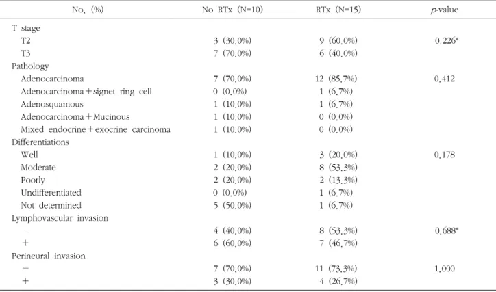
There were no statistical differences between the two groups in clinical characteristics (Table 1) and pathological characteristics (Table 2). The median age of the RTx group was younger than the no RTx group (56.0±9.4 vs. 63.0±
15.2, p =0.183).
The median follow-up duration was 25.6 months (range,
4.0∼165.9 months). We analyzed the patterns of failure
and overall (OS) and disease-free survivals (DFS) for
comparing the RTx group (N=15) and the no RTx (N=10)
group. The median DFS of the RTx group (21.6±9.1
months) was longer than the no RTx group (6.6±5.2
months), however, there was no statistically significant
difference ( p =0.451; Table 3). The patient number who
developed recurrence within 1 year was 3 (20.0%) in RTx
Table 1. Comparison of the clinical characteristics between no RTx group and RTx group
No. (%) No RTx (N=10) RTx (N=15) p-value
Gender Male Female Age (years) Tumor size (cm) Operations LC‡ OC§
OC+LN dissection Extended chole¶ OC+hepatectomy
Extended chole+Whipple’s OC+colon resection
3 (30.0%) 7 (70.0%) 63.0±15.2 3.0±1.5
1 (10.0%) 0 (0.0%) 0 (0.0%) 4 (40.0%) 4 (40.0%) 1 (10.0%) 0 (0.0%)
7 (46.7%) 8 (53.3%) 56.0±9.4 5.0±2.4
1 (6.7%) 1 (6.7%) 3 (20.0%) 7 (46.7%) 1 (6.7%) 1 (6.7%) 1 (6.7%)
0.678*
0.183† 0.262†
0.211
*Fisher’s exact test; †Mann-Whitney test; ‡LC=laparoscopic cholecystectomy; §OC=open cholecystectomy; ¶Extended chole=open cholecystectomy+liver wedge resection+LN dissection
Table 2. Comparison of the pathological characteristics between no RTx group and RTx group
No. (%) No RTx (N=10) RTx (N=15) p-value
T stage T2 T3 Pathology
Adenocarcinoma
Adenocarcinoma+signet ring cell Adenosquamous
Adenocarcinoma+Mucinous
Mixed endocrine+exocrine carcinoma Differentiations
Well Moderate Poorly
Undifferentiated Not determined Lymphovascular invasion −
+
Perineural invasion −
+
3 (30.0%) 7 (70.0%)
7 (70.0%) 0 (0.0%) 1 (10.0%) 1 (10.0%) 1 (10.0%)
1 (10.0%) 2 (20.0%) 2 (20.0%) 0 (0.0%) 5 (50.0%)
4 (40.0%) 6 (60.0%)
7 (70.0%) 3 (30.0%)
9 (60.0%) 6 (40.0%)
12 (85.7%) 1 (6.7%) 1 (6.7%) 0 (0.0%) 0 (0.0%)
3 (20.0%) 8 (53.3%) 2 (13.3%) 1 (6.7%) 1 (6.7%)
8 (53.3%) 7 (46.7%)
11 (73.3%) 4 (26.7%)
0.226*
0.412
0.178
0.688*
1.000
*Fisher’s exact test
group and 5 (50.0%) in no RTx group.
The median OS of the RTx group (30.5±3.7 months)
was also longer than the no RTx group (14.2±4.0 months).
One-, 2-, and 5-yr OS were 86.7%, 70.9% and 26.6% in the
Table 3. Comparisons of the disease free survival and overall survival between no RTx group and RTx group
Group No RTx (N=10) RTx (N=15) p-value
Median disease free survival (mo) Disease free survival (%)
1-year 2-year 5-year
Median overall survival (mo) Overall survival rates (%) 1-year
2-year 5-year
6.6±5.2
50.0%
40.0%
40.0%
14.2±4.0
60.0%
40.0%
40.0%
21.6±9.1
79.0%
49.2%
41.0%
30.5±3.7
86.7%
70.9%
26.6%
0.451
0.507
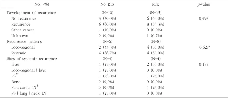
Table 4. Comparison of the pattern of failure between no RTx group and RTx group
No. (%) No RTx RTx p-value
Development of recurrence No recurrence
Recurrence Other cancer Unknown Recurrence patterns Loco-regional Systemic
Sites of systemic recurrence Liver
Loco-regional+liver PS†
Bone
Para-aortic LN‡ PS+lung+neck LN
(N=10) 3 (30.0%) 6 (60.0%) 1 (10.0%) 0 (0.0%)
(N=6) 2 (33.3%) 4 (66.7%) (N=4) 1 (25.0%) 1 (25.0%) 1 (25.0%) 0 (0.0%) 0 (0.0%) 1 (25.0%)
(N=15) 6 (40.0%) 8 (53.3%) 0 (0.0%) 1 (6.7%) (N=8) 4 (50.0%) 4 (50.0%) (N=4) 2 (50.0%) 0 (0.0%) 1 (25.0%) 0 (0.0%) 1 (25.0%) 0 (0.0%)
0.497
0.627*
0.175
*Fisher’s exact test; †PS=peritoneal seeding; ‡LN=lymph node
RTx group and 60.0%, 40.0% and 40.0% in the no RTx group, respectively. There were no statistical differences in OS between the two groups ( p =0.507). Subgroup analysis was performed to identify the benefit from adding chemotherapy to radiotherapy. Patients who received chemoradiotherapy had shorter median OS than patients treated with radiotherapy alone (25.9±26.0 months vs.
30.5±4.4 months, p =0.923).
The recurrence pattern was described in Table 4. In the no RTx group, 6 patients (60.0%) showed postoperative recurrences; the first site of failure was loco-regional in 2
patients (33.3%) and systemic in 4 (66.7%). In the RTx group, 8 of 14 evaluable patients (53.3%) experienced recurrences; loco-regional failure in 4 (50.0%) and systemic metastasis in 4 (50.0%). In subgroup analysis, recurrence was developed in 3 of 7 patients treated with chemora- diotherapy and in 5 of 7 radiotherapy alone patients.
In the RTx group, 12 out of 15 patients (80.0%) experien-
ced acute complications. Most of the side effects were
tolerable. Most common side effect was gastrointestinal
trouble such as nausea (5 patients, 38.5%) and anorexia (2
patients, 15.4%). Leukopenia was observed in 1 patient
(7.7%); this patient restarted the radiotherapy after rest for some period and the other patient should stop the concurrent chemotherapy but maintained the radiotherapy.
Discussion
Surgical treatment is the only potentially curable modality for GB cancer, currently. However, unfortunately, the majority of patients present advanced lesions and have no chance of a curative resection.
9And moreover, even if patients undergo a curative resection they harbor frequently poor prognosis factors like as positive lymph node, higher T stage, old age and non-papillary histology those increase the chance of failure.
10,11Because of this aggressive nature of GB cancer, individualized multimodality approach is required to be optimized.
The rationale for postoperative radiation therapy is to sterilize tumor cell areas surrounding the tumor that could be left after surgery.
9,12Radiosensitive nature of gallbladder cancer is evidenced by numerous studies reporting tumor size reduction after radiotherapy for unresectable disea- se.
13,14However, because of the rarity of GB cancer, actual benefit of adjuvant therapy has not been well established.
7,8There are some reports about the efficacy of adjuvant radiotherapy for GB cancer. Kresl et al. reported an improved 5-year survival (33%) of GB cancer patients underwent adjuvant radiation therapy following optimal surgical resection. They included 21 patients who received 50.4∼60.8 Gy of radiation with concurrent chemotherapy after surgery.
5Czito et al. reported 22 patients with GB cancer who received adjuvant therapy consisting of 5-FU chemotherapy and a median radiation dose of 45 Gy with an overall 5-year survival of 37%.
6Reports announced the benefit of adjuvant radiotherapy in node positive GB cancer is few. Wang et al.
11suggested greatest net benefit from adjuvant radiotherapy in patients with higher stage (T2 or greater) and lymph node-positive disease. Balachandran et al.
15reported adjuvant chemora- diotherapy made significant survival improvement in node
positive patients. In their study, patients who underwent adjuvant chemoradiotherapy (N=31) showed 18 months median survival and 25% 5-year survival, whereas, patients who did not undergo chemoradiotherapy (N=25) showed 7 months median survival and 0% 5-year survival ( p =0.005).
However, their data included non-curative resection, and node positive patients had more R1 resection and it could reflect the efficacy of chemoradiotherapy in non-curable surgical patients. Mogica et al.
16reported adjuvant radiation therapy for patients who presented with regional disease (positive lymph nodes) and demonstrated an improved overall median survival from 5 to 16 months with the use of radiation therapy (no RTx (n=277) vs. RTx (n=127), p
<0.0001). Gold et al.
7also reported the median OS for patients receiving adjuvant chemoradiotherapy vs. surgery only was 4.8 years and 4.2 years, respectively (log-rank test, p =0.56). After adjusting for prognostic factors in the multivariate analysis, the administration of adjuvant che- moradiotherapy resulted in significantly improved OS (hazard ratio for death, 0.30; 95% confidence interval, 0.13
∼0.69; p =0.004).
We restricted the study group to patients with positive lymph node metastasis after curative resection. In our study, the median DFS and median OS were longer in RTx group than no RTx group. Five-year OS of RTx group was similar with no RTx group, whereas, 1- and 2 - year OS were superior in RTx group (1-year OS, 86.7% vs. 60.0%
and 2-year OS, 70.9% vs. 40.0%). The recurrence rate within 1 year after surgery was lower in RTx group than no RTx group (20.0% vs. 50.0%). From this result, we guessed the effect of radiotherapy in terms of delay time to recurrence and this effect improved the short term DFS and OS of GB cancer until 2 years after surgery.
In our study, RTx group has slightly younger age
compared with no RTx group ( p =0.183). This is maybe due
to more aggressive application of treatment modality to
young patients than old aged patients. Some reports
suggested age was important independent prognostic factor
of GB cancer and the survival rates decreased with
increasing age.
17-19Therefore, the effect of younger age to superior survival in RTx group cannot be ruled out.
There is no uniform agreement regarding radiation dose, modality and timing of therapy.
16About the timing of radiation therapy, Kraybill et al.
20noted a trend towards long - term survival in the patients who received radiation therapy within 2 months after surgery compared with delayed radiation with an absolute improvement of about 30% in 5 years. Several reports have suggested the relation between radiation dose higher than 40 Gy and improved survival.
21,22In our study, radiation therapy was started within 2 months after surgery and all patients received radiation of 40 Gy and greater.
Based on the recurrence patterns, it is difficult to advocate the use of radiotherapy alone. Jarnagin et al.
23observed that GB cancer had a higher incidence of distant metastases as a first site of failure compared with hilar cholangiocarcinoma. Therefore, the addition of chemothe- rapy would be essential for improving results.
6,9,14How- ever, in our study, the patients who underwent adjuvant chemoradiotherapy (N=8) showed short median OS than the patients who underwent only radiotherapy (25.9±26.0 months vs. 30.5±4.4 months, p =0.923) despite the two groups showed no statistical differences in the stages.
Because of small number of patients and variety of chemotherapy regimens, we supposed our results could not reflect the effect of chemoradiation, sufficiently.
Current National Comprehensive Cancer Network 2007 guidelines stated although there is limited clinical trial data to support a standard regimen, all patients with stage higher than T1N0 should be considered for adjuvant therapy.
11Although, our study has a drawbacks such as the small number of patients in each group and retrospective study, our results support these suggestions. Therefore, we are continued to consider adjuvant radiotherapy for GB cancer with lymph node metastasis after curative resection.
Conclusion
Our data showed adjuvant radiotherapy after curative resection for GB cancer with lymph node metastasis might have some beneficial effect on survival of patients. And adjuvant radiotherapy seemed to delay time to recurrence.
The reason of no statistical significance in this study was regarded as small number of the patients. Therefore, further study with large number of patients can be helpful to evaluate the effect of radiotherapy for gallbladder cancer.
References
1. Gourgiotis S, Kocher HM, Solaini L, Yarollahi A, Tsiambas E, Salemis NS. Gallbladder cancer. Am J Surg 2008;196:
252-264.
2. Reid KM, Ramos-De la Medina A, Donohue JH. Diagnosis and surgical management of gallbladder cancer: a review. J Gastrointest Surg 2007;11:671-681.
3. Duffy A, Capanu M, Abou-Alfa GK, et al. Gallbladder cancer (GBC): 10-year experience at Memorial Sloan-Kettering Cancer Centre (MSKCC). J Surg Oncol 2008;98:485-489.
4. Mahantshetty UM, Palled SR, Engineer R, Homkar G, Shrivastava SK, Shukla PJ. Adjuvant radiation therapy in gall bladder cancers: 10 years experience at Tata Memorial Hospital. J Cancer Res Ther 2006;2:52-56.
5. Kresl JJ, Schild SE, Henning GT, et al. Adjuvant external beam radiation therapy with concurrent chemotherapy in the management of gallbladder carcinoma. Int J Radiat Oncol Biol Phys 2002;52:167-175.
6. Czito BG, Hurwitz HI, Clough RW, et al. Adjuvant external-beam radiotherapy with concurrent chemotherapy after resection of primary gallbladder carcinoma: a 23-year experience. Int J Radiat Oncol Biol Phys 2005;62:1030-1034.
7. Gold DG, Miller RC, Haddock MG, et al. Adjuvant therapy for gallbladder carcinoma: the Mayo Clinic Experience. Int J Radiat Oncol Biol Phys 2009;75:150-155.
8. Houry S, Barrier A, Huguier M. Irradiation therapy for gallbladder carcinoma: recent advances. J Hepatobiliary Pancreat Surg 2001;8:518-524.
9. de Aretxabala X, Roa I, Berrios M, et al. Chemoradiotherapy in gallbladder cancer. J Surg Oncol 2006;93:699-704.
10. Misra S, Chaturvedi A, Misra NC, Sharma ID. Carcinoma of the gallbladder. Lancet Oncol 2003;4:167-176.
11. Wang SJ, Fuller CD, Kim JS, Sittig DF, Thomas CR Jr, Ravdin PM. Prediction model for estimating the survival benefit of
adjuvant radiotherapy for gallbladder cancer. J Clin Oncol 2008;26:2112-2117.
12. Yoshimitsu K, Honda H, Kuroiwa T, et al. Liver metastasis from gallbladder carcinoma: anatomic correlation with cholecystic venous drainage demonstrated by helical computed tomography during injection of contrast medium in the cholecystic artery. Cancer 2001;92:340-348.
13. Smoron GL. Radiation therapy of carcinoma of gallbladder and biliary tract. Cancer 1977;40:1422-1424.
14. Kopelson G, Harisiadis L, Tretter P, Chang CH. The role of radiation therapy in cancer of the extra-hepatic biliary system:
an analysis of thirteen patients and a review of the literature of the effectiveness of surgery, chemotherapy and radiothe- rapy. Int J Radiat Oncol Biol Phys 1977;2:883-894.
15. Balachandran P, Agarwal S, Krishnani N, et al. Predictors of long-term survival in patients with gallbladder cancer. J Gastrointest Surg 2006;10:848-854.
16. Mojica P, Smith D, Ellenhorn J. Adjuvant radiation therapy is associated with improved survival for gallbladder carcinoma with regional metastatic disease. J Surg Oncol 2007;96:8-13.
17. Ong SL, Garcea G, Thomasset SC, et al. Ten-year experience
in the management of gallbladder cancer from a single hepatobiliary and pancreatic centre with review of the literature. HPB (Oxford) 2008;10:446-458.
18. Kayahara M, Nagakawa T, Nakagawara H, Kitagawa H, Ohta T. Prognostic factors for gallbladder cancer in Japan. Ann Surg 2008;248:807-814.
19. Manfredi S, Benhamiche AM, Isambert N, Prost P, Jouve JL, Faivre J. Trends in incidence and management of gallbladder carcinoma: a population-based study in France. Cancer 2000;89:757-762.
20. Kraybill WG, Lee H, Picus J, et al. Multidisciplinary treatment of biliary tract cancers. J Surg Oncol 1994;55:239-245.
21. Houry S, Schlienger M, Huguier M, Lacaine F, Penne F, Laugier A. Gallbladder carcinoma: role of radiation therapy.
Br J Surg 1989;76:448-450.
22. Bosset JF, Mantion G, Gillet M, et al. Primary carcinoma of the gallbladder. Adjuvant postoperative external irradiation.
Cancer 1989;64:1843-1847.
23. Jarnagin WR, Ruo L, Little SA, et al. Patterns of initial disease recurrence after resection of gallbladder carcinoma and hilar cholangiocarcinoma: implications for adjuvant therapeutic strategies. Cancer 2003;98:1689-1700.