163
한우 송아지의 소바이러스성 설사바이러스 지속감염률 조사
배유찬*·김하영·박중원·윤순식·우계형·이오수·강문일 국립수의과학검역원
(게재승인: 2007년 5월 7일)
Prevalence for persistent infection with bovine viral diarrhea virus in Korean native calves
You-Chan Bae*, Ha-Young Kim, Jung-Won Park, Soon-Seek Yoon, Gye-Hyeong Woo, O-Soo Lee, Mun-Il Kang
National Veterinary Research and Quarantine Service, Anyang 430-824, Korea (Accepted: May 7, 2007)
Abstracts : Bovine viral diarrhea (BVD) is very important disease in cattle industry with a worldwide distribution. Detection and elimination of persistently infected calves with bovine viral diarrhea virus (BVDV) was valuable strategy for BVD eradication because those calves were main source for transmission. We surveyed persistent infection with BVDV by reverse transcription polymerase chain reaction (RT-PCR) and immunohistochemistry (IHC) using whole blood and skin. Five hundred thirty nine Korean native calves were tested. Four calves (0.7%) were positive for BVDV antigen for both tests. Those calves remained positive for follow-up by RT-PCR and IHC. Therefore they were identified as persistently infected with BVDV. We confirmed that immunohistochemistry using skin biopsy samples was very useful tool to detect persistently infected calves with BVDV. As far as we know, this would be first report on persistent infection with BVDV in Korea.
Key words : bovine viral diarrhea virus, immunohistochemistry, Korean native calf, persistent infection
서 론
소바이러스성설사
(bovine viral diarrhea, BVD)
는전세 계적으로소에서경제적으로많은피해를주고있는중 요한질병이다[1, 5, 9].
이질병은RNA
바이러스이며Togaviridae, pestivirus
에속하는소바이러스성설사바이 러스(bovine viral diarrhea virus, BVDV)
에의해발생하 며호흡기형,
소화기-
점막형,
유산형,
지속감염형등매우다양한임상증상을나타낸다
[1, 3, 6].
특히생산성에대한피해는우군당
40,000~100,000
달러에이르는것으로보고된바있다
[9].
지속감염형은자궁내의태아가임신
4
개월령이전에BVDV
에감염되면태아가이바이러스에 면역관용을보임으로서항체형성이되지않으면서살아있는동안
지속적으로바이러스를배출하는임상형이다
[1].
지속 감염형 송아지는정상적으로출생하는경우도 있으나 허약하거나저체중상태로태어나기도한다[1, 2].
이 들지속감염우가BVD
의주요전파요인으로확인되어 이들을색출하여 제거하는 것이BVD
로인한 피해를줄이는데 매우중요한 정책이 되고 있다
[5, 12, 21].
BVD
지속감염우는 보통3-4
주간격으로2
회이상백 혈구나 혈청에서바이러스가분리되는송아지를 가리 킨다[4].
외국의경우
BVD
지속감염우감염률은보통우군별로
0.1-2%
로다양하게보고되었으며다수의모우가임신
4
개월전에BVDV
에감염된우군의경우25-30%
의 높은감염률을보인경우도있다[1, 3, 4, 8-11, 13-15, 17, 18, 20-22].
*Corresponding author: You-Chan Bae
National Veterinary Research and Quarantine Service, Busan 602-833, Korea [Tel: +82-51-603-0641, Fax: +82-51-603-0649, E-mail: [email protected]]
최근지속감염우를진단하기위해귀피부생검조직 을이용하여 면역조직화학염색
(immunohistochemistry, IHC),
항원Enzyme Linked Immunosorbent Assay(ELISA)
를활용한논문들이보고되고있다
[6, 8, 16].
그러나국내에서는
BVD
지속감염률을 조사한연구결과가없는실정이다
.
따라서본연구에서는농장에서BVD
에의한피해를줄이기위한기초자료로활용하기위해국내송 아지에서의
BVDV
지속감염률을조사하였다.
재료 및 방법
송아지
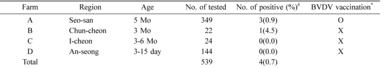
Table 1
과같이충남서산,
강원춘천,
경기이천,
경 기안성소재4
개한우농장에서3
일에서6
개월령의송아지총
539
두를선정하였다.
시료채취는2005
년8
월부터
2006
년6
월까지실시하였으며1
차역전사중합효소연쇄반응
(reverse transcription polymerase chain reaction,
RT-PCR)
검사에서양성반응을보인 개체에대해서는IHC
로확인검사하였고,
양성개체에대해서는4
주후2
차
RT-PCR
과IHC
를실시하였다.
시료 채취
“V”
자형의샘플채취기(ear notcher)
를이용하여송아지귓바퀴의끝부분에서약
3
×4 mm
의피부를채취하였다
(Fig. 1).
채취즉시10%
중성완충포르말린에고정하고일반적인조직처리과정을거쳐
hematoxylin&eosin
(H & E)
염색을한후병리조직학적인병변의유무를관찰하였고일부는
IHC
에사용하였다.
또한경정맥에서 전혈을채취하여항응고제가처리된튜브에넣은후RT- PCR
에사용하였다.
RNA 추출 및 RT-PCR
RNeasy mini kit(Qiagen, Germany)
를사용하여제조사 의 지시에 따라 전혈에서RNA
를 추출하였다. PCR
primer
는Radwan
등이사용한것과동일한것을사용하였다
[19]. PCR
반응은 추출한RNA 5 ul
와primer
(5'UTR1,2) 1 ul(20 pmol)
를onestep RT-PCR kit(Qiagen, Germany)
에첨가하여총량이25 ul
가되게하였다. 50
oC
에서
30
분간반응시켜cDNA
를합성한후94
oC
에서1
분
, 55
oC
에서1
분, 72
oC
에서1
분씩35
회반복반응시 킨다음최종72
oC
에서10
분간반응시켰다. PCR
반응 이완료된후각반응액10 ul
를취하여ethidium bromide
가함유된
1.5% agarose gel
에서전기영동을실시한다음자외선하에서특이밴드유무를관찰하였다
.
IHC
일반적인조직처리과정을거쳐파라핀포매한시료를
3-4 um
두께로박절하여silane
으로코팅된슬라이드에부착시킨후탈파라핀과정을거쳐
0.1% protease 14
(Sigma, USA)
로37
oC
에서10
분간항원을노출시킨후protein block(Dako, Denmark)
을10
분간반응시켰다.
일차항체는
Anti-BVDV monoclonal antibody(IDEXX Laboratories, USA)
를1 : 1,000
으로 희석하여실온에서30
분간 반응시킨다음LSAB 2 system-AP(Dako, Den- mark)
를 반응시켰으며Liquid Permanent Red Chro- mogen(Dako, Denmark)
을이용하여발색시켰다. Mayer’s hematoxylin(Dako, Denmark)
으로대조염색한후광학현미경으로관찰하였다
.
A Seo-san 5 Mo 349 3(0.9) O
B Chun-cheon 3 Mo 22 1(4.5) X
C I-cheon 3-6 Mo 24 0(0.0) X
D An-seong 3-15 day 144 0(0.0) X
Total 539 4(0.7)
# positive for both RT-PCR and IHC at initial and follow up test.
* O : BVDV vaccinated, X : No BVDV vaccinated.
Fig. 1. Sampling of pinna by ear notcher ( “ V ” shape).
결 과
RT-PCR
총
4
개농장539
두의한우전혈에대한RT-PCR
결과Fig. 2
와같이4
두가246 bp
의BVDV
특이유전자가증폭되어양성으로판정되었다
.
또한양성개체에대해서는
4
주후RT-PCR
을실시했을때도양성으로확인되어지속감염률은
0.7%
로확인되었다.
IHC
지속감염우로확인된
4
두는면역조직화학염색법에서1
차검사와4
주후2
차검사에서양성반응을보였다.
지속감염우로판명된
4
두의귀피부에대한병리조직검사결과특이한병변이관찰되지않았으며
(Fig. 3),
면역조직화학염색결과
BVDV
항원은표피층의각질세포와모 낭의상피세포에서확인되었다(Fig. 4).
고 찰
BVD
에의한경제적피해가크기때문에독일을비롯한몇몇유럽에서는이질병을근절하기위한국가적또 는지역적차원의방역정책이수립되어운용되고있다
[12].
이들방역정책의성공적추진을위해효과적인진단법을사용하는것은매우중요하다
[12]. BVDV
항원을검출하는방법으로는바이러스분리법
,
항원ELISA
법
, IHC
법, RT-PCR
법이활용되고있으며최근에는귀피부생검조직을이용한항원
ELISA
법과IHC
법이많 이사용되고있는추세이다[5, 6, 8, 9, 12, 16].
Table 1
의B
농장의경우지속감염률이4.5%
로비교적높게나타났는데이는검사두수가
22
두로서적었기 때문으로생각되며보다정확한감염률을파악하기위 해서는더많은두수에대한검사가필요하리라고판단 된다.
이번연구결과지속감염우로확인된개체는해당 목장에통보하여도태를권유하여다른송아지에전염 되는것을차단하도록하였다.
이번연구가국내송아지의
BVD
지속감염률에대한첫보고인반면미국을포함한북미지역과유럽에서는 다수의연구보고가있었다
[3, 4, 8, 11, 13, 14, 18, 20-
22].
그러나동일한대륙안에서도지속감염률은우군별Fig. 2. Photography of BVDV RNA amplification. Note the specific bands (246 bp).
Fig. 3. Ear skin from a calf (#05R169-28) persistently infected with BVDV. No histopathological lesion was observed H & E.
×200.
Fig. 4. Ear skin from a calf (#05R169-28) persistently
infected with BVDV. Immunohistochemical staining. Note
BVDV antigen within epidermal keratinocytes (short arrow),
follicular epithelium (long arrow). Alkaline phosphatase
technique, permanent red chromogen, and hematoxylin
counterstain.
×400.
0.4% 1.4%
까지다양하게나타났다
.
이처럼지역별로우군별로감염률의차이가 나타나는점은우군의구성
,
우사의구조
,
백신프로그램및사양관리가다르기때문으로추정 하고있다[9, 10].
IHC
으로BVDV
지속감염우에대한항원분포를조사한결과신경계
,
림프계,
소화기계,
호흡기계,
피부등 전신장기에서검출된다고보고되었다[2].
그이유는이바이러스가혈액을통해전신에퍼지기때문으로보고
있다
[2]. BVDV
지속감염우의경우호흡기,
소화기증상이나위축을보이는개체도있으나일부개체들은임 상증상이나 병리조직학적병변이관찰되지않은 개체 도있으므로임상증상이나조직학적병변만으로 지속
감염우여부를판별하는것은곤란하다
[1, 2].
이번연구결과지속감염우로판명된
4
두의송아지도특이한임 상증상이없었고 피부에 대한병리조직검사결과특이한병변이관찰되지않았으나
1
차및2
차RT-PCR
법과IHC
에서양성으로판명된 사례들이다.
따라서 외관적으로건강한송아지라할지라도
BVD
지속감염우를색출하기 위한검사에 반드시포함되어야 할것으로 판 단된다
.
BVDV IHC
결과BVDV
항원이표피층의각질세포와모낭의상피세포에서확인된점은이전연구자들이보 고한지속감염우의항원분포와일치하였다
[6, 16].
그러나
BVD
급성감염우에대한IHC
결과도지속감염우의항원분포부위와일치하므로항원분포만으로지속 감염우와급성감염우를판별하는것은어려운실정이 다
[7, 16]. IHC
법은이번연구에서RT-PCR
법과검사결과가모두일치하여
BVD
지속감염우의진단에효과적인진단법임을 확인하였기때문에향후국내에서도활 발하게사용될것으로보인다
.
또한이번한우의BVD
지속감염률조사를계기로향후홀스타인도감염률조 사를통해지속감염우를도태하여이질병으로인한피 해를줄이는것이필요하리라생각한다
.
결 론
국내사육중인한우의
BVD
지속감염률을조사하기위해
IHC
법과RT-PCR
을이용하여539
두의송아지피 부와전혈을검사하였다.
검사결과4
두(0.74%)
가BVD
지속감염우로판명되어해당농장에도태를권유하여
이질병으로인한피해를줄이도록하였다
. IHC
법은향후국내에서
BVD
지속감염우진단에유용하게활용될수있을것으로사료된다
.
깊이감사드린다