Copyright ⓒ 2012, The Microbiological Society of Korea
Virus Inactivation during the Manufacture of a Collagen Type I from Bovine Hides
Jung Eun Bae1, Chan Kyung Kim1, Sungpo Kim2, Eun Kyung Yang2, and In Seop Kim1*
1Department of Biological Sciences and Biotechnology, Center for Biopharmaceuticals Safety Validation, Hannam University, Daejeon 305-811, Republic of Korea
2R&D Center, Bioland Co. Ltd, Cheongwon 363-885, Republic of Korea
소 가죽 유래 Type I Collagen 생산 공정에서 바이러스 불활화
배정은1․김찬경1․김성포2․양은경2․김인섭1*
1한남대학교 생명·나노과학대학 생명시스템과학과 & 바이오의약품안전성검증센터
2(주)바이오랜드 조직공학연구소
(Received October 29, 2012 / Accepted December 12, 2012)
Most types of collagen used for biomedical applications, such as cell therapy and tissue engineering, are derived from animal tissues. Therefore, special precautions must be taken during the production of these proteins in order to assure against the possibility of the products transmitting infectious diseases to the recipients. The ability to remove and/or inactivate known and potential viral contaminants during the manufacturing process is an ever-increasingly important parameter in assessing the safety of biomedical products. The purpose of this study was to evaluate the efficacies of the 70% ethanol treatment and pepsin treatment at pH 2.0 for the inactivation of bovine viruses during the manufacture of collagen type I from bovine hides. A variety of experimental model viruses for bovine viruses including bovine herpes virus (BHV), bovine viral diarrhea virus (BVDV), bovine parainfluenza 3 virus (BPIV-3), and bovine parvovirus (BPV), were chosen for the evaluation of viral inactivation efficacy. BHV, BVDV, BPIV-3, and BPV were effectively inactivated to undetectable levels within 1 h of 70% ethanol treatment for 24 h, with log reduction factors of ≥5.58, ≥5.32, ≥5.11, and ≥3.42, respectively. BHV, BVDV, BPIV-3, and BPV were also effectively inactivated to undetectable levels within 5 days of pepsin treatment for 14 days, with the log reduction factors of ≥7.08, ≥6.60, ≥5.60, and ≥3.59, respectively. The cumulative virus reduction factors of BHV, BVDV, BPIV-3, and BPV were ≥12.66, ≥11.92, ≥10.71, and ≥7.01. These results indicate that the production process for collagen type I from bovine hides has a sufficient virus-reducing capacity to achieve a high margin of virus safety.
Keywords: bovine collagen, ethanol treatment, pepsin treatment, virus safety
*For correspondence. E-mail: [email protected]; Tel.: +82-42-629-8754;
Fax: +82-42-629-8751
Collagen is a term given to a group of naturally occurring proteins found in animals, especially in the flesh and connective tissues of mammals. It is the main component of connective tissue, and is the most abundant protein in mammals, making up about 25% to 35% of the whole-body protein content (Harkness, 1961; Di Lullo et al., 2002;
Shoulder and Ranies, 2009). Collagen, in the form of elongated fibrils, is mostly found in fibrous tissues such as tendon, ligament and skin, and is also abundant in cornea, cartilage, bone, blood vessels, the gut, and intervertebral disc. The
fibroblast is the most common cell which produces collagen (Franchi et al., 2007). Collagen plays an essential role in providing a scaffold for cellular support, thereby affecting cell attachment, migration, proliferation, differentiation, and survival.
Collagen has a wide variety of applications, from food to medical, and it is regarded as one of the most useful biomaterials. The excellent biocompatibility and safety of collagen, due to its biological characteristics, such as biodegradability and weak antigenicity, have made collagen the primary resource in medical applications. The main applications of collagen as drug delivery systems are collagen shields in ophthalmology, sponges for burns/wounds, mini-pellets and tablets for protein delivery, gel formulation in combination
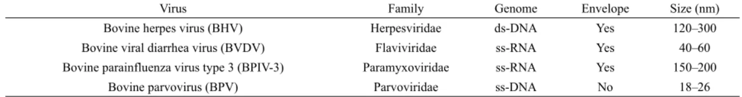
Virus Family Genome Envelope Size (nm)
Bovine herpes virus (BHV) Herpesviridae ds-DNA Yes 120–300
Bovine viral diarrhea virus (BVDV) Flaviviridae ss-RNA Yes 40–60
Bovine parainfluenza virus type 3 (BPIV-3) Paramyxoviridae ss-RNA Yes 150–200
Bovine parvovirus (BPV) Parvoviridae ss-DNA No 18–26
Table 1. Salient features of viruses used for the evaluation of virus inactivation
with liposomes for sustained drug delivery, as a controlling material for transdermal delivery, and as nanoparticles for gene delivery and basic matrices for cell culture systems. It is also used for tissue engineering, including skin replacement, bone substitutes, and artificial blood vessels and valves (Lee et al., 2001; Malafaya et al., 2002, 2007; Willerth and Sakiyama-Elbert, 2007; Ma et al., 2011).
The major obstacle to widespread use of animal-derived material is the potential for viral contamination (Hodde and Hiles, 2002; Bae et al., 2010a). Therefore, the ability to remove and/or inactivate known and potential viral contaminants during the manufacturing process has become an important parameter in assessing the safety of the product (International Organization for Standardization, 1998, 2002; Pruss et al., 2003; Forest et al., 2007; Bae et al., 2009, 2010b)
Bioland Corp. in Korea is currently producing a collagen type I from bovine fresh hides. In order to utilize bovine hides as the raw material for collagen extraction, bovine fresh hides obtained from slaughter are de-haired and de-fleshed by dissecting adjacent tissues. This results in the isolation of the collagen-rich corium layer as the base material for collagen extraction. The de-haired and de-fleshed bovine hides are soaked in 70% ethanol for 24 h in order to inactivate contaminated microorganisms such as viruses, bacteria, and fungi. The ethanol-treated hides are suspended in an acid solution of pH 2.0 and are mechanically dispersed at 4℃.
Pepsin is then added to the acidic fluid with the hide-to-enzyme ratio of 10:1 in order to solubilize collagen from the hides, and the mixture is then incubated with stirring at 20℃ for 14 days.
The resulting solubilized collagen is filtered by filter units to remove insoluble debris. Sodium chloride is then added to the filtrate to provide a concentration of 2 M and the resulting precipitate is collected by centrifugation at 15,000×g for 30 min. The precipitate is dissolved in 10-3 M HCl at 4℃ and is then filtered. Sodium chloride is added to the filtrate at a concentration of 4 M. After 2 h, the mixture is centrifuged at 15,000×g for 30 min. The precipitate is then separated from the supernatant fluid and is dissolved in 10-3 M HCl at 4℃. The resulting fraction is a highly purified and telopeptide-poor solution of collagen. The manufacturing process contains the viral inactivation step of 70% ethanol treatment. Also it has a
further non-specific virus reducing process through the addition of pepsin at pH 2.0. Until now, there has been no report of a viral validation study to evaluate the safety of collagen derived from bovine tissue.
In this study, we have evaluated the efficiency of viral inactivation during the manufacture of collagen type I from bovine hides. Bovine herpes virus (BHV), bovine viral diarrhea virus (BVDV), bovine parainfluenza virus type 3 (BPIV-3), and bovine parvovirus (BPV) were chosen as the model viruses for the evaluation of virus safety of bovine-derived medical products (Table 1). The viruses used in this study were selected to represent viruses with a range of biophysical and structural features, which might also present themselves as unknown or unidentified contaminants in the starting material, and display a significant resistance to physical or chemical agents (International Organization for Standardization, 1998; Bae et al., 2010a).
For the propagation and titration of BHV (ATCC VR188), BVDV (ATCC VR534), BPIV-3 (ATCC VR281), and BPV (ATCC VR767), Madin-Derby bovine kidney (MDBK) cell (ATCC CRL-22), Embryonic bovine trachea (EBTr) cells (ATCC CCL-44), Vero cells (ATCC CCL-81), and EBTr cells (ATCC CCL-44) were used, respectively, as described in previous reports (Bae et al., 2010a). The cells were grown in high-glucose Dulbecco’s modified Eagle’s medium (HG DMEM) containing 2% fetal bovine serum.
An aliquot from each sample from the virus inactivation studies, as well as an appropriate control were collected, and immediately titrated in 7-fold serial dilutions to the end point using a quantal 50% tissue culture infectious dose (TCID50) assay (Kärber, 1931). For titration of BHV, BVDV, BPIV-3, and BPV, indicator cell monolayers in 24-well culture plates were infected, using at least eight replicates of 0.25 ml of the appropriate dilution of each sample or of the positive control.
Negative control wells were mock-infected using at least eight replicates of 0.25 ml of culture medium. The plates were then incubated at 35℃ for approximately 1 h, and the wells were fed with 1 ml of tissue culture medium. After 7–14 days incubation, the wells were examined for cytopathic effect (CPE).
As a part of the virus validation protocol, cytotoxicity, interference, and load titer tests were performed. The cytotoxicity tests were performed on those samples generated
Sample Total virus titer (log10 TCID50)
BHV BVDV BPIV-3 BPV
Spiked starting material 7.92 7.66 7.45 5.76
1 h after 70% ethanol treatment NDa (≤ 2.34)b ND (≤ 2.34) ND (≤ 2.34) ND (≤ 2.34)
6 h after 70% ethanol treatment ND (≤ 2.34) ND (≤ 2.34) ND (≤ 2.34) ND (≤ 2.34)
24 h after 70% ethanol treatment ND (≤ 2.34) ND (≤ 2.34) ND (≤ 2.34) ND (≤ 2.34)
Log reduction factor ≥ 5.58 ≥ 5.32 ≥ 5.11 ≥ 3.42
a No infectious virus was detected.
b These values were calculated using a theoretical minimum detectable level of infectious virus, with a 95% confidence level (Kärber, 1931).
Table 2. Inactivation of viruses during 70% ethanol treatment
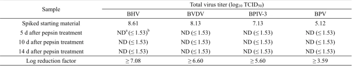
Sample Total virus titer (log10 TCID50)
BHV BVDV BPIV-3 BPV
Spiked starting material 8.61 8.13 7.13 5.12
5 d after pepsin treatment NDa (≤ 1.53)b ND (≤ 1.53) ND (≤ 1.53) ND (≤ 1.53)
10 d after pepsin treatment ND (≤ 1.53) ND (≤ 1.53) ND (≤ 1.53) ND (≤ 1.53)
14 d after pepsin treatment ND (≤ 1.53) ND (≤ 1.53) ND (≤ 1.53) ND (≤ 1.53)
Log reduction factor ≥ 7.08 ≥ 6.60 ≥ 5.60 ≥ 3.59
aNo infectious virus was detected.
bThese values were calculated using a theoretical minimum detectable level of infectious virus, with a 95% confidence level (Kärber, 1931).
Table 3. Inactivation of viruses during pepsin treatment at pH 2.0
for virus titration in the virus spiking experiments, to control for any possible cytotoxic effects on the indicator cells which might interfere with the virus titration. The interference tests were performed to determine whether the starting materials for virus spiking studies exerted an inhibitory effect on the ability of the cell lines to permit the detection of the virus. The load titer assays were performed to determine precisely the point at which spiking the virus into the starting material resulted in a loss in the virus titer.
Virus spiking experiments were conducted using the validated scale-down processes. Evaluation of virus inactivation was performed by spiking 1 g of air-dried bovine hide at each step of the process with 1 ml of an aliquot of the virus stock. After subjecting the sample to the processing step, the appropriate fractions were collected in order to determine the presence of infectious virus. The virus log reduction factor was defined as the log10 of the ratio of the virus loads in the spiked starting and post process materials, as previously described (International Conference on Harmonisation, 1998; Bae et al., 2010a). All virus inactivation experiments were carried out in duplicate and mean values are given.
The effectiveness of 70% ethanol treatment for 24 h for the inactivation of viruses was determined. For each virus, four samples of air-dried bovine hide were spiked with the appropriate stock virus solution, at a ratio of 1 ml of virus per 1 g bovine hide. The samples were incubated at room temperature for 10 min to allow for the adsorption of the virus solution. Following
the 10 min incubation, 5 ml of virus culture media was added to one of the virus-spiked bovine hide samples, which was thoroughly mixed with a vortex mixer in order to withdraw viruses from the virus-spiked bovine hide. A portion of the sample was immediately titrated. The remaining bovine hides were treated with 70% ethanol at 4°C for 1, 6, or 24 h. Viruses were extracted from the bovine hide with 5 ml of virus culture media and then immediately titrated. All the viruses tested were completely inactivated to undetectable levels within 1 h of treatment (Table 2). BHV was completely inactivated from an initial titer of 7.92 log10 TCID50 to undetectable levels with the log reduction factor of ≥5.58. BVDV was completely inactivated from an initial titer of 7.66 log10 TCID50 to undetectable levels with the log reduction factor of ≥5.32. BPIV-3 was completely inactivated from an initial titer of 7.45 log10 TCID50 to undetectable levels with the log reduction factor of ≥5.11. BPV was completely inactivated from an initial titer of 5.76 log10 TCID50 to undetectable levels with the log reduction factor of ≥ 3.42. Ethanol exhibits rapid broad-spectrum antimicrobial activity against viruses as well as bacteria and fungi. Although little is known about the specific mode of virucidal action of ethanol, it is generally believed that ethanol causes lipid-envelope damage and the rapid denaturation of proteins, with subsequent interference with infection of virus to host cell (McDonnell, 2007b).
Previous reports have shown that lipid-enveloped viruses are more susceptible to alcohols than non-enveloped viruses (Kim et al., 2000; Engelenburg et al., 2002; Eterp et al., 2009)
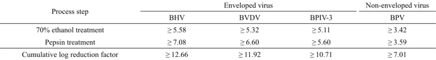
Process step Enveloped virus Non-enveloped virus
BHV BVDV BPIV-3 BPV
70% ethanol treatment ≥ 5.58 ≥ 5.32 ≥ 5.11 ≥ 3.42
Pepsin treatment ≥ 7.08 ≥ 6.60 ≥ 5.60 ≥ 3.59
Cumulative log reduction factor ≥ 12.66 ≥ 11.92 ≥ 10.71 ≥ 7.01
Table 4. Cumulative log reduction factors of viruses achieved during the manufacturing process of collagen type I from bovine hides
The efficacy of pepsin treatment at pH 2.0 for 14 days in inactivating viruses was evaluated. For each virus, four samples of air-dried bovine hide were each spiked with the appropriate stock virus solution at a ratio of 1 ml of virus per 1 g bovine hide. The samples were incubated at room temperature for 10 min to allow for the adsorption of the virus solution. Following the 10 min incubation, 5 ml of virus culture media was added to one of the virus-spiked bovine hide samples, which was thoroughly mixed with a vortex mixer in order to withdraw viruses from the virus-spiked bovine hide. A portion of the sample was immediately titrated. The remaining bovine hides were treated with pepsin at 20°C for 5, 10, or 14 days. Viruses were extracted from the bovine hide with 5 ml of virus culture media and were then immediately titrated. All the viruses tested were completely inactivated to undetectable levels during treatment (Table 3). BHV was completely inactivated from an initial titer of 8.61 log10 TCID50 to undetectable levels with the log reduction factor of ≥7.08. BVDV was completely inactivated from an initial titer of 8.13 log10 TCID50 to undetectable levels with the log reduction factor of ≥6.60.
BPIV-3 was completely inactivated from an initial titer of 7.13 log10 TCID50 to undetectable levels with the log reduction factor of ≥5.60. BPV was completely inactivated from an initial titer of 5.12 log10 TCID50 to undetectable levels with the log reduction factor of ≥3.59. Low pH treatment is a well-known process to inactivate viruses (Kim et al., 2001). The first mechanism of virus inactivation during pepsin treatment at pH 2.0 may be the virucidal effect of low pH. Low pH treatment also shows broad-spectrum antimicrobial activity against viruses as well as bacteria and fungi (McDonnell, 2007a).
Lowering the environmental pH may disrupt the structure and function of essential surface and intracellular macromolecules.
Digestion of essential surface proteins by pepsin may be a secondary mechanism of virus inactivation during pepsin treatment at pH 2.0.
The cumulative virus reduction factor for a manufacturing process is determined from the sum of the individual virus reduction factors based on individual process involving different physicochemical methods (International Conference on Harmonisation, 1998). The cumulative virus reduction factors achieved for the different viruses by the process steps
evaluated in this study are presented in Table 4. The cumulative log reduction factors, ≥12.66 for BHV, ≥11.92 for BVDV, ≥ 10.71 for BPIV-3, and ≥7.01 for BPV, are several magnitudes greater than the potential virus load of current bovine hides.
Accordingly, these results indicate that process steps in manufacturing collagen type I are capable of inactivating a wide range of viruses that represent a broad spectrum of physicochemical attributes. Regulatory guidelines recommend incorporating multiple orthogonal methods for viral clearance;
that is, methods that have independent (unrelated) clearance mechanisms. Therefore, since the mechanisms of virus inactivation in each of these steps are different to one another, it is concluded that the overall process of collagen type I production is robust in reducing the virus load. This is the first systematic evaluation of virus clearance during the process of manufacturing collagen type I from bovine hides.
적 요
세포치료제 또는 조직공학제제에 사용되는 동물 유래 콜라겐 은 원료물질 유래 바이러스가 오염될 가능성이 있기 때문에 생 산과정 중 바이러스가 오염되지 않도록 하여야 한다. 이를 위해 콜라겐 생산공정은 오염될 가능성이 있는 바이러스들을 불활화 하거나 제거하는 과정을 포함하여야 하며, 바이러스 불활화/제 거 능력은 제품의 안전성을 보증하는 중요한 지표로 사용된다.
본 연구의 목적은 소 가죽을 원료로 하여 type I 콜라겐을 생산하 는 공정에서 소 유래 바이러스들의 불활화/제거 효능을 평가하 는 데 있다. 이를 위해 70% 에탄올 처리 공정과 펩신 처리 공정 (pH 2)에서 바이러스 불활화 효과를 평가하였다. 바이러스 불활 화 효과 평가를 위해 bovine herpes virus (BHV), bovine viral diarrhoea virus (BVDV), bovine parainfluenza 3 virus (BPIV-3), bovine parvovirus (BPV)를 모델 바이러스로 선정하였다. 바이 러스 불활화를 위해 24시간 동안 70% 에탄올을 처리하는 공정 에서 BHV, BVDV, BPIV-3, BPV 모두 처리 1시간 안에 검출 한계 이하로 불활화되었으며, 바이러스 로그 감소 값은 각각
≥5.58, ≥5.32, ≥5.11, ≥3.42이었다. 또한 소 조직으로부터 콜라 겐을 추출하기 위한 14일간의 펩신 처리 공정에서 BHV, BVDV, BPIV-3, BPV 모두 처리 5일 안에 검출한계 이하로 불 활화되었으며, 바이러스 로그 감소 값은 각각 ≥7.08, ≥6.60,
≥5.60, ≥3.59이었다. 두 공정에서 BHV, BVDV, BPIV-3, BPV 의 누적 바이러스 로그 감소 값은 각각 ≥12.66, ≥11.92, ≥10.71,
≥7.01이었다. 이상의 결과에 의하면, 소 가죽 유래 type I 콜라겐 제조공정은 바이러스 안전성 보증을 위한 충분한 바이러스 불활 화 능력을 가지고 있는 것으로 판단된다.
Acknowledgements
This research was financially supported by the Ministry of Knowledge Economy (MKE) and Korea Industrial Technology Foundation (KOTEF) through the Human Resource Training Project for Strategic Technology.
References
Bae, J.E., Kim, J., Ahn, J., Choi, D.M., Jeong, H.S., Lee, D.H., and Kim, I.S. 2010a. Virus inactivation process for manufacturing of human acellular dermal matrix. Kor. J. Microbiol. Biotechnol. 38, 168–176.
Bae, J.E., Kim, C.K., and Kim, I.S. 2009. Inactivation of infectious microorganisms by disinfection and sterilization processes for human amniotic membrane grafts. Kor. J. Microbiol. 45, 346–353.
Bae, J.E., Kim, C.K., Kim, S., Yang, E.K., and Kim, I.S. 2010b. Process development of a virally-safe acellular bovine amniotic membrane for biological dressing. Kor. J. Microbiol. Biotechnol. 38, 420–427.
Di Lullo, G.A., Sweeney, S.M., Körkkö, J., Ala-Kokko, L., and San Antonio, J.D. 2002. Mapping the ligand-binding sites and disease-associated mutations on the most abundant protein in the human, type I collagen. J. Biol. Chem. 277, 4223–4231.
Engelenburg, F.A.C., Terpstra, F.G., Schuitemaker, H., and Moorer, W.R. 2002. The virucidal spectrum of a high concentration alcohol mixture. J. Hospital Infect. 51, 121–125.
Eterpi, M., McDonnell, G., and Thomas, V. 2009. Disinfection efficacy against parvoviruses compared with reference viruses. J. Hospital Infect. 73, 64–70.
Forest, P., Morfin, F., Bergeron, E., Dore, J., Bensa, S., Wittmann, C., Picot, S., Renaud, F.N., Freney, J., and Gagnieu, C. 2007. Validation of a viral and bacterial inactivation step during the extraction and purification process of porcine collagen. Biomed. Mater. Eng. 17, 199 –208.
Franchi, M., Trirè, A., Quaranta, M., Orsini, E., and Ottani, V. 2007.
Collagen structure of tendon relates to function. Sci. World J. 7, 404–
420.
Harkness, R.D. 1961. Biological functions of collagen. Biol. Rev. 36, 399–463.
Hodde, J. and Hiles, M. 2002. Virus safety of a porcine-derived medical device: evaluation of a viral inactivation method. Biotechnol. Bioeng.
79, 211–216.
International Conference on Harmonisation. 1998. Guidance on viral safety evaluation of biotechnology products derived from cell lines of human or animal origin. Federal Resister 63, 51074–51084.
International Organization for Sandardization. 1998. Sterilization of medical devices-Microbiological methods. Part 2: Test of sterility performed in the validation of a sterilization process. Geneva, Switzerland.
International Organization for Standardization. 2002. Sterilization of health care products - Radiation sterilization-Substantiation of 25 kGy as a sterilization dose for small or infrequent production batches.
Geneva, Switzerland.
Kärber, J. 1931. Beitrag zur kollectiven Behandlung pharmakologische Reihenversuche. Arch. Exp. Path. Pharmak. 162, 480–483.
Kim, I.S., Choi, Y.W., Lee, S.R., Cho, H.B., Eo, H.G., Woo, H.S., Chang, C.E., and Lee, S. 2001. Improvement of virus safety of a human intravenous immunoglobulin by low pH incubation. J. Microbiol.
Biotechnol. 11, 619–627.
Kim, I.S., Eo, H.G., Chang, C.E., and Lee, S. 2000. Partitioning and inactivation of viruses by cold ethanol fractionation and pasteurization during manufacture of albumin from human plasma.
J. Microbiol. Biotechnol. 10, 858–864.
Lee, C.H., Singla, A., and Lee, Y. 2001. Biomedical applications of collagen. Int. J. Pharm. 221, 1–22.
Ma, J., Holden, K., Zhu, J., Pan, H., and Li, Y. 2011. The application of three-dimensional collagen-scaffolds seeded with myoblasts to repair skeletal muscle defects. J. Biomed. Biotechnol. Article ID 812135.
Malafaya, P.B., Silva, G.A., Baran, E.T., and Reis, R.L. 2002. Drug delivery therapies I : General trends and its importance on bone tissue engineering applications. Curr. Opin. Solid Sate Mat. Science 6, 283–
295.
Malafaya, P.B., Silva, G.A., and Reis, R.L. 2007. Natural-origin polymers as carriers and scaffolds for biomolecules and cell delivery in tissue engineering applications. Adv. Drug Deliv. Rev. 59, 207–233.
McDonnell, G.E. 2007a. Antisepsis, disinfection, and sterilization, pp.
79–83. ASM Press, Washington, D.C., USA.
McDonnell, G.E. 2007b. Antisepsis, disinfection, and sterilization, pp.
90–92. ASM Press, Washington, D.C., USA.
Pruss, A., Kao, M., Garrel, T., Frommelt, L., Gurtler, L., Benedix, F., and Pauli, G. 2003. Virus inactivation in bone tissue transplants (femoral heads) by moist heat with the 'Marburg bone bank system’.
Biologicals 31, 75–82.
Shoulders, M.D. and Raines, R.T. 2009. Collagen structure and stability.
Annu. Rev. Biochem. 78, 929–958.
Willerth, S.M. and Sakiyama-Elbert, S.E. 2007. Approaches to neural tissue engineering using scaffolds for drug delivery. Adv. Drug Deliv.
Rev. 59, 325–338.