83
Prevalence and genotypes of pestivirus in Korean goats Dong-Kun Yang1,*, Chang-Hee Kweon
1, Byoung-han Kim
1, Cheong-Up Choi
1, Mun-Il Kang
1,
Bang-Hun Hyun
1, In-Jin Hwang
1, Cheong-San Lee
2, Kyoung-Oh Cho
31
National Veterinary Research and Quarantine Service, Anyang 430-824, Korea
2
Chungbuk Livestock and Veterinary Research Institute, Cheongwon 363-931, Korea
3
Chonnam National University, Gwangju 500-757, Korea
(Accepted: January 17, 2008)
Abstract : In total, 1,142 serum samples were collected from 223 goat flocks rising in five different regions of Korea. These samples were screened for the presence of border disease virus (BDV) antibodies using an enzyme linked immunosorbent assay. Of the 1,142 samples, we found 47 bovine viral diarrhea virus (BVDV) positive cases (4.1%). These positive serum samples were also examined further by using the virus neutralization test against BDV. In addition, samples were tested for both BVDV and classical swine fever virus (CSFV). All of the samples that were seropositive for BDV also demonstrated positive antibody titers against BVDV and CSFV. Due to their common antigenicity, we also determined further the prevalence and carried out virus neutralization test against three pestiviruses: 314 of the goat samples were screened using reverse transcription polymerase chain reaction with primer pairs specific to common pestivirus genome regions. Overall, 1.6% (5/314) of the samples tested was positive for pestivirus. Based on the nucleotide sequence data and the phylogenetic analysis, three isolates were characterized as BVDV type 1 and two isolates as BVDV type 2. However, none of the isolates could be classified as BDV.
These results indicate that BVDV-1 and BVDV-2 are the pestivirus strains circulating among Korean goat populations.
Keywords : genotype, goat, pestivirus, RT-PCR.
Introduction
Border disease (BD), hairy shaker disease, is a congenital disease, which is found in both sheep and goats, causing abortion, stillbirths, and/or weak offspring.
Lambs infected with border disease virus (BDV) may exhibit tremors, alterations of the central nervous system, abnormal body conformations, and hairy fleeces [10]. Although persistently infected (PI) animals are the main source of virus transmission in cattle because they continuously shed large amounts of the virus particles into the environment, the persistent infection in goat kids seems to be more difficult to establish [3, 6]. Vertical transmission, however, plays an important role in the epidemiology of the disease [5].
BDV occurs worldwide in all ruminant-raising countries in which bovine viral diarrhea virus (BVDV) infections are endemic [5, 7, 10]. BDV belongs to the
family Flaviviridae, has 12.3 kb of its genome size and is one of four groups in the genus
Pestivirus. Pestivirus groups have been identified in sheep and goats in many countries [8-10], and viruses in this genus include classical swine fever virus (CSFV), bovine viral diarrhea virus (BVDV-1, BVDV-2), and a tentative giraffe strain [13]. Recently, three major genotypes have been identified within BDV and are designated as subgroups BDV-1, BDV-2, and BDV-3 [5]. In addition, the subgroup BDV-4 has also been reported in Pyrenean chamois (
Rupicapra pyrenaica pyrenaica) [1]. BDV is antigeni- cally and serologically closely related to other pestivirus groups, which makes identification of these viruses very difficult [12]. By using 5’ noncoding region (NCR) analysis, these pestivirus groups have been segregated into several genotypes [5, 8]. Based on the comparison of sequences derived from three genetic regions (i.e., 5’ NCR, N
pro, and E2), BVDV-1 has been subdivided
*Corresponding author: Dong-Kun Yang
National Veterinary Research and Quarantine Service, Anyang 430-824, Korea
[Tel:+82-31-467-1731, Fax: +82-31-467-1795, E-mail: [email protected]]
into at least 11 BVDV-1 subgroups [11, 14, 17]. In addition, the typing of these viruses was the same regardless of whether sequences from the 5’ NCR or N
procoding region were used [17].
Seroepidemiological surveys and genetic analyses of pestiviruses circulating among Korean goats are important for determining and implementing prevention measures. Therefore, between 2004 and 2006, we investigated the current prevalence of antibodies against BDV in Korean goat populations and determined the predominant pestiviruses in the populations.
Materials and Methods
Viruses and cells
The BDV strain Aveyon (France), BVDV strain NADL (USA), and CSFV strain LOM (Japan) were used for the virus neutralization (VN) tests. Black goat fetal lung (BGFL) primary cells, Madin-Darby bovine kidney epithelial cells (MDBK), and porcine kidney (PK-15) cells were cultured in
α-minimum essential medium (MEM) containing antibiotics (100 IU/ml penicillin and 100 µg/ml streptomycin), antimycotic (0.25 µg/ml amphotericin B) and 5% fetal bovine serum (FBS; Gibco BRL, USA). All FBS and media prepara- tions were previously tested to ensure the absence of pestivirus or antibodies to pestivirus groups.
Serological tests
In total, 1,142 goat serum samples were obtained from 223 farms between 2004 and 2006. To screen BDV specific antibodies in the serum samples, an enzyme linked immunosorbent assay (ELISA) kit (Svanova Biotech, Sweden) was used according to the manufac- turer’s instructions. To check cross-neutralizing reactions among the pestivirus groups, the VN tests against BDV, BVDV, and CSFV were carried out in 96-well microtiter plates using BGFL primary cells, MDBK cells, and PK-15 cells, respectively. The microtiter plates were incubated for 5 days at 37
oC in 5% CO
2.The VN titers to BDV and BVDV were expressed as the reciprocal of the highest serum dilution that completely inhibited cytopathic effects in the wells.
The VN antibody titer to CSFV was determined using a neutralizing peroxidase linked assay [4]. The serum dilutions ranged from 1 : 2 to 1 : 2,048 and an antibody titer of
≥1 : 2 was considered positive.
RNA extraction and RT-PCR
Each viral RNA was extracted from 314 goat samples (195 plasma, 106 homogenized tissues, 5 feces, and 8 nasal discharges) using an RNA extraction kit (Bioneer, Korea) according to the manufacturer’s instructions. A conventional RT-PCR using the pan- pestivirus primer pair V324/V326 for the detection of pestivirus 5’ NCR was applied for genetic detection of pestivirus groups [8, 16]. The PCR was carried out in a reaction mixture containing 10 µl of denaturated RNA, 10 µl of 5
×buffer (12.5 mM MgCl
2), 2 µl of enzyme mix (reverse transcriptase and
Taqpolymerase), 1 µl of each primer (50 pmol), and 26 µl of distilled water (Qiagen, Germany) for a final volume of 50 µl.
The cycling profile was run as follows: cDNA synthesis at 42
oC for 30 min; followed by 35
oC cycles with denaturation at 94
oC for 30 sec, annealing at 50
oC for 30 sec and extension at 72
oC for 30 sec; and a final extension at 72
oC for 10 min. The PCR products (288 bp) were visualized using electrophoresis on an 1.5%
agarose gel containing ethidium bromide. Purified PCR products obtained using a gel extraction kit (Qiagen, Germany) were ligated with pGEM-T easy vector (Promega, USA). Plasmid DNA was isolated from amplified
Escherichia coliand recombinant plasmids were identified using
EcoRI restriction enzyme digestion (Promega, USA).
Sequencing and phylogenetic analysis
Sequencing reactions were performed by using recombinant plasmids and the ABI PRISM Big Dye Terminator Cycle Sequencing Kit (Perkin-Elmer, USA).
Phylogenetic analysis (245 bp) was carried out on 5’
NCR nucleotide sequence data from 5 clones and 46 reference pestivirus strains. The sequence data of the reference strains were obtained from GenBank (National Center for Biotechnology Information, NCBI). Phylo- genetic tree and sequence pair distances of the nucleotides were obtained using the DNAStar software program (DNAStar, USA). Homology analysis was performed using DNASIS software (Hitachi Software, Japan).
Statistical analysis
VN titers of BDV, BVDV, and CSFV were compared
by statistical analysis on log-transformed data, using
one-way ANOVA followed by Tukey test.
Results
Seroprevalence of BDV in goats
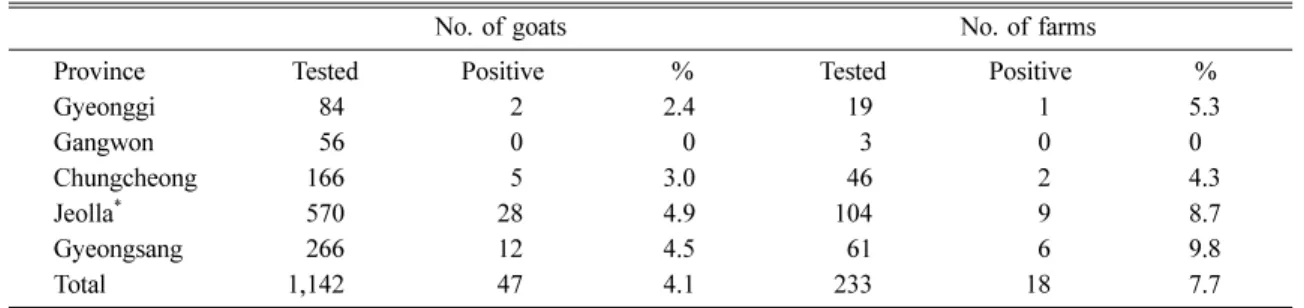
Of the 1,142 goat sera tested, 47 (4.1%) showed a positive ELISA result to BDV. These positive sera were obtained from 18 of the 233 farms, indicating farm prevalence of 7.7%. Major differences were not found in individual prevalence by regions; these values ranged from 0% in Gangwon to 4.9% in Jeolla province. Farm prevalences in the different provinces ranged from 0% in Gangwon to 9.8% in Gyeongsang (Table 1). Comparative neutralizing test studies on 28 of the 47 seropositive samples against BDV, BVDV, and CSFV showed a twofold or higher average titer to BVDV comparing to BDV and CSFV (Fig. 1). The Tukey post-hoc test revealed that the significantly higher log-transformed VN titers of BVDV as compared to CSFV, but not to BDV.
Genetic typing of goat pestiviruses
The RT-PCR using specific primers of pestivirus could amplify the 5’ NCR gene from the samples. The PCR products of the 5’ NCR gene were detected with 288 base pairs on 1.5% agarose gel (Fig. 2). Of the five isolates (designated as 0601D, 0602J, 0603J, 0604P, and 0605J) obtained from the 314 goat samples, two isolates were originated from Saanen goats that were raised for milk production and three isolates were from native Korean goats. The 5’ NCR genes of the five isolates were sequenced and all data were deposited in GenBank under accession numbers DQ973183- DQ973187. Phylogenetic analysis classified these viruses into two different BVDV genotypes; isolates 0603J, 0604P, and 0605J were clustered within the BVDV-1 subgenotype 1b and isolates 0601D and 0602J were clustered within the BVDV-2 genotype (Fig. 3). Isolates
0603J, 0604P, and 0605J showed higher nucleotide homology (93.8-96.7%) with strains of the BVDV-1 type than those (72.2-75.3%) of the 890 (BVDV-2), LE31C2 (BDV) and Alfort (CSFV) strains. Isolates 0601D and 0602J showed higher nucleotide identity (94.7-98.7%) with strains of the BVDV-2 type than Table 1. Seroprevalence of border disease virus antibodies tested by ELISA in goats from 5 regions of Korea
No. of goats No. of farms
Province Tested Positive % Tested Positive %
Gyeonggi 84 2 2.4 19 1 5.3
Gangwon 56 0 0 3 0 0
Chungcheong 166 5 3.0 46 2 4.3
Jeolla
*570 28 4.9 104 9 8.7
Gyeongsang 266 12 4.5 61 6 9.8
Total 1,142 47 4.1 233 18 7.7
*
Approximately 150,000 goats have been raised in this area for meat production.
Fig. 1. Comparison of average neutralizing antibody titers of 28 positive samples against BDV, BVDV and CSFV, respectively. All data points are average VN titer
±SD. The statistical analysis was carried out using one-way ANOVA followed by Tukey test
*p< 0.05 vs. CSFV.
Fig. 2. Detection of pestivirus genes using RT-PCR on 1.5%
agarose gel. The expected gene size was 288 bp. M: 100
bp DNA ladder, Lane 1-13: samples, +: NADL strain,
−:
negative sample.
those of the Osloss (BVDV-1 type), Alfort (CSFV), and C27 (BDV) strain (shared nucleotide identity of 70.2 to 75.5%).
Discussion
The sero-prevalence rates of BDV infection can vary from 5 to 50% in sheep and goats depending upon the country and the region within the country [5, 10, 15].
Border disease-like symptoms such as ataxia, fever, and death of two-week-old kids have been observed in southern regions of Korean peninsula since 1998.
As a result of retrospective investigations of BD-like syndrome, a BVDV-2 strain of pestivirus was confirmed as the causative agent of BD-like disease in goat
species in Korea [8]. In the current study, we investigated the nationwide seroprevalence of pestivirus groups in Korean goats for the first time. We found that the overall prevalence rate (4.1% seropositive) of BDV in Korean goats was similar to the rates found in Namibian goats (4.6% seropositive to BVDV), but lower than those found in goats of Austria (11.5%) [6, 9]. The low seroprevalence rates found in Korea may be due to the presence of isolated farms and the low rates of livestock trade. Since no vaccine has been used to prevent pestivirus infections in Korea, this antibody- positive rate is considered to be the result of natural pestivirus exposure.
In this study, we also performed a molecular
epidemiological examination that sought to identify the
Fig. 3. Phylogenetic tree showing the genotypes of BVDV isolates. The tree was generated from comparative alignmen
of sequences from 245 bp of the 5’NCR of the pestivirus genome. The multiple sequences were constructed by the neighbo
joining method. Genotypes were given on the right of tree. Abbreviations: Korea (KOR), Australia (AUS), Germany (GER)
Canada (CAN), Slovenia (SLO), Belgium (BEL), Japan (JPN), China (CHI), Italy (ITA), Sweden (SWE), United State o
America (USA), United Kingdom (UK).
pestivirus groups circulating in goat populations in Korea. Five isolates (1.63%; 5/314) were obtained from 195 plasma samples. Sequence analysis identified three of these isolates as the BVDV-1 genotype and two as the BVDV-2 genotype. Interestingly, two isolates (0601D and 0602J) obtained from British Saanen goats (which had been imported from Australia) were classified as the BVDV-2 genotype. Recently, it was reported that both BVDV-1 and BVDV-2 genotype were identified in cattle populations of Korea and BVDV-1 was the main genotype [18]. These BVDV infections were confirmed by comparative cross-neutralization assays on the 28 seropositive samples. Based on genetic typing, the pestiviruses are further classified into eight pestivirus genotypes: BVDV-1, BVDV-2, CSFV, BDV- 1, BDV-2, BDV-3, BDV-4, and giraffe [1, 2]. Cases of goat infection by BVDV-1 and BVDV-2 have been reported in a few countries [8, 12]. Krametter-Froetscher
et al.