Journal of Bacteriology and Virology 2007. Vol. 37, No. 3 p.147 – 152
Genetic Typing of Bovine Viral Diarrhea Viruses (BVDV) Circulating in Korea
Dong-Kun Yang1*, Byoung-Han Kim2, Chang-Hee Kweon1,Jeong-Kyu Park1, Ha-Young Kim3, Byung-Jae So1 and In-Joong Kim2
1Animal Disease Diagnosis Division, 2Virology Division, 3Pathology Division, National Veterinary Research and Quarantine Service, Anyang, Gyeonggi-do 430-824, Republic of Korea
Received : May 15, 2007 Accepted : July 25, 2007
To characterize the genetic diversity of bovine viral diarrhea viruses (BVDV) circulating in Korea, 11 BVDV isolates were obtained from 467 field samples collected during 2005~2006 in Korea. All of the BVDV isolates were identified as non-cytopathic (non-cp) BVDV biotypes. The 5' noncoding region (NCR) genes of the isolates were sequenced and analyzed. In total, ten BVDV isolates were typed as BVDV-1 by comparing the genomic sequences to the 5' NCR. One isolate (05R169) showed 98.6% nucleotide sequence identity with the BVDV-2 reference strain and was therefore typed as BVDV-2. Our results indicate that BVDV-1 is the main genotype circulating in the cattle population of Korea.
Key Words: BVDV, Genotype, Isolation, Cattle
INTRODUCTION
Bovine viral diarrhea virus (BVDV) is a pestivirus belonging to the family Flaviviridae. BVDV acts as the causative agent of bovine viral diarrhea-mucosal disease (BVD-MD) in cattle and is a common bovine pathogen that causes significant economic losses worldwide (4,5).
Other examples of pestiviruses with veterinary importance include classical swine fever virus and border disease virus in sheep. BVDV infection can lead to various clinical sym- ptoms including diarrhea, fever, respiratory disease, and abortion or early embryonic death of fetuses in pregnant cattle. BVDV has a genome size of 12.5 kb, and on the basis of 5' noncoding region (NCR) gene analysis, appears
to be segregated into two genotypes: BVDV-1 and BVDV-2 (10,11). Based on recent comparisons of sequences derived from three genetic regions (i.e., 5' NCR, a coding region for the autoprotease Npro, and a coding region for the structural protein E2), the BVDV-1 genotype has been subdivided into at least 11 BVDV-1 genetic subgroups (9,10,15). In addition to BVDV-1, BVDV-2 has been identified in many countries including Korea (1,2,6~8,12,16) and is associated with more severe clinical symptoms such as hemorrhagic diarrhea and abortion (8,11). During the first trimester of gestation, viral infection in dams may produce persistently infected (PI) calves due to immunotolerance effects. PI animals are the main source of viral transmission because they continu- ously shed large amounts of virus into the environment and are permanent carriers of BVDV. In addition, the presence of PI animals leads to an increased risk of mucosal disease (3). In this study, we investigated which genotypes of Korean BVDV isolates were circulating in cattle populations during 2005~2006.
147
*Corresponding author: Dong-Kun Yang. National Veterinary Research and Quarantine Service, 480 Anyang 6-dong Gyeonggi-do, 430-824, Republic of Korea.
Phone: +82-31-4671792, Fax: +82-31-4671868, e-mail: [email protected]
**This work was supported by a grant from the NVRQS, Republic of Korea.
MATERIALS AND METHODS 1. Virus and cells
BVDV, NADL (National Animal Disease Laboratory) strain of reference virus and field isolates were propagated in Madin-Darby Bovine Kidney (MDBK) cells cultured in α-minimum essential medium (MEM: Gibco BRL, Grand Island, NY, USA) with antibiotics (penicillin, 100 IU/ml, streptomycin, 100 µg/ml), 5% fetal bovine serum (FBS:
Gibco BRL) free of BVDV and antibodies against BVDV.
The virus infected cell cultures were maintained in α-MEM supplemented with 5% FBS.
2. Virus isolation
To isolate BVDV, samples were obtained from organ suspensions of 46 aborted fetuses and homogenized tissues of 37 cattle that displayed clinical signs of BVD. Tissue samples were membrane-filtered and later used in the BVDV isolation procedure. All samples were obtained from tissues that were submitted to the Pathology Division, National Veterinary Research and Quarantine Service (NVRQS), Korea, between January 2005 and June 2006.
To detect PI calves with BVDV, a total of 384 plasma samples were collected from the calves which were between 3 days and 6 months old in 4 provinces (Table 1). The samples were initially examined for the detection of BVDV genome by using RT-PCR; PCR-positive samples were further used to isolate BVDV. Specifically, MDBK mono- layers in 24-well microplates were washed twice with PBS and then inoculated with 100 µl of the membrane-filtered samples. Following adsorption, the inoculated samples were replaced with 1 ml MEM containing 3% FBS. These micro- plates were incubated for 7 days and then screened for cytopathic effects and examined using an immunofluore- scence antibody (IFA) test. All isolates were cloned three times using the limit dilution method, propagated on MDBK cells, and examined using an IFA test.
3. RNA extraction and RT-PCR
RNA was isolated from 250 µl of BVDV-infected cell
culture supernatants using an RNA isolation kit (Bioneer, Deajeon, Korea) according to the manufacturer's instructions.
Conventional RT-PCR using the pan-pestivirus primer pair V324/V326 for the 5' NCR of the pestivirus was used for genetic detection (14). PCR was carried out in a reaction mixture containing 10 µl of denaturated RNA, 10 µl of 5×
buffer (12.5 mM MgCl2), 2 µl of enzyme mix (reverse transcriptase and Taq polymerase), 1 µl of each primer (50 pmol) and 26 µl of distilled water (Qiagen, Hilden, Germany) for a 50 µl final volume. The cycling profile was run as follows: cDNA synthesis at 42℃ for 30 min; followed by 35 cycles with denaturation at 94℃ for 30s, annealing at 50℃ for 30s and extension at 72℃ for 30s; and a final extension at 72℃ for 10 min. The PCR products were visualized using electrophoresis on 1.5% agarose gel con- taining ethidium bromide. Purified PCR products using the gel extraction kit (Qiagen) were ligated with pGEM-T easy vector (Promega, Madison, WI, USA). Plasmid DNA was isolated from amplified Escherichia coli, and recombinant plasmids were identified using EcoRI restriction enzyme digestion (Promega).
4. Sequencing and phylogenetic analysis
Sequencing reactions were performed using recombinant plasmids and the ABI PRISM Big Dye Terminator Cycle Sequencing Kit (Perkin-Elmer, Wellesley, MA, USA). Phy- logenetic analysis was performed on the 5' NCR nucleotide sequence data from 11 Korean isolates and 30 reference BVDV strains. Sequence data for the reference strains were obtained from GenBank (National Center for Biotechno- logy Information, NCBI). Phylogenetic trees and sequence pair distances of the nucleotides were obtained using the DNASTAR software program (DNAStar, Madison, WI, USA). Homology analysis was performed using DNASIS software (Hitachi software, Tokyo, Japan).
RESULTS
1. Virus isolation
In total, 11 BVDVs were isolated from 7 provinces (Table 1). Six BVDV isolates (designated 05R169, 05R203,
05R204, 06R447, 06Z71, and 06Z127) were obtained from the 384 plasma samples, three isolates were obtained from tissue samples of cattle demonstrating clinical symptoms (05D26, 05D73, 05Q174), and two isolates were obtained from aborted fetuses (05Q122, 05D137; Table 1). All the isolates were identified as non-cp BVDV biotypes by no appearance of cytopathic effect and IFA test positive using BVDV specific monoclonal antibody (IDEXX, Westbrook, Maine, USA). The BVDV-specific fluorescences were noticed in the cytoplasms of the virus infected cells (Fig. 1).
Six BVDV isolates were isolated from native Korean cattle (Table 1).
2. Identification and genotyping of the isolates by RT-PCR and sequence analysis
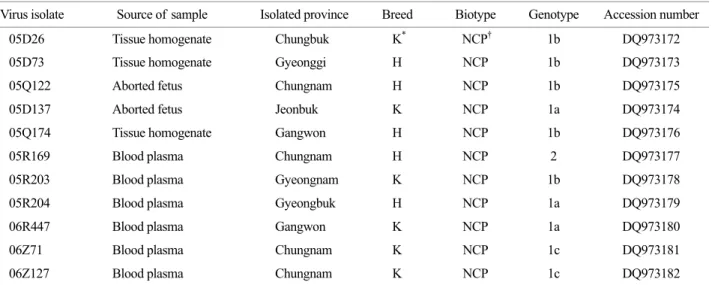
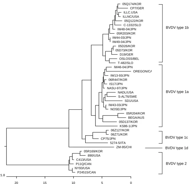
The RT-PCR using specific primers of BVDV could amplify 5' NCR gene of the isolates. The PCR products of 5'NCR were detected with 288 base pairs on 1.5% agarose gel (Fig. 2). The nucleotides of these 11 BVDV isolates were sequenced, and all data were deposited in GenBank under accession numbers DQ973172-DQ973182. Phylo- genetic analysis classified these viruses into two different genotypes (BVDV-1 and BVDV-2) as described previously (Fig. 3). We found that isolates 05D137, 05R204, and 06R447 were clustered with subgenotype 1a; isolates 05D26, 05D73, 05Q122, 05Q174, and 05R203 were clustered with Table 1. Bovine viral diarrhea viruses isolated from cattle in Korea and GenBank accession numbers of their 5'NCR nucleotide sequences
Virus isolate Source of sample Isolated province Breed Biotype Genotype Accession number 05D26 Tissue homogenate Chungbuk K* NCP† 1b DQ973172 05D73 Tissue homogenate Gyeonggi H NCP 1b DQ973173
05Q122 Aborted fetus Chungnam H NCP 1b DQ973175
05D137 Aborted fetus Jeonbuk K NCP 1a DQ973174
05Q174 Tissue homogenate Gangwon H NCP 1b DQ973176
05R169 Blood plasma Chungnam H NCP 2 DQ973177
05R203 Blood plasma Gyeongnam K NCP 1b DQ973178
05R204 Blood plasma Gyeongbuk H NCP 1a DQ973179
06R447 Blood plasma Gangwon K NCP 1a DQ973180
06Z71 Blood plasma Chungnam K NCP 1c DQ973181
06Z127 Blood plasma Chungnam K NCP 1c DQ973182
K: Korean native cattle, H: Holstein dairy cattle. † NCP: non-cytopathic.
Figure 1. Immunofluorescence of 05R169 isolate by IFA test using monoclonal antibody against BVDV in MDBK cell.
500 bpÆ 288 bpÆ
M 1 2 3 4
Figure 2. RT-PCR products of BVDV isolates were detected on 1.5% agarose gel. The expected size was 288 bp. M; 100 bp DNA ladder, Lane 1; 05D26, Lane 2; 05D73, Lane 3; NADL strain, Lane 4; negative sample.
subgenotype 1b; and isolates 06Z71 and 06Z127 were segregated into subgenotype 1c. In contrast, isolate 05R169 obtained from a sample of Chungnam Province was clustered with the genotype 2 virus (Fig. 3).
DISCUSSION
BVD occurs worldwide in most cattle-producing coun- tries, and the prevalence of BVDV antibody-positive herds
is on average approximately 55% (4). Vilcek et al. (13) reported that virus typing was the same whether sequences from the 5'NCR or Npro coding regions were used. In this study, we examined the genetic diversity of Korean BVDV isolates by identifying the 5'NCR nucleotide sequences of 11 field isolates. Sequence analysis showed that ten of the BVDV isolates were classified as the BVDV-1 genotype and only one was classified as the BVDV-2 genotype.
These results indicate that the type 1 BVDV genotype is
BVDV type 1b
BVDV type 1a
BVDV type 2
0 21.8
5 10
15 20
05Q174/KOR CP7/GER ILLC.USA ILLNC/USA
05Q122/KOR C-1332/SLO IW48-04/JPN 05R203/KOR IW44-03/JPN IW49-04/JPN
05D26/KOR 05D73/KOR
D19/GER OSLOSS/BEL T-482/SLO IW46-04/JPN
OREGON/CA IW13-93/JPN
06R447/KOR IS17/JPN NASU-97/JPN
NADL/USA S-ALT6/SWE
SD1/USA IW43-03/JPN
NOSE/JPN
05R204/KOR BEGA/AUS 05D137/KOR
KS86-1/JPN 06Z127/KOR 06Z71/KOR CP75/JPN
5274-5/ITA ZM-95/CHI 05R169/KOR
890/USA C413/USA P11Q/CAN NY93/USA
P24515/CAN
BVDV type 1c BVDV type 1d
Figure 3. Phylogenetic tree of the 5' non-coding regions (NCR) of BVDV strains and isolates. The tree was generated from comparative alignment of sequences from 245 bp of the 5'NCR of the BVDV genome. The multiple sequences were constructed by the neighbor joining method. Genotypes were given on the right of tree. Abbreviations: Korea (KOR), Australia (AUS), Germany (GER), Canada (CAN), Slovenia (SLO), Belgium (BEL), Japan (JPN), China (CHI), Italy (ITA), Sweden (SWE), United State of America (USA).
the predominant virus circulating in the cattle population of Korea. In addition, the phylogenetic tree analysis indicated that isolates 05D137, 05R204, and 06R447 belong to the BVDV-1a subtype, whereas isolate 05R169 clustered with the BVDV-2 genotype. Moreover, isolate 05R169 is very closely related to strain 890, which is the North American type 2a reference strain. In fact, the genetic similarity of the 5'NCR nucleotide sequence between isolate 05R169 and strain 890 was 98.6%.
Ridpath et al. (14) reported that BVDV-1 is generally used in vaccine production, and BVDV-2 is isolated predo- minantly from fetal bovine sera. This BVDV-2 isolate is particularly common in PI calves delivered from dams vaccinated against BVDV and cattle that have died from hemorrhagic syndrome (an acute form of BVDV). Since little information is currently available regarding PI calves and BVD-MD cases in Korea, further studies are required to investigate the pathological and clinical characteristics of BVDV-2 isolates.
In addition, local vaccines against BVDV are produced from BVDV-1 strains only in Korea. For the prevention of cattle from 2 genotypes of BVDV infection, it is necessary to develop effective BVDV vaccine using both BVDV-1 and BVDV-2 strains.
Acknowledgements
We thank Dr. You-Chan Bae for providing the clinical samples used for virus isolation and Ms. Choi SS for technical assistance.
REFERENCES
1) Becher P, Orlich M, Shannon AD, Horner G, Konig M, Thiel HJ: Phylogenetic analysis of pestiviruses from domestic and wild ruminants. J Gen Virol 78:
1357-1366, 1997.
2) Beer M, Wolf G, Kaaden OR: Phylogenetic analysis of the 5'-untranslated region of german BVDV type II isolates. J Vet Med B Infect Dis Vet Public Health 49:
43-47, 2002.
3) Brock KV, Redman DR, Vickers ML, Irvine NE:
Quantitation of bovine viral diarrhea virus in embryo
transfer flush fluids collected from a persistently infected heifer. J Vet Diagn Invest 3: 99-100, 1991.
4) Houe H: Epidemiology of bovine viral diarrhea virus.
Vet Clin North Am Food Anim Pract 11: 521-547, 1995.
5) Houe H: Epidemiological features and economical importance of bovine virus diarrhoea virus (BVDV) infections. Vet Microbiol 64: 89-107, 1999.
6) Jones LR, Zandomeni R, Weber EL: Genetic typing of bovine viral diarrhea virus isolates from Argentina.
Vet Microbiol 81: 367-375, 2001.
7) Kim IJ, Hyun BH, Shin JH, Lee KK, Lee KW, Cho KO, Kang MI: Identification of bovine viral diarrhea virus type 2 in Korean native goat (Capra hircus). Virus Res 121: 103-106, 2006.
8) Park JS, Moon HJ, Lee BC, Hwang WS, Yoo HS, Kim DY, Park BK: Comparative analysis on the 5'-untranslated region of bovine viral diarrhea virus isolated in Korea. Res Vet Sci 76: 157-163, 2004.
9) Pellerin C, van den Hurk J, Lecomte J, Tussen P:
Identification of a new group of bovine viral diarrhea virus strains associated with severe outbreaks and high mortalities. Virology 203: 260-268, 1994.
10) Ridpath JF, Bolin SR: Differentiation of types 1a, 1b and 2 bovine viral diarrhea virus (BVDV) by PCR.
Mol Cell Probes 12: 101-106, 1998.
11) Ridpath JF, Bolin SR, Dubovi EJ: Segregation of bovine viral diarrhea virus into genotypes. Virology 205: 66-74, 1994.
12) Vilcek S, Durkovic B, Kolesárová M, Greiser-Wilke I, Paton D: Genetic diversity of international bovine viral diarrhoea virus (BVDV) isolates: identification of a new BVDV-1 genetic group. Vet Res 35: 609-615, 2004.
13) Vilcek S, Durkovic B, Kolesárová M, Paton DJ:
Genetic diversity of BVDV: consequences for classifi- cation and molecular epidemiology. Prev Vet Med 72:
31-35, 2005.
14) Vilcek S, Herring AJ, Herring JA, Nettleton PF, Lowings JP, Paton DJ: Pestiviruses isolated from pigs, cattle and sheep can be allocated into at least three genogroups using polymerase chain reaction and restriction endonuclease analysis. Arch Virol 136: 309 -323, 1994.
15) Vilcek S, Paton DJ, Durkovic B, Strojny L, Ibata G,
Moussa A, Loitsch A, Rossmanith W, Vega S, Scicluna MT, Paifi V: Bovine viral diarrhoea virus genotype 1 can be separated into at least eleven genetic groups. Arch Virol 146: 99-115, 2001.
16) Wakeley PR, Turner JL, Ibata G, King DP, Sandvik T, Howard P, Drew TW: Characterisation of a type 2 bovine viral diarrhoea virus isolated from cattle in the UK. Vet Microbiol 102: 19-24, 2004.