http://dx.doi.org/10.14405/kjvr.2015.55.3.185
185
<Original Article>
Serosurveillance and establishment of a reverse transcription-polymerase chain reaction assay for bovine parainfluenza virus type 5
Dong-Kun Yang*, Sung-Suk Choi, Beom-Joo Lee, Ha-Hyun Kim, Hyun-Ye Jo Viral disease division, Animal and Plant Quarantine Agency, Anyang 430-757, Korea (Received: June 20, 2015; Revised: August 14, 2015; Accepted: August 24, 2015)
Abstract : Bovine parainfluenza virus type 5 (bPIV5) was isolated from cattle with downer cow syndrome in 2012, and included both respiratory and neurotropic pathogens from a variety of animals. In the current study, we conducted serosurveillance using sera obtained from seven Korean farms and optimized a reverse transcription-polymerase chain reaction (RT-PCR) assay to detect bPIV5. The overall seropositive rate for Korean cattle was 21.4% (163/760). A farm located near the city of Milyang in Gyeoungnam province had a markedly elevated seropositive rate for bPIV5 compared to that of the other six farms. The regional seropositive rates were 4.2% (8/192) for Haman, 19.5% (18/
55) for Hwasung, 73.9% (65/88) for Milyang, 26.0% (50/192) for Namwon, 1.0% (1/96) for Uljin, 13.5% (13/96) for Yeongju, and 32.7% (8/41) for Yongin. The sensitivity and specificity of three RT-PCR primer sets used to amplify the conserved fusion gene of bPIV5 were also evaluated. An RT-PCR assay using the bPIVFR3 primer set was 10- fold more sensitive than the assays using the two other primer sets and did not result in non-specific amplification.
These results demonstrated that the bPIFR3 primer set can be used to detect bPIV5.
Keywords : bovine, parainfluenza virus 5, reverse transcription-polymerase chain reaction, serosurveillance
Introduction
The family Paramyxoviridae is composed of several gen- era and gives rise to a number of important human and ani- mal diseases, including measles, human parainfluenza viruses (hPIV), respiratory syncytial virus (RSV), Newcastle disease virus, and parainfluenza virus type 5 (PIV-5/SV-5) [11].
Parainfluenza virus 5 (PIV5), previously known as simian virus 5, belongs to the Rubulavirus genus within the subfam- ily Paramyxovirinae, and includes a number of respiratory and neurotropic pathogens found in a variety of animals, as well as humans [12]. PIV5 has a non-segmented, negative-strand RNA genome of approximately 15,000 nucleotides and con- tains seven genes that encode the following viral proteins:
nuclepcapsid (N), phosphor (P), matrix, fusion (F), small hydrophobic, hemagglutinin-neuraminidase (HN) and large protein [15, 17]. Human PIV5 (hPIV5) has been isolated from clinical samples proven the neutralizing antibody against PIV5 [8, 9]. Canine PIV5 (cPIV5) is considered to be one of the important viral agents causing canine respiratory disease, including bronchitis and bronchiolitis, in combination with certain bacterial agents [2, 3]. In addition, porcine PIV5 has been isolated from the lung of fetuses within sows infected with porcine reproductive and respiratory syndrome virus
(PRRSV), and is considered to be a mild pathogen in pigs [13]. Bovine PIV5 (bPIV5) infection was reported in Korean cattle showing neurological symptoms and the biological/
molecular characterization of bPIV5 isolates was reported in 2014 [20].
Several methods of detecting antibodies to PIV are avail- able, including enzyme-linked immunosorbent assay (ELISA), hemagglutination inhibition (HI), complement fixation, and hemadsorption inhibition assays (HADI) [10]. ELISA is by far the most sensitive and can be applied to a large number of serum samples, but it is the least specific assay and detects antibody titers against other type of hPIV. In addition, no species-specific ELISA to detect antibodies against PIV5 has been developed. The HADI assay, which is based on the bio- logical properties of PIV5, can be applied to detect antibod- ies, but requires a cell culture system, erythrocytes, and a skilled technician [19]. The HI test is considered the stan- dard method for detecting PIV5 antibodies [3]. Therefore, in this study, the HI test was used to screen for bPIV5 antibod- ies and confirm bPIV5 infection in Korean cattle.
We performed a seroepidemiological survey of bPIV5 and also established a diagnostic tool, an optimized reverse tran- scription-polymerase chain reaction (RT-PCR) assay, to facil- itate detection of bPIV5 infection in cattle. These contributions
*Corresponding author
Tel: +82-31-467-1783, Fax: +82-31-467-1797 E-mail: [email protected]
for a bPIV5 RT-PCR assay.
Materials and Methods
Collection of sera and preparation of bPIV5
In total, 760 blood samples were collected from Korean cattle on seven farms from four provinces in South Korea.
Clotted blood samples were separated by centrifugation, and the sera were stored at –20oC until use. Bovine PIV5 (strain QIA-B1201) isolated from a farm field in 2012 was propa- gated in Vero cells. Vero cells were regularly maintained in α-minimum essential medium (α-MEM) supplemented with 5% fetal bovine serum, penicillin (100 IU/mL), streptomycin (10 µg/mL), and amphotericin B (0.25 µg/mL). To propagate bPIV5, Vero cells grown in α-MEM were washed three times with phosphate-buffered saline (PBS), and then inocu- lated. After adsorption, α-MEM was added, and the cells were incubated until a 90% cytopathic effect was observed in the infected Vero cells. After harvesting, the antigen was used in the HI test and for detecting antigens via RT-PCR.
The Akabane virus, Aino virus, Chuzan virus, bovine ephem- eral fever virus (BEFV), bovine viral diarrhea virus (BVDV), and infectious bovine rhinotracheitis virus (IBRV) were used as reference strains to test the specificity test of the RT-PCR assay.
Hemagglutination assay (HA) and HI test
The HA test was performed by preparing a serial twofold dilution of bPIV5 (strain QIA-B1201) into 50 µL of PBS (pH 7.2) with 50 µL of 0.6% chicken erythrocytes. The HA titer was expressed as the reciprocal of the highest dilution of bPIV5 showing complete hemagglutination. The HI test was performed in 96-well microplates according to a modified method. Briefly, to remove non-specific inhibitors, 20 µL of serum were mixed with 180 µL of 25% kaolin and incubated for 30 min. After pipetting, the kaolin was removed by cen- trifugation at 2,500× g for 15 min in a microfuge. The result-
serum. After incubation for 1 h at room temperature, 50 µL of 0.6% chicken erythrocytes were added, and the micro- plates were incubated at 37oC for 1 h. The HI titer was expressed as the reciprocal of the highest dilution of serum showing complete inhibition of hemagglutination. Serum samples showing an HI titer ≥ 1 : 20 were considered positive.
Selection of primers and RT-PCR
Three different primer pairs were designed based on the highly conserved F gene from strain QIA-B1201 (GenBank accession no. KR061998). Viral RNA was extracted from QIA-B1201 using an RNA extraction kit (Bioneer, Korea) according to the manufacturer’s instructions. The extracted RNA was eluted in 50 µL of RNase- and DNase-free water.
The RT-PCR assay was performed with specific primers (PIV5F1, PIV5R1, PIV5F2, PIV5R2, PIV5F3, and PIV5R3) that amplify the F gene of bPIV5 (Table 1). RT-PCR was car- ried out in a reaction mixture containing 10 µL of denatured RNA, 1 µL of each primer (50 pmol), 10 µL of 5× buffer (12.5 mM MgCl2), 2 µL of dNTP mix, 2 µL of enzyme mix (reverse transcriptase and Taq polymerase), and 24 µL of dis- tilled water (Qiagen, Hilden, Germany). The cycling profile consisted of cDNA synthesis at 42oC for 30 min, followed by 35 cycles at 95oC for 30 sec, 52oC for 30 sec, and 72oC for 45 sec, with a final extension at 72oC for 5 min. The PCR products were visualized using gel electrophoresis on 1.8%
agarose gels containing ethidium bromide.
Results
Serosurveillance of bovine PIV5
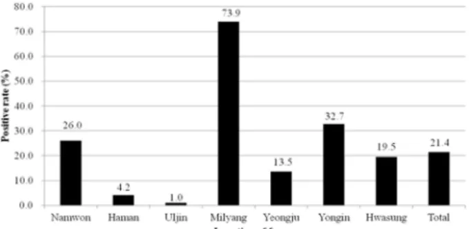
The results of bPIV5 serosurveillance in Korean cattle were shown in Figures 1 and 2. An HI titer of ≥ 1 : 20 was considered to be positive for bPIV5. The overall bPIV5 seropositive rate in Korean cattle was 21.4% (163/760). One farm located near the city of Milyang in Gyeoungnam prov- ince showed a markedly elevated seropositive rate compared
Table 1. List of the oligonucleotide primers used for RT-PCR assays against the F gene of bPIV5
Primer designated Oligonucleotide sequence (5'-3') nucleotide position Size of amplicon
bPIVF1 GGCAGGAGCAGGCAGCCTTG 9942–61*
bPIVR1 CTCACCGATCGGCTGTAGGAG 9255–235 213
bPIVF2 CGG,AGG,AGACGCCGGTTTGC 9291–311
bPIVR2 GGTACTCCCCAGTAGGATCC 9693–674 402
bPIVF3 CCTTCTCTCCAGTGGTTGGG 1069–1089
bPIVR3 CACCGCAGCTAGATTCTGGG 1368–1359 299
*GenBank accession no. KR061998. RT-PCR, reverse transcription-polymerase chain reaction; F, fusion protein; bPIV5, bovine parainflu- enza virus type 5.
to the remaining six farms. Seropositive rates according to region were 4.2% (8/192) in Haman, 19.5% (18/55) in Hwa- sung, 73.9% (65/88) in Milyang, 26.0% (50/192) in Nam- won, 1.0% (1/96) in Uljin, 13.5% (13/96) in Yeongju, and 32.7% (8/41) in Yongin (Fig. 1). At the highly seropositive farm in Milyang, the most frequent HI titer was 1 : 20 (22.7%) (Fig. 2).
Sensitivity and specificity
An RT-PCR assay for the detection of bPIV5 was carried out using three primer sets to optimize assay sensitivity. The bPIV5 culture supernatant (QIA-B1201; 107.0 TCID50/mL) was subjected to a sensitivity test. The eluted RNA was seri- ally diluted 10-fold to 10−5 and then used for one-step RT- PCR. Sensitivity of the RT-PCR assay was expressed as infectivity titer equivalent. As shown in Figure 3, the detec- tion limit of the RT-PCR assays using primer sets bPIVFR1 and 2 was 2 × 10³ TCID50/reaction in each assay, whereas the sensitivity of the assay using primer set bPIVFR3 was 2 × 10² TCID50/reaction. Overall, the bPIVFR3 primer set was 10-fold more sensitive than bPIVFR1 or bPIVFR2. As shown in Figure 4, RT-PCR using the bPIVFR1 primer set resulted in non-specific reactions in Aino virus, BEFV, and Chuzan virus, and amplified a band of roughly the same size as bPIV5 in IBRV. However, RT-PCR with primer sets bPIVFR2 and 3 did not result in any non-specific reaction.
Although RT-PCR with any of the three primer sets was able
to amplify bPIV5 with high sensitivity, only bPIVFR3 was acceptable in terms of both sensitivity and specificity.
Discussion
Since the first hPIV was reported in the late 1950s, four major hPIV serogroups have been identified based on anti- genic and genetic characteristics. Serogroups hPIV1 and 3 gave rise to Respirovirus, and hPIV2 and 4 have been clus- tered into Rubulavirus. These hPIVs have caused respiratory illness in children less than five years of age [21]. Viral anti- gens commonly associated with respiratory disease in cattle include IBRV, bovine respiratory syncytial virus (BRSV), and parainfluenza virus type 3 (PIV3) [16]. It is notable that PIV3 alone does not usually produce severe disease in cattle.
However, combination with certain bacterial agents, such as Mannheimia and Mycoplasma, promotes the formation of the enzootic pneumonia complex. PIV3 infection is thought to be stress-related, perhaps due to mixing of animals of different age groups, transport stress, or winter housing under poor hygienic conditions. However, PIV5, known as cPIV2 in a veterinary context, is less well understood in cattle through- out the world.
The seroprevalence of cPIV2 in dogs has been estimated to range from 10.4% in Canada to 28.9% in Czechoslovakia [7, 22]. It is reported that about 29% of human serum samples had VN antibodies against hPIV5, suggesting that some humans have been exposed to hPIV5 from dogs [6]. How- ever, no serosurvey data are available regarding bPIV5 in cattle populations in any part of the world. In the present study, 12.4% (163/760) of Korean cattle from seven farms were seropositive for bPIV5. The cattle population (n = 672) Fig. 1. Distribution of seropositivity for bovine PIV5 according
to the farm.
Fig. 2. Frequency distribution of HI titers against bovine PIV5 on Milyang farm. Titers < 1 : 20 were considered negative.
Fig. 3. Sensitivity test using three primer sets against bovine PIV5. (A) bPIVFR1. (B) bPIVFR2. (C) bPIVFR3. M, 1-kb DNA ladder; Lane 1–6, 10-fold dilution of bPIV5.
Fig. 4. Specificity test using three primer sets against bovine PIV5. (A) bPIVFR1. (B) bPIVFR2. (C) bPIVFR3. M, 1kb DNA ladder; Lane 1, Akabane virus; Lane 2, Aino virus; Lane 3, bovine ephemeral fever virus; Lane 4, bovine viral diarrhea virus; Lane 5, Chuzan virus; Lane 6, infectious bovine rhinotra- cheitis virus; Lane 7, bovine PIV5; Lane 8, negative.
that the virus had circulated within the cattle population. In addition, most of the cattle on the farm had experienced a respiratory illness and some cattle were found to be persis- tently infected with BVDV. We assume that close contact amongst the farmer, dogs, and cattle may be a contributing factor to the exposure of cattle to bPIV5 from dogs or humans. Most of the dogs reared on cattle farms are inocu- lated with a multivalent vaccine containing cPIV2. A previ- ous study reported that PIV5 was detected in dogs at post- inoculation day 3 using an RT-PCR assay [6]. Many diseases in humans are associated with hPIV5 and antibodies against hPIV5 are detected in human populations [14].
Virus isolation by cell culture and direct immunofluores- cent antibody assay are the most commonly used technique for detecting PIV5. However, the above-mentioned methods have significant limitations in terms of sensitivity [18]. There- fore, RT-PCR was explored as a means to overcome limita- tions on sensitivity and specificity and provide rapid results.
Conserved sequences within the N, P, F, and HN regions have been used for amplifying PIVs [4, 13]. However, the use of a poor primer set can result in low sensitivity or false- positive reactions in the RT-PCR assay. In the present study, after considering the length of the amplified fragment, three primer sets based on the F gene of QIA-B1201 were used to evaluate sensitivity and specificity using 10-fold serial dilu- tions of viral RNA extracted from QIA-B1201. The results showed that the bPIVFR3 primer set was 10-fold more sensi- tive (2 × 10² TCID50/reaction) than the bPIVFR1 or bPIVFR2 primer set, and was able to amplify only the F gene of bPIV5 without any non-specific reaction. The detection limit was consistent with previous reports [1, 4, 6]. Cell lines used in laboratories can be persistently infected with PIV5 and these cells typically do not show detectable cytopathic effects [5].
Therefore, the RT-PCR assay using the bPIVFR3 primer set may be useful in the diagnosis of PIV5 infection in animals and when checking cell lines in laboratories.
In conclusion, based on the serosurveillance of bPIV5, a small number of Korean cattle have been exposed to bPIV5, which may cause respiratory disease under poor hygienic conditions. The RT-PCR assay described here was both sen- sitive and specific for the detection of bPIV5. However, we did not attempt to apply this RT-PCR assay to a large num- ber of field samples. Therefore, further studies are required to determine whether this RT-PCR assay provides robust results from several types of specimen.
Acknowledgments
This work was supported financially by a grant (F1543083-2014-2015-02) from Animal, and Plant Quaran-
1. Aguilar JC, Pérez-Breña MP, García ML, Cruz N, Erdman DD, Echevarría JE. Detection and identification of human parainfluenza viruses 1, 2, 3, and 4 in clinical samples of pediatric patients by multiplex reverse transcription- PCR. J Clin Microbiol 2000, 38, 1191-1195.
2. Ajiki M, Takamura K, Hiramatsu K, Nakai M, Sasaki N, Konishi S. Isolation and characterization of parainfluenza 5 virus from a dog. Nihon Juigaku Zasshi 1982, 44, 607- 618.
3. Appel MJ, Percy DH. SV-5-like parainfluenza virus in dogs. J Am Vet Med Assoc 1970, 156, 1778-1781.
4. Bellau-Pujol S, Vabret A, Legrand L, Dina J, Gouarin S, Petitjean-Lecherbonnier J, Pozzetto B, Ginevra C, Freymuth F. Development of three multiplex RT-PCR assays for the detection of 12 respiratory RNA viruses. J Virol Methods 2005, 126, 53-63.
5. Chatziandreou N, Stock N, Young D, Andrejeva J, Hagmaier K, McGeoch DJ, Randall RE. Relationships and host range of human, canine, simian and porcine isolates of simian virus 5 (parainfluenza virus 5). J Gen Virol 2004, 85, 3007-3016.
6. Chen Z, Xu P, Salyards GW, Harvey SB, Rada B, Fu ZF, He B. Evaluating a parainfluenza virus 5-based vaccine in a host with pre-existing immunity against parainfluenza virus 5. PLoS One 2012, 7, e50144.
7. Ellis J, Anseeuw E, Gow S, Bryan H, Salb A, Goji N, Rhodes C, La Coste S, Smits J, Kutz S. Seroepidemiology of respiratory (group 2) canine coronavirus, canine parainfluenza virus, and Bordetella bronchiseptica infections in urban dogs in a humane shelter and in rural dogs in small communities. Can Vet J 2011, 52, 861-868.
8. Goswami KK, Lange LS, Mitchell DN, Cameron KR, Russell WC. Does simian virus 5 infect humans? J Gen Virol 1984, 65, 1295-1303.
9. Johnson JB, Capraro GA, Parks GD. Differential mechanisms of complement-mediated neutralization of the closely related paramyxoviruses simian virus 5 and mumps virus. Virology 2008, 376, 112-123.
10. Julkunen I. Serological diagnosis of parainfluenza virus infections by enzyme immunoassay with special emphasis on purity of viral antigens. J Med Virol 1984, 14, 177-187.
11. Lamb RA, Parks GD. Paramyxoviridae: The viruses and their replication. In: Knipe DM, Howley PM (eds.). Fields Virology. 5th ed. pp. 1449-1496. Lippincott Williams &
Wilkins/Wolters Kluwer Business, Philadelphia, 2007.
12. Lamb RA, Paterson RG, Jardetzky TS. Paramyxovirus membrane fusion: lessons from the F and HN atomic structures. Virology 2006, 344, 30-37.
13. Lee YN, Lee C. Complete genome sequence of a novel porcine parainfluenzavirus 5 isolate in Korea. Arch Virol 2013, 158, 1765-1772.
14. Randall RE, Young DF. Comparison between parainfluenza virus type 2 and simian virus 5: monoclonal antibodies reveal major antigenic differences. J Gen Virol 1988, 69, 2051-2060.
15. Schmitt AP, He B, Lamb RA. Involvement of the cytoplasmic domain of the hemagglutinin-neuraminidase protein in assembly of the paramyxovirus simian virus 5. J Virol 1999, 73, 8703-8712.
16. Stott EJ, Thomas LH, Collins AP, Crouch S, Jebbett J, Smith GS, Luther PD, Caswell R. A survey of virus infections of the respiratory tract of cattle and their association with disease. J Hyg (Lond) 1980, 85, 257-270.
17. Sun D, Luthra P, Xu P, Yoon H, He B. Identification of a phosphorylation site within the P protein important for mRNA transcription and growth of parainfluenza virus 5. J Virol 2011, 85, 8376-8385.
18. Syrmis MW, Whiley DM, Thomas M, Mackay IM, Williamson J, Siebert DJ, Nissen MD, Sloots TP. A sensitive, specific, and cost-effective multiplex reverse transcriptase-PCR assay for the detection of seven common respiratory viruses in respiratory samples. J Mol Diagn
2004, 6, 125-131.
19. van der Logt JT, Heessen FW, van Loon AM, van der Veen J. Hemadsorption immunosorbent technique for determination of mumps immunoglobulin M antibody. J Clin Microbiol 1982, 15, 82-86.
20. Yang DK, Nah JJ, Kim HH, Choi SS, Bae YC, Park JW, Song JY. Isolation of novel bovine parainfluenza virus type 5 (bPIV5) and its incidence in Korean cattle. Korean J Vet Res 2014, 54, 107-112.
21. Yin HS, Wen X, Paterson RG, Lamb RA, Jardetzky TS.
Structure of the parainfluenza virus 5 F protein in its metastable, prefusion conformation. Nature 2006, 439, 38- 44.
22. Zuffa T, Krobot P. Detection of antibodies against infectious viral laryngotracheitis and parainfluenza 2 in dogs bred in Czechoslovakia. Vet Med (Praha) 1987, 32, 689-694.