Dexamethasone 에 의한 RANKL 유도성 파골세포 분화 촉진 효과
노아롱새미·천링·박효정·양미혜
*
·이정민·임미정#숙명여자대학교 약학대학, *경북대학교 자연과학대학 생명공학부
(Received January 8, 2009; Revised March 10, 2009; Accepted April 6, 2009)
The Stimulatory Effect of Dexamethasone on RANKL-induced Osteoclastogenesis
A Long Sae Mi No, Ling Chen, Hyojung Park, Mihye Yang*, Jung Min Lee and Mijung Yim
#College of Pharmacy, Sookmyung Women's University, Seoul 140-742, Korea
*College of Natural Sciences, Kyungpook National University, Daegu 702-701, Korea
Abstract
— We explored the effects of dexamethasone on osteoclast precursors using BMMs. Dexamethasone inhibited the proliferation of BMMs. Furthermore, it stimulated the osteoclast formation via NFATc1 activation in the presence of RANKL. Since dexamethasone targeted the early stage of osteoclast formation, we investigated its effect on mRNA expres- sion of GR and IFN-
β. Whereas dexamethasone had no effects on GR expression in the presence of RANKL, it reduced the expression of IFN-
β, suggesting that dexamethasone increased RANKL-induced osteoclast formation by modulating IFN-
β.
Keywords □
glucocorticoid, dexamethasone, osteoclast, IFN-
β골량이감소함으로써나타나는골다공증은척추가굽어키가 작아지거나골절을유발하여삶을질을저하시킨다
.
국민건강영 양조사에따르면1998
년2.8
명이던골다공증유병률은2002
년11.5
명으로무려4
배나증가하였으며,
고령화에따라골다공증환자는점차증가하는추세이다
.
건강한뼈는일생동안뼈가지 속적으로형성되고파괴되며,
이를뼈의재형성이라한다.
1)뼈의재형성은새로운뼈를만드는조골세포
(osteoblast)
와오래된뼈를파괴하는파골세포
(osteoclast)
에의해평형을유지한다.
조골 및파골세포는밀접한관계를맺고있어,
파골세포의분화는조골세포에의해엄격하게조절된다
.
1,2)즉,
조골세포는파골세포분화인자인
M-CSF(Macrophage Colony-Stimulating Factor, or CSF-1)
및RANKL(Receptor Activator of Nuclear Factor- kappaB Ligand, OPGL, ODF, or TRANCE)
을통해파골세포의분화를조절함으로써체내골형성과골파괴의동적인평형을유 지한다
.
2-5)Glucocorticoid(GC)
는치료목적으로투여되었을때체내에서소염또는면역억제와같은다양한면역반응을유발하는것이
알려져있다
.
6-8)따라서GC
는염증및면역부전질환에빈번하게사용되고있다
.
그러나장기간의GC
투여는골다공증을유 발하는것이널리알려져있으며,
이러한골손실의부작용으로인해
GC
의사용이제한되고있는실정이다.
9-12)GC
에의한골다공증유발은골형성부전에서기인하는것으로생각되어왔다
.
실제로조골세포에대한GC
의영향은 in vitro및 in vivo에서많이보고되었으며
,
in vitro에서는GC
가조골세포의분화와기능에중요한역할을담당하는
Runx2
와collagen I
의발현을차단하는것으로나타났다.
13)또한GC
는조골세포표면의
RANKL
과M-CSF
발현을증가시키며, RANKL
의decoy receptor
인osteoprotegerin(OPG)
의발현을감소시킨다.
14,15)GC
의장기투여모델동물을사용한in vivo실험에서는
GC
에의한조골세포의아포토시스가가속화된다는것이밝혀졌다
.
10,16)이상다수의연구결과는
GC
사용에의한골손실의발병기전 으로오랫동안인지되어 왔다.
그러나최근들어GC
가파골세포의수명을증가시키는것으로나타나는등17)파골세포에 직접적으로미치는영향이보고되기시작하였다
.
이는GC
에 의한골손실이조골세포에의한골형성부전뿐아니라파골세포 에의한골파괴이상과도연관되어있음을시사하는것이다.
그러나
GC
의파골세포에대한직접적인영향은아직충분히밝 혀지지않았고,
특히파골세포분화에대한GC
의효과는보고#본논문에관한문의는저자에게로
(
전화) 02-710-9572 (
팩스) 02-710-9871
(E-mail) [email protected]
된바가거의없다
.
이에본연구자는마우스골수세포를이용해GC
가파골세포분화에미치는영향을조사하였기에보고하는바이다
.
실험 방법
마우스골수세포의배양
ICR mouse(6~9
주,
수컷)
를경추탈골한후70%
에탄올로소 독하였다.
경골부분의피부를절개하여부착근육을떼어냈다.
경골원심부를절단하고슬개골을탈골시켜경골을적출하였다
.
뼈양끝을조금잘라한쪽끝에
25G
의주사바늘을꽂고 α-MEM
을흘려보내골수세포를시험관에모았다
.
원심분리한후 α- MEM
에현탁하고2
배의Gey's solution
을가해적혈구를제거했다
.
원심분리한후10% FBS
가함유된α-MEM
으로재현탁 했다.
파골세포의분화유도
초기배양한골수세포를
M-CSF 10 ng/m
l로하룻밤배양한후부유세포를
M-CSF 30 ng/m
l로3
일간추가배양했다.
접착세포를 회수 후
1
×10
5cells/well
에RANKL 50 ng/m
l과M-CSF 30 ng/m
l존재하에서4
일간배양했다.
배양이끝난세포는10%
formalin
으로10
분간고정한후ethanol-aceton(1 : 1)
로1
분간재고정하여
TRAP(tartrate-resistant acid phosphatase) staining
을 했다. 3
개이상의핵을가진TRAP+
세포를다핵파골세포로판정했다
.
Western blot
분석시료를처리한세포를
lysate buffer
로용해하고원심분리하 였다.
여기서얻은상등액을10% SDS-PAGE
를이용해전기영 동하고이를nitrocellulose membrane
으로이전시켰다.
이를5%
skim milk
가함유된TBS
용액으로blocking
하고, 1
차항체로서anti-NFatc1(1 : 1000, Santa cruiz)
또는 β-actin(1 : 4000, Sigma)
항체와각각반응시켰다
. PBST
로5
회세정하고HRP(Horseradish peroxidase)
가결합된2
차항체와 반응시킨후ECL Advance (Amersham. CO.)
로발색시켜분석하였다.
RNA
분리및RT-PCR
분석Total RNA
추출은Easy-blue(Intron, Biochemistry. INC.)
를 이용하였다. cDNA
는1
µg
의total RNA
를oligodT primer, 10 mM dNTP, 1 unit RNase inhibitor
그리고4 unit Script reverse transcriptase(Fermentas, Life science)
로42
oC
에서60
분처리하여합성한후, 70
oC
에서10
분가열함으로써반응을중지시켰다
. Polymerase chain reaction(PCR)
의조건과사용한primer(5'
→3')
의서열은다음과같다.
세포증식측정
3-(4,5-dimethylthiazol-2-yl)-2,5-diphenyltetazolium bromide (MTT)
정량은Mosmann
의방법을변형하여실시하였다. 1
×10
4cells/well
농도로96 well plate
에분주한세포에시료를처리하고일정시간동안배양하였다
.
배양액을버리고PBS
로wash
한 후, MTT
용액(0.5 mg/m
l)
을100
µl/well
첨가하여호일로싼상태에서
5
시간동안배양하고, Solubilization buffer(10% SDS in 0.01 M HCl)
를100
µl/well
첨가하고다시호일로쌌다. 16~17
시간동안배양한후
, 570 nm
에서흡광도를측정하였다.
통계처리
실험결과는평균
+
표준편차로표기하였고, Student's
t-test
로분석하여 p값이
0.05
미만일때통계적으로유의하다고판단하였다
.
실험 결과 및 고찰
파골세포분화에미치는
dexamethasone
의촉진적효과마우스골수세포를
M-CSF
로처리하여3
일간배양하면Bone Marrow Macrophages(BMM)
가 생성되며,
이는M-CSF
와RANKL
존재하에서파골세포로분화한다. Dexamethasone
이파골세포분화에직접적으로미치는영향을조사하기위해먼저
BMM
의증식에미치는영향을조사하였다. BMM
을M-CSF
존 재하에서dexamethasone
으로처리하여 최대6
일간배양하고, day 0, 3, 4, 5, 6
에그증식정도를MTT assay
로측정하였다. Dexamethasone
은측정한시점에서BMM
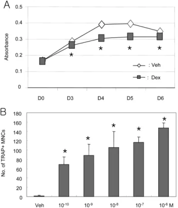
의증식을모두유의 성 있게 감소시켰다(Fig. 1A).
이 결과는dexamethasone
이BMM
의증식을억제하고분화를촉진할가능성을제시하는것이므로
,
다음은dexamethasone
이파골세포의분화에미치는영 향을조사하였다.
이를위해골수세포를M-CSF
와RANKL
존재하에서
10
−10M~10
−6M
의dexamethasone
으로농도별로처리 한후4
일간배양하였다. Dexamethasone
은골수세포로부터파골세포로의분화를농도의존적으로촉진하는것을알수있다
(Fig. 1B).
이상의결과로dexamethasone
은BMM
의증식을억 제하고파골세포로의분화를촉진하는것으로밝혀졌다.
Primer
서열PCR
조건Cycle
GR F: CGCTCAGTGTTTTCTAATGG
R: ATCAGGAGCAAAGCATAGCA 94
oC 30
초, 55
oC 30
초, 72
oC 30
초cycle 28
IFN-
βF: CTTCTCCACCACAGCCCTCTC
R: CCCACGTCAATCTTTCCTCTT 94
oC 30
초, 53
oC 30
초, 72
oC 30
초cycle 35
β
-actin F: TGTGATGGTGGGAATGGGTCAG R: TTTGATGTCACGCACGATTTCC 94
oC 30
초, 58
oC 30
초, 72
oC 1
분cycle 22
Dexamethasone
이파골세포분화의어느단계를촉진하는지 조사하기위해골수세포의M-CSF
와RANKL
배양액에dexa- methasone
을day 0~4
까지 날짜별로 처리하였다. Dexame- thasone
은4
일간의배양기간중day1
까지처리되었을때(D0-1)
가장효율적으로 파골세포분화를촉진하였으며
,
이는dexa- methasone
이4
일간처리되었을때(D0~4)
와유사한정도를보였다
(Fig. 2).
그러나dexamethasone
의파골세포분화에대한촉 진적효과는day 2(D0~2)
와day 3(D0~3)
까지처리되었을때소멸하였다
.
이에대한원인은현재로서는알수없으며,
추후추가적인실험을통해밝혀지기를기대한다
.
파골세포 분화 촉진에 관여하는
dexamethasone
의 작용기전규명
파골세포분화에관여하는다양한유전자가밝혀졌으며
,
그중에서 특히
Nuclear Factor of Activated T cells(NFAT)c1
은파골세포의분화를 조절하는중요한전사인자로 알려져있다
. Dexamethasone
이파골세포분화를촉진하였으므로, dexame- thasone
의처리가NFATc1
의발현을변화시키는지여부를항NFATc1
항체를이용한western blotting assay
로조사하였다. BMM
을48
시간처리하였을때dexamethasone
은RANKL
존재하에서
NFATc1
의발현을유의성있게증가시켰으며(Fig. 3),
이는
dexamethasone
이파골세포로의분화를촉진하는것과일치 하는결과이다.
따라서dexamethasone
은NFATc1
을매개한파골세포분화를촉진하는것으로생각되었다
.
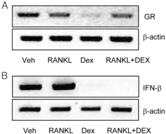
Dexamethasone
은glucocorticoid receptor(GR)
를통해 신호 를 전달하며, GR
은nuclear receptor superfamily of ligand- dependent transcription factors
에속하는것으로알려져있다.
18)Dexamethasone
에의한파골세포로의분화촉진이GR
발현과 연관되어있는지알아보기위해GR
특이적서열을이용해RT-
Fig. 1 −
Effects of dexamethasone on the proliferation of mouse bone-marrow macrophage (BMM) cells. A, BMM cells were cultured with 30 ng/m l M-CSF in the absence or presence of dexamethasone (100 nM) for indicated times.
Cell proliferation was determined by MTT assay as described in materials and methods. B, BMM cells were cultured with 30 ng/m l M-CSF and 50 ng/m l RANKL in the presence of various concentration of dexamethasone for 4 days. Cells were then fixed and stained for TRAP. TRAP- positive (+) multinucleated cells (MNCs) were counted.
The experiments were performed 3 times, and the reproducibility was confirmed. Values are the mean±SD of triplicate cultures in a representative experiment. Veh:
vehicle, *: p <0.05, significantly different from vehicle.
Fig. 2 −
Stage-specific effects of dexamethasone on osteoclast formation. BMM cells were cultured with 30 ng/m l M-CSF and 50 ng/ml RANKL for 4 days. Dexamethasone (100 nM) was added during the indicated times. Cells were then fixed and stained for TRAP. TRAP-positive (+) multi- nucleated cells (MNCs) were counted. The experiments were performed 3 times, and the reproducibility was confirmed. Values are the mean±SD of triplicate cultures in a representative experiment. Veh: vehicle, *: p <0.05, significantly different from vehicle.
Fig. 3 −
Effects of dexamethasone on the expression of NFATc1.
BMM cells were cultured for 48 hrs and the expression of
NFATc1 was determined by western blotting assay. The
experiments were performed 3 times, and the reproduci-
bility was confirmed.
PCR assay
를수행하였다.
BMM
은GR mRNA
를발현하며,
발현정도는dexamethasone
을
48
시간처리하였을때큰폭으로감소하였다.
그러나RANKL
존재하에서
dexamethasone
처리는GR
발현에큰영향을미치지않았다
(Fig. 4A).
이상의결과로,
파골세포에대한dexame- thasone
의분화촉진효과는GR
발현과밀접하게연관되지않음을알수있다
.
파골세포의분화를조절하는다양한분자중
interferon-
β(IFN-
β
)
는파골세포의분화초기에억제적효과를나타낸다고알려져있다
.
19)Dexamethasone
을BMM
초기에처리하였을때파골세 포로의분화를촉진하는효과를보였으므로,
이에IFN-
β의발현 이관여하는것은아닌지조사하고자IFN-
β특이적서열로RT- PCR assay
를수행하였다. BMM
을RANKL
로처리하였을때기 존에알려진바와같이IFN-
β의발현은증가하였다(Fig. 4B).
그 러나dexamethasone
또는RANKL
과dexamethasone
의처리는IFN-
β의 발현을 현저하게 감소시켰다.
이상의 결과는dexamethasone
이RANKL
에의한IFN-
β의발현을감소시킴으로써파골세포로의분화를촉진시키는것을의미한다
.
본연구는마우스골수세포에
dexamethasone
을단독처리하였을때파 골세포분화에미치는영향과그작용기전을분석하였으며,
이는기존의논문20-22)과는다른차별성을가진다고할수있다
.
결 론
Dexamethasone
은마우스골수세포의파골세포로의분화를농 도의존적으로촉진하였으며,
이는분화에필수적인전사인자NFATc1
의활성화를매개하는것으로밝혀졌다. Dexamethasone
의파골세포분화촉진은분화초기에작용하였으며
,
이의작용기전으로
dexamethasone
에의한마우스골수세포의증식억제 와IFN-
β의발현억제가제시되었다.
감사의 말씀
본연구는숙명여자대학교
2008
학년도교내연구비지원에의 해수행되었으므로이에감사드립니다.
참고문헌
1) Takahashi, N., Akatsu, T., Udagawa, N., Sasaki, T., Yamaguchi, A., Moseley, J. M., Martin, T. J. and Suda, T. : Osteoblastic cells are involved in osteoclast formation. Endocrinology
123, 2600 (1988).
2) Suda, T., Takahashi, N., Udagawa, N., Jimi, E., Gillespie, M. T.
and Martin, T. J. : Modulation of osteoclast differentiation and function by the new members of the tumor necrosis factor receptor and ligand families. Endocr. Rev.
20, 345 (1999).
3) Wong, B. R., Rho, J., Arron, J., Robinson, E., Orlinick, J., Chao, M., Kalachikov, S., Cayani, E., Bartlett, F. S. 3rd, Frankel, W. N., Lee, S. Y. and Choi, Y. : TRANCE is a novel ligand of the tumor necrosis factor receptor family that activates c-Jun N- terminal kinase in T cells. J. Biol. Chem.
272, 25190 (1997).
4) Yasuda, H., Shima, N., Nakagawa, N., Yamaguchi, K., Kinosaki, M., Mochizuki, S., Tomoyasu, A., Yano, K., Goto, M., Murakami, A., Tsuda, E., Morinaga, T., Higashio, K., Udagawa, N., Takahashi, N. and Suda, T. : Osteoclast differentiation factor is a ligand for osteoprotegerin/osteoclastogenesis-inhibitory factor and is identical to TRANCE/RANKL. Proc. Natl. Acad. Sci.
USA
95, 3597 (1998).
5) Lacey, D. L., Timms, E., Tan, H. L., Kelley, M. J., Dunstan, C. R., Burgess, T., Elliott, R., Colombero, A., Elliott, G., Scully, S., Hsu, H., Sullivan, J., Hawkins, N., Davy, E., Capparelli, C., Eli, A., Qian, Y. X., Kaufman, S., Sarosi, I., Shalhoub, V., Senaldi, G., Guo, J., Delaney, J. and Boyle, W. J. : Osteoprotegerin ligand is a cytokine that regulates osteoclast differentiation and activation. Cell
93, 165 (1998).
6) Pitzalis, C., Pipitone, N. and Perretti, M. : Regulation of leukocyte-endothelial interactions by glucocorticoids. Ann.
N.Y. Acad. Sci.
966, 108 (2002).
7) Langenegger, T. and Michel, B. A. : Drug treatment for rheumatoid arthritis. Clin. Orthop. Relat. Res.
366, 22 (1999).
8) Riccardi, C., Zollo, O., Nocentini, G., Bruscoli, S., Bartoli, A., D'Adamio, F., Cannarile, L., Delfino, D., Ayroldi, E. and Migliorati, G. : Glucocorticoid hormones in the regulation of cell death. Therapie
55, 165 (2000).
Fig. 4 −
Effects of dexamethasone on the mRNA expression of GR
and IFN-
β. BMM cells were cultured for 48 hrs and the
mRNA expression of GR (A) or IFN-
β(B) was determined
by RT-PCR assay. The experiments were performed 3
times, and the reproducibility was confirmed.
9) Canalis, E. and Delany, A. M. : Mechanisms of glucocorticoid action in bone. Ann. N.Y. Acad. Sci.
966, 73 (2002).
10) Weinstein, R. S. : Glucocorticoid-induced osteoporosis. Rev.
Endocr. Metab. Disord.
2, 65 (2001).
11) Patschan, D., Loddenkemper, K. and Buttgereit, F. : Molecular mechanisms of glucocorticoid-induced osteoporosis. Bone.
29, 498 (2001).
12) Manolagas, S. C. and Weinstein, R. S. : New developments in the pathogenesis and treatment of steroid-induced osteoporosis.
J. Bone. Miner. Res.
14, 1061 (1999).
13) Pereira, R. M., Delany, A. M. and Canalis, E. : Cortisol inhibits the differentiation and apoptosis of osteoblasts in culture.
Bone.
28, 484 (2001).
14) Hofbauer, L. C., Gori, F., Riggs, B. L., Lacey, D. L., Dunstan, C. R., Spelsberg, T. C. and Khosla, S. : Stimulation of osteoprotegerin ligand and inhibition of osteoprotegerin production by glucocorticoids in human osteoblastic lineage cells: potential paracrine mechanisms of glucocorticoid- induced osteoporosis. Endocrinology
140, 4382 (1999).
15) Rubin, J., Biskobing, D. M., Jadhav, L., Fan, D., Nanes, M. S., Perkins, S. and Fan, X. : Dexamethasone promotes expression of membrane-bound macrophage colony-stimulating factor in murine osteoblast-like cells. Endocrinology
139, 1006 (1998).
16) Weinstein, R. S., Jilka, R. L., Parfitt, A. M. and Manolagas, S. C. : Inhibition of osteoblastogenesis and promotion of
apoptosis of osteoblasts and osteocytes by glucocorticoids. J.
Clin. Invest.
102, 274 (1998).
17) Jia, D., O'Brien, C. A., Stewart, S. A., Manolagas, S. C. and Weinstein, R. S. : Glucocorticoids act directly on osteoclasts to increase their life span and reduce bone density. Endocrinology
147