Effects of Green Tea Extract on the p53 Pathway in the MCF-7 Breast Cancer Cell Line
Inseok Kwak*
Department of Biological Science, Silla University, Busan 617-738, Korea
Received October 12, 2018 /Revised October 24, 2018 /Accepted October 29, 2018
The effects of a green tea extract (GTE) were examined using the MCF-7 human breast cancer cell line. Cell viability assays using 3-(4,5-Dimethylthiazol-2-yl)-2,5-diphenyltetrazolium bromide (MTT) as- says revealed that GTE had a significant cytotoxic effect on MCF-7 cells, depending on the concen- tration of GTE. Western blotting of p53 and its related proteins, p21/cip1 and CDK2, after GTE treat- ment revealed that a significant and concentration dependent increase in p53 protein in response to GTE. The levels of p21/cip1 proteins were also increased at low GTE concentrations were significantly increased even at the highest GTE concentrations. However, the level of CDK2 was significantly de- creased by treatment with high concentrations of GTE. These results indicate that treatment with GTE increased the p53 level in MCF-7 cells, and this activation of p53 markedly elevated the levels of p21/cip1proteins, which, in turn, inhibited CDK2 expression in the MCF-7 cells. The inhibition of CDK2 expression might then affect cell cycle progression. Subsequent FACS analysis indicated that GTE treatment the gradually increased progression of the MCF-7 to the G1 phase. These results clear- ly demonstrate that the anti-tumor effect of GTE in MCF-7 cells is regulated by p53 arrest of the MCF-7 cells at the G1 stage of cell cycle.
Key words : Breast cancer, green tea extract, MCF-7, p53, p21
*Corresponding author
*Tel : +82-51-999-6307, Fax : +82-51-999-5176
*E-mail : [email protected]
This is an Open-Access article distributed under the terms of the Creative Commons Attribution Non-Commercial License (http://creativecommons.org/licenses/by-nc/3.0) which permits unrestricted non-commercial use, distribution, and reproduction in any medium, provided the original work is properly cited.
Journal of Life Science 2018 Vol. 28. No. 11. 1316~1320 DOI : https://doi.org/10.5352/JLS.2018.28.11.1316
서 론
녹차(green tea: GT)는 발효시키지 않은 차나무 종(Camellia.
sinensis.)의 잎을 사용하여 만든 음료이며, GT추출물은 식물유 래 기능성 식이 보조제로 시판된다. GT의 효능은 동맥경화와 당뇨병 등의 예방에 효과가 보고되었고, 기초적인 연구를 통 해 여러 종류의 암에 대한 효능이 많이 밝혀져 있다[5, 8, 18].
유방암은 우리나라의 여성에게 발생빈도가 가장 높은 악성 종양 중 하나로, 2010년에는 16,398명의 유방암 환자가 발생하 였다는 보고가 있다[9]. GT추출물 중 GT의 떫은맛을 내는 성 분의 일종인 Epigallocatechin-3-gallate (EGCG)는 유방암, 전 립선 암, 폐암, 위암, 난소 암 등 에서 광범한 항암 작용을 하는 것으로 알려졌다[1, 2, 7]. EGCG는 세포 사멸 및 세포주기 정 지 유도뿐 아니라 암이 전이에 필요한 혈관 신생 과정을 방해 하는 등 암의 진행을 멈추게 하는 항암 작용을 하는 것으로 알려졌다[11, 16]. 또한 EGCG는 유방암 세포에서 유방암 세포 의 침투능력을 방해하는 등의 항암기전이 확인되었다[3, 19].
그러나 세포 차원이 아닌 유방암 환자 차원에서 GT의 섭취량 과 유방암의 발생률과의 연관 관계는 아직 확실하지 않다. 유 방암 발생률이 GT를 마시는 아시아 국가들에서 훨씬 낮고 5,617건의 유방암환자의 메타 분석을 통해 GT 섭취 증가와 유방암 발병률의 반비례 관계에 있고 재발률이 의미 있게 낮 아 졌다는 보고가 있다[14]. 그러나 다른 연구에 의하면 약 43,639 여성에서 GT를 마시는 것과 유방암 발병률 사이에 연 관 관계가 없다는 연구도 있다[8]. 그러나 GT의 항암효과 중 유방암에 관한 연구 중 종양억제 유전자로 잘 알려진 p53에 관련된 연구는 많지 않다. p53는 많은 종류의 인체 암에서 p53 의 돌연변이가 발견되어 종양억제자의 일종임이 밝혀졌다 [10]. P53는 DNA에 변형을 초래하는 자극이 있을 때, p53의 기능이 활성화 되어 세포주기를 정지시켜서[16, 17]. 세포의 DNA가 손상을 입었는지 점검한 후, 손상된 DNA를 복구하는 과정에 관여한다[4, 12]. 본 연구는 GT추출물의 유방암에 대한 작용기전을 유방암 세포에서 연구하여, GT추출물이 종양억제 인자인 p53의 진행경로에 관여함을 규명하여 유방암 연구 및 치료를 위한 기본 자료를 제공하려 한다.
재료 및 방법
시료조제
녹차는 전라남도 보성녹차 연구소에서 판매되는 녹차 잎을 구입하여 건조 한 후, 2-3 mm의 크기로 분쇄한 후 추출용 시료 로 사용하였다. 70℃에서 MeOH로 4시간 처리 후 진공조건에
Fig. 1. Cytotoxicity of MCF-7 cells by different concentrations of green tea extract. MCF-7 cells were treated with GT extract (3, 6, 9, 12, and 20 ug/ml) and 50% of DMSO was used as control (Ct: 0) for 16 hr and cell viability was measured by the MTT assay.
서 용매를 제거한 후, 30.3 g의 MeOH 추출물을 얻었다.
MeOH 추출물을 3% HCl 처리하고, CH2Cl2 가용성 층을 제거 후, CH2Cl2와 함께 추출 한 후 pH 9.0으로 조정 한 후, 10,000 rpm에서 1시간 원심 분리한 후, 상등 액은 여과한 후, 1 mg/ml의 농도로 희석하여 50%의 DMSO에 녹여 사용하였다.
세포배양
인체 유방암 유래 세포주인 MCF-7은 American Type Cul- ture Collection (Rockville, MD, USA)에서 분양 후 사용하였 다. 세포배양에 사용된 Dulbecco's Modified Eagle's Medium:
Nutrient Mixture과 Ham's F12, fetal bovine serum과 페니실 린-스트렙토마이신 등 은 Gibco/BRL (Gaitherburg, MD, USA) 에서 구입하였다. MCF-7 세포는 3×10⁶ cells/ml로 세포의 농 도를 유지하면서 37℃ CO2 배양기에서 배양하였다.
Western blot Analysis
세포를 차가운 PBS로 세척한 후 lysis buffer (50 mM Tris-HCl, 0.5% NP-40, 50 mM NaCl, 5 mM EDTA, 30 mM NaF, 0.1 mM Na3VO4, 1 mM PMSF, pH 7.4)를 첨가하여 단백 질을 추출한 다음, 13,000 rpm에서 30분간 원심분리 후 상층 액을 cell lysate로 사용하였다. BCA protein assay kit (Pierce, Rockford, IL, USA)를 사용하여 단백질 농도를 측정하였다.
Sodium dodecyl sulfate-polyacryl amide gel electrophoresis (SDS-PAGE) 사용하여 단백질을 분리한 후, nitrocellulose membrane에 transfer한 후, 5% non-fat milk를 함유한 Tris buffer (10 mM Tris-HCl, pH 7.4, 0.1 M NaCl, 0.1% Tween 20) 용액에서 1시간 동안 상온에서 blocking하여, 비 특이적 단백질에 대한 결합을 차단하였다. p53, p21/Cip1 그리고 Cdk2에 대한 1차 항체를 각각 첨가하여 1시간 상온에서 반응 하였다. 각 항체들에 대한 2차 항체인 anti-mouse IgG 또는 anti-rabbit IgG로 각각 1시간씩 상온에서 반응시켰다. 각 반응 사이에 TBS-T로 10분씩 3회 세척하였다. 각 항체에 상응하는 단백질의 확인은 enhanced chemiluminescence (ECL)을 사용 하였다.
세포 생존율 측정
유방암 유래 MCF-7 세포는 3×10⁴ cells/well 농도로 분주하 여, 37℃ CO2 배양기에서 20시간 이상 안정화된 세포를 사용 하였다. GT추출물을 50%의 DMSO에 주어진 농도에 따라 희 석한 후 처리하고 16시간 동안 배양한 후 MCF-7 세포의 세포 생존율(Cell viability)을 MTT분석법으로 측정하였다. CellTiter 96Ⓡ Cell Proliferation Assay kit (Promega, Madison, WI, USA)의 염색 액을 첨가한 후 4시간 동안 상온에서 반응시킨 후, 반응정지 용액을 넣고 1시간 반응 후 570 nm에서 ELISA Reader (Molecular Devices, Sunnyvale, CA, USA)에서 흡광 도를 측정하였다.
세포 주기 분석
MCF-7 세포를 1×10⁶ cells/ml의 농도로 분주하고 24시간 안정화시킨 후, GT 추출물을 농도에 따라 처리하였다. 16시간 동안 GT추출물에 노출한 후 PBS로 washing하고 75%의 차가 운 EtOH에서 24시간 세포를 고정한 후 3,000 rpm에서 3분 원심 분리 후, 상등 액을 제거하고, RNase A를 포함한 propi- dium- iodide 용액에 30분 동안 암실에서 반응하였다. 염색된 세포의 DNA 함량은 flow cytometer (Becton Dickinson, France)를 사용하여 분석하였다
결 과
GT의 세포 생존율 효과
GT추출물의 처리가 MCF-7의 세포의 생존에 미치는 영향 을 MTT법을 사용하여 측정한 후 그 결과를 Fig. 1에 나타내었 다. GT추출물의 농도가 0, 3, 6, 9, 12 및 20 ug/ml을 처리 후 세포 독성을 측정한 결과, 3 ug/ml의 GT추출물을 처리한 경우 16시간 내에 세포 생존율이 10% 정도 감소한 반면, 9 ug/ml의 GT추출물을 처리한 경우 50% 정도의 생존율이 관찰 되었다. 20 ug/ml의 GT추출물은 80% 정도의 세포 치사율이 관찰되어(Fig. 1), 이 연구 결과는 GT추출물은 MCF-7 세포의 세포 생존율에 GT 농도 의존적 세포 독성을 나타내며, 이는 GT추출물이 MCF-7 세포에 강한 항암효과가 있음을 보여준 다.
GT의 세포주기 관련 단백질 p53, p21/cip1과 CDK2 변화 다양한 농도의 GT추출물이 p53를 포함한 세포주기 관련 단백질에 미치는 영향을 Western blot으로 조사하였다(Fig. 2).
p53 단백질은 MCF-7 세포에서 GT추출물의 농도를 3 ug/ml 처리한 경우 16시간 내에 p53 단백질의 양이 약간 증가함을
Fig. 2. Effect of green tea extracts on the level of p53, p21/cip1 and CDK2 in MCF-7 cells. MCF-7 cells were treated with GT extract (0, 3, 9 and 20 ug/ml) for 16 hr and the ex- pression of p53, p21/cip1 and CDK2 protein was ana- lyzed by Western blot.
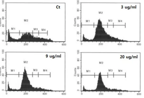
Table 1. The distribution of MCF-7 cells in the cell cycle after treating with 0 and 3, 9 and 20 ug/ml of GT extract GT (μg/ml)
Stage 0 3 9 20
G0 G1 S G2
21.43 48.41 13.54 13.71
8.11 73.51 10.89 7.70
8.19 75.88 7.69 8.26
8.06 78.60 6.35 7.05
Fig. 3. The distribution of MCF-7 cells in the cell cycle after treating with green tea extract for 16 hr. MCF-7 cells were treated with 0 and 3, 9 and 20 ug/ml of GT extract and cells were evaluated for DNA con- tents by flow cytometric analysis.
보였으나, 9 ug/ml 처리한 경우에는 p53 단백질의 양이 현저 하게 증가함이 관찰되었다. 그러나 세포 생존율 실험에서 높 은 세포 독성을 보였던 고농도(20 ug/ml)의 GT추출물을 처리 하였을 때는 9 ug/ml 처리한 시료보다는 약간 감소함을 보였 다(Fig. 2). 이와 같은 결과는 GT추출물에 의해 세포가 손상되 었을 경우 유전적 변화를 방지하기 위해 p53 단백질이 활성화 되었음을 나타내며, p53의 발현이 현저히 증가한 결과, p53의 target 유전자인 p21/cip1 단백질 발현을 유도한 것으로 나타 났다 [19]. GT추출물에 의한 p21/cip1 단백질의 발현은 낮은 농도(3 ug/ml)의 GT추출물에서 약간 증가하였고, 고농도(20 ug/ml)에서도 증가하는 양상을 나타내어, p53 단백질과는 다 른 패턴이 관찰되었다. 그러나 GT추출물에 의한 CDK2 단백 질의 발현 양상은 p 53나 p21/cip1 과는 다른 패턴이 관찰되었 다. 즉 CDK2의 단백질의 양은 낮은 농도의 GT추출물 처리에
서는 약간 증가하나, 높은 농도(9-20 ug/ml)에서는 CDK2 발 현이 급격한 감소되는 것이 관찰되었다(Fig. 2).
GT추출물 처리에 의한 세포 주기 분석(FACS)
녹차추출물이 세포의 세포주기에 어떤 영향을 미치는지 확 인하기 위하여 GT추출물을 16시간 처리한 후 FACS 분석을 통하여 관찰하였다(Fig. 3, Table 1). MCF-7 세포에서 GT추출 물에 의한 세포 생존율 결과를(Fig. 1) 참고하여 0, 3. 9 및 20 ug/ml의 GT추출물을 처리한 후, 세포주기인 G0, G1, S과 G2 를 비교하였다, 그 결과를 control 그룹과 과 비교 하였을 때, 세포주기의 G0단계가 감소됨이 관찰되었고, 다음 단계인 G1 단계는 현저하게 증가됨이 관찰되었다(Table 1). G1 다음의 단계인 S단계나 G2단계는 감소됨이 관찰되었다(Fig. 3). 이 결 과는 MCF-7 세포에 미치는 GT추출물의 영향은 유방암 세포 를 세포주기의 G1단계에서 정지(arrest)하여, 세포 주기의 진 행을 중단하는 작용을 함을 확인할 수 있었다.
Discussion
p53는 Tumor suppressor (종양 억제 인자)로 알려진 단백 질로, p53은 세포의 성장과 분열을 조절하는 세포주기에서 중 요한 역할을 수행한다[13, 16]. Cyclin은 세포주기를 조절하는
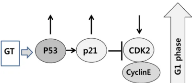
Fig. 4. Schematic representation of the effects of green tea ex- tract on the cell cycle of MCF-7 cells. The treatment of GT extract increases p53 and p21/cip1 in MCF-7 cells.
GT inhibits the CDK2 expression and induces growth arrest at the G1 stage of cell cycle in MCF-7 cells.
단백질 군으로 Cyclin의 주기적인 농도 변화로 cyclin-de- pendent kinase(CDK)를 활성화한다[6, 13]. 세포주기의 진행 은 cyclin과 cyclin dependent kinase의 Cyclin/CDK 복합체에 의해 조절된다[6, 13, 19]. 또한 CDK 억제인자(cyclin-depend- ent kinase inhibitor)는 Cyclin/CDK 복합체에 결합하여, 인산 화 효소를 불활성화하여 세포주기의 진행을 억제한다[6, 15].
p53 기능은 세포주기 진행 여부를 결정하는 check point 중 에서 G1/S1 check point 기전이 잘 알려져 있다[13, 16]. 전사 인자로서 p53은 세포의 DNA가 손상된 경우, 그 기능이 활성 화 되어 CDK 억제인자인 p21/cip1를 포함한 유전자들의 전 사를 활성화시킨다[16, 19]. p21은 cyclin-dependent kinase in- hibitor-1 (cip1)로 불리는 단백질로 p21/cip1는 CDK 복합체 중 cyclin E-CDK2 활성을 억제하며 세포 주기의 G1기에서 S기로 변화과정에 억제제로서 작용한다[6, 16, 19]. 많은 연구 에서 인체 암에서 P53의 돌연변이가 발견되었으나, 유방암에 미치는 GT의 영향 중 p53의 역할에 관한 연구는 잘 알려져 있지 않다. 본 연구에서는 GT추출물의 처리로 유방암 유래 세포가 보이는 반응과 항암 및 종양억제 조절인자로 알려져 있는 p53과 p21/cip1 및 CDK2 단백질의 변화 패턴을 관찰하 여(Fig. 2), 세포주기 관련된 신호전달 과정을 연구하였다(Fig.
4). GT추출물은 MCF-7 세포에서 p53 단백질의 양을 증가시키 며, 증가된 p53는 그 하위(downstream) 유전자인 p21/cip1를 증가시킨다(Fig. 2) [15]. 증가된 p21/cip1은 CDK 억제인자로 잘 알려져 있어, CDK2를 감소시킨다(Fig. 2). 감소된 CDK2는 cyclin-CDK2의 활성화를 억제하며 그 결과 세포 주기의 진행 을 중단시킨다(Fig. 4). GT추출물의 처리는 유방암 유래 MCF- 7 세포의 생존을 현저히 감소하여 항암효과가 있음을 보여주 며(Fig. 1), Western blot 결과에서 관찰된 p53의 현저한 변화 는 GT추출물의 항암효과는 p53 의존성 임을 입증하였다(Fig.
2). 또한 유방암 세포에 미치는 GT추출물의 효과는 p21/cip1 과 CDK2 등 세포 주기에 관여하는 단백질에도 영향을 미치는 것으로 관찰되었으며(Fig. 2), 이 또한 p53 의존성임을 밝혔다.
이는 유방암 유래 MCF-7 세포에 GT추출물의 처리는 세포주 기의 FACS 분석에서 유방암 세포를 G1단계에서 arrest시키는
것으로 관찰된 결과(Table 2)와 일치한다. 이 실험의 결과는 EGCG의 처리는 세포주기 관련 단백질발현의 활성화로 세포 사멸(apoptosis)를 유도한다는 사실과 일치한다[11, 14]. 녹차 추출물 같은 천연 추출 물질의 세포 내 유전자/단백질 조절에 관한 기전에 관한 연구에 의한(Fig. 4.) 세포의 신호전달 과정 의 이해는 새로운 치료제의 연구 개발에 도움을 줄 수 있다.
References
1. Bettuzzi, S., Brausi, M., Rizzi, F., Castagnetti, G., Peracchia, G. and Corti, A. 2006. Chemoprevention of human prostate cancer by oral administration of green tea catechins in vol- unteers with high grade prostate intraepithelial neoplasia.
Cancer Res. 66, 1234-1240.
2. Butt, M. S. and Sultan, M. T. 2009. Green tea: nature’s de- fense against malignancies. Crit. Rev. Food Sci. Nutr. 49, 463-473.
3. Caffo, O., Doglioni, C., Veronese, S., Bonzanini, M., Marche- tti, A., Buttitta, F., Fina, P., Leek, R., Morelli, L., Palma, P.
D., Harris, A. L. and Barbareschi, M. 1996. Prognostic value of p21WAF1 and p53 expression in breast carcinoma: An immuno-histochemical study in 261 patients with long term follow-up. Clin. Cancer Res. 2, 1591-1599.
4. Chen, Q. M., Bartholomew, J. C., Campisi, J., Acosta, M., Reagan, J. D. and Ames, B. N. 1998. Molecular analysis of H2O2-induced senescent-like growth arrest in normal hu- man fibroblasts: p53 and Rb control G1 arrest but not cell replication. Biochem. J. 332, 43-50.
5. Clement, Y. 2009. Can green tea do that? A literature review of the clinical evidence. Prev. Med. 49, 83-87.
6. Fung, T. K. and Poon, R. Y. 2005. A roller coaster ride with the mitotic cyclins. Semin. Cell Dev. Biol. 16, 335-342.
7. Henning, S. M., Wang, P., Carpenter, C. L. and Heber, D.
2013. Epigenetic effects of green tea polyphenols in cancer.
Epigenomics 5, 729-741.
8. Iwasaki, M., Inoue, M., Sasazuki, S., Sawada, N., Yamaji, T., Shimazu, T., Willett, W. C. and Tsugane, S. 2010. Green tea drinking and subsequent risk of breast cancer in a pop- ulation-based cohort of Japanese women. Breast Cancer Res.
12, R88.
9. Kim, Z., Min, S. Y., Yoon, C. S., Jung, K. W., Ko, B. S., Kang, E., Nam, S. J., Lee, S. and Hur, M. H. 2015. The basic facts of Korean breast cancer in 2012: Results from a nationwide survey and breast cancer registry database. J. Breast Cancer 18, 103-111.
10. Kern, S. E., Kinzler, K. W., Bruskin, A., Jarosz, D., Friedman, P., Prives, C. and Vogelstein, B. 1991. Identification of p53 as a sequence-specific DNA-binding protein. Science 252, 1708-1711.
11. Khan, N., Afaq, F., Saleem, M., Ahmad, N. and Mukhtar, H. 2006. Targeting multiple signaling pathways by green tea polyphenol (−)-epigallocatechin-3-gallate. Cancer Res.
66, 2500-2505.
12. May, P. and May, E. 1999. Twenty years of p53 research:
초록:유방암 세포 주 MCF-7에서의 녹차 추출물이 p53 경로에 미치는 영향
곽인석*
(신라대학교 의생명과학대학 생명과학과)
녹차(GT) 추출물의 효과를 인간 유방암 유래 세포인 MCF-7 세포를 사용하여 조사 하였다. GT추출물의 세포 독성 효과를 MTT 방법을 사용하여 관찰한 결과, MCF-7 세포는 현저한 세포 독성 효과를 보였고, 이 독성 효과는 GT추출물 농도 의존적으로 증가하였다. p53과 관련 단백질인 p21/cip1과 CDK2의 연관성을 조사하기 위해 GT추 출물 처리 후 웨스턴 분석법을 통해 이들 단백질의 발현을 조사하였다. GT추출물 처리 후, MCF-7 세포에서 p53 단백질의 양은 농도에 따라 현저하게 증가 하였다. p21/cip1 단백질의 발현은 낮은 농도의 GT추출물에서 증가되 며, 고농도에서도 감소하지 않았다. 그러나 CDK2의 단백질의 양은 높은 농도의 GT추출물에서 CDK2 발현의 급 격한 감소가 관찰되었다. 이 결과는 GT추출물의 처리는 MCF-7 세포에서 p53와 p21/cip1를 증가시켜, 그 결과로 활성화 된 p21/cip1는 CDK2의 발현을 억제 함을 나타내고 있다. GT추출물이 MCF-7 세포의 세포주기에 어떤 영향을 미치는지 확인하기 위하여 FACS 분석으로 관찰한 결과, MCF-7 세포에서 세포주기의 G1 단계가 점차 증 가하는 결과를 보였다. 이 결과는 GT추출물의 유방암 세포에서의 항암 효과는 세포주기의 G1 단계에서 MCF-7 세포를 정지시키는 p53에 의해 조절된다는 사실을 명확하게 보여 주고 있다.
structural and functional aspects of the p53 protein. Onco- gene 18, 7621-7636.
13. Morgan, D. O. 1995. Principles of CDK regulation. Nature 374, 131-134.
14. Ogunleye, A. A., Xue, F. and Michels, K. B. 2010. Green tea consumption and breast cancer risk or recurrence: a meta-analysis. Breast Cancer Res. Treat. 119, 477-484.
15. Rey, M., Fernandez, P. L., Jares, P., Rey, M. J., Fernández, P. L., Jares, P., Muñoz, M., Nadal, A., Peiró, N., Nayach, I., Mallofré, C., Muntané, J., Campo, E., Estapé, J. and Cardesa, A. 1998. P21WAF1/CIP1 is associated with cyclin D1 CCND1 expression and tubular differentiation but is in- dependent of p53 overexpression in human breast carcinoma.
J. Pathol. 184, 265-271.
16. Salnikow, K., Costa, M., Figg, W. D. and Blagosklonny, M.
V. 2000. Hyperinducibility of hypoxia-responsive genes without p53/p21-dependent checkpoint in aggressive pros- tate cancer. Cancer Res. 60, 5630-5634.
17. Schwartz, D. and Rotter, V. 1998. P53-dependent cell cycle control: response to genotoxic stress. Semin. Cancer Biol. 8, 325-336.
18. Shirakami, Y., Shimizu, M. and Moriwaki, H. 2012. Cancer chemoprevention with green tea catechins: from bench to bed. Curr. Drug Targets. 13, 1842-1857.
19. Wakasugi, E., Kobayashi, T., Tanaki, Y., Ito, Y., Miyashiro, I., Komoike, Y., Takeda, T., Shin, E., Takatsuka, Y., Kikkawa, N., Monden, T. and Monden, M. 1997. p21 and p53 protein expression in breast cancer. Am. J. Clin. Pathol. 107, 684-691.