http://dx.doi.org/10.4174/astr.2015.88.2.55 Annals of Surgical Treatment and Research
Effects of combination therapy of docetaxel with selenium on the human breast cancer cell lines MDA-MB-231 and MCF-7
Sang O Park, Young Bum Yoo1, Yong Hun Kim2, Kwang Je Baek, Jung-Hyun Yang1, Pil Cho Choi3, Jeong Hun Lee4, Kyeong Ryong Lee, Kyoung Sik Park1
Departments of Emergency Medicine and 1Surgery, Konkuk University Medical Center, Konkuk University School of Medicine, Seoul, 2Department of Surgery, Konkuk University Chungju Hospital, Chungju, 3Department of Emergency Medicine, Kangbuk Samsung Hospital, Sungkyunkwan University School of Medicine, Seoul, 4Department of Emergency Medicine, Dongguk University Ilsan Hospital, Dongguk University College of Medicine, Goyang, Korea
INTRODUCTION
Breast cancer is the most common invasive cancer and is the second most common cause of deaths in women. Breast cancer is usually treated surgically, followed by chemotherapy
and immunotherapy or radiation therapy, or both in cases of advanced cancer [1,2]. Metastatic breast cancer is the most serious and incurable disease with a 2- to 3-year median overall survival [3]. In advanced breast cancer patients, chemotherapy is generally recommended in patients with hormone receptor Purpose: The anticancer property and cytoprotective role of selenium in chemotherapy have been reported. However, the combination effects of selenium on chemotherapy for advanced breast cancer have not yet been clearly defined. The purpose of this study was to investigate the combined effects of selenium on chemotherapy using docetaxel on breast cancer cell lines.
Methods: Under adherent culture conditions, two breast cancer cell lines, MDA-MB-231 and MCF-7, were treated with docetaxel at 500pM and selenium at 100nM, 1μM, or 10μM. Changes in cell growth, cell cycle duration, and degree of apoptosis after 72 hours in each treated group were evaluated.
Results: In the MDA-MB-231 cells, the combination therapy group (docetaxel at 500pM plus selenium at 10μM) showed a significantly decreased percentage of cell growth (15% vs. 28%; P = 0.004), a significantly increased percentage of late apoptosis (63% vs. 26%; P = 0.001), and an increased cell cycle arrest in the G2/M phase (P = 0.001) compared with the solitary docetaxel therapy group. Isobologram analysis demonstrated the synergistic effect of the combination therapy in the MDA-MB-231 cells. However, in the MCF-7 cells, no significant differences in the percentage of cell growth apoptosis, the percentage of apoptosis, and the pattern of cell cycle arrest were noted between the combination therapy groups and the solitary docetaxel therapy group.
Conclusion: Our in vitro study indicated that the combination of selenium with docetaxel inhibits cell proliferation through apoptosis and cell arrest in the G2/M phase in MDA-MB-231 breast cancer cells.
[Ann Surg Treat Res 2015;88(2):55-62]
Key Words: Breast neoplasms, Selenium, Docetaxel, Combination drug therapy
Received June 17, 2014, Revised September 6, 2014, Accepted September 25, 2014
Corresponding Author: Kyoung Sik Park
Department of Surgery, Konkuk University Medical Centre, Konkuk University School of Medicine, 120 Neungdong-ro, Gwangjin-gu, Seoul 143-729, Korea
Tel: +82-2-2030-7697, Fax: +82-2-2030-7346 E-mail: [email protected]
Copyright ⓒ 2015, the Korean Surgical Society
cc Annals of Surgical Treatment and Research is an Open Access Journal. All articles are distributed under the terms of the Creative Commons Attribution Non- Commercial License (http://creativecommons.org/licenses/by-nc/3.0/) which permits unrestricted non-commercial use, distribution, and reproduction in any medium, provided the original work is properly cited.
negative tumors, endocrine-resistant disease of the luminal subtype, and rapidly proliferating disease [4,5]. Generally, cyclophosphamide and adriamycin are used for chemotherapy, and sometimes, a taxane drug such as docetaxel is added [5,6].
Docetaxel is an antimitotic chemotherapeutic agent used for treating patients, who have locally advanced or metastatic breast cancer and have undergone unsuccessful anthracycline- based chemotherapy [5-7]. Docetaxel exerts its cytotoxicity by promoting tubulin polymerization and stabilizing microtubule assembly, while preventing physiological microtubule depolymerization [4]. This action significantly decreases free tubulin needed for microtubular formation and inhibits mitotic cell division between metaphase and anaphase, thus preventing further cell progeny. Clinically, docetaxel may inhibit the proliferation of breast cancer cells, but the drug results in several adverse effects in normal cells [8].
Selenium is well known as an essential micronutrient for human health [9] and has been recently regarded as a potent nutritional anticancer element. In breast cancer, various ecolo- gical studies and case-control studies have found an inverse rela tionship between selenium levels and cancer risk and mortality [10,11]. An experimental study showed that the administration of selenium reduced the rate of breast cancer cell growth in an in vitro setting [12]. Selenium administration of 0.8-μg/g body weight was associated with an 80%–93%
reduction in the rate of breast cancer cell growth, without apparent adverse effects on the host [12].
Some cancer patients experience several adverse effects after radiotherapy and chemotherapy as well as develop chronic lym- phedema resulting from cellular processes during oxidative normal cell damage in the human body [13]. Selenium has recently been suggested as an alternative remedy against che- motherapy and radiotherapy-associated side effects as well as its effects on lymphedema [14-19].
Selenium supplementation may be beneficial in advanced breast cancer patients undergoing chemotherapy, considering its anticancer properties and added potential to control side effects and lymphoedema caused by chemotherapy. However, the combination effects of selenium in chemotherapy for advanced breast cancer have not yet been clearly defined.
Therefore, we hypothesized that a combination therapy with selenium may be beneficial for inhibiting breast cancer cell growth in an experimental setting of conventional chemotherapy with docetaxel for advanced breast cancer. The aim of this study was to investigate the combination effects of selenium in chemotherapy with docetaxel in breast cancer cells, MDA-MB-231 and MCF-7, in comparison with solitary chemotherapy therapy.
METHODS
Cell culture
Two human breast cancer cell lines (MDA-MB-231 and MCF- 7) were obtained from American Type Cell Culture (ATCC, Bethesda, MD, USA). The experimental cells were cultured in tissue culture dishes (Cat no: 353003, Falcon, San Jose, CA, USA) in Dulbecco’s modified Eagle’s medium (DMEM) containing 10% fetal bovine serum (FBS). Cultured cells (5 × 10⁵) were grown at 37°C in a humidified atmosphere containing 5% CO2.
Assessment of cell growth after treatment
For each cell line, 5 × 10⁵ cells were seeded in DMEM con- taining 10% FBS. After 24 hours, the cells were washed with phosphate buffered saline (PBS) and cultured in fresh media.
First, 500pM was determined as a therapeutic docetaxel dose for the MDA-MB-231 and MCF-7 cells for 72 hours.
Next, selenium at 100nM, 1μM, or 10μM was added to the MDA-MB-231 and MCF-7 cells for 72 hours to confirm the independent effects of selenium on cell proliferation and survival at different concentrations before we evaluated the effect of selenium combination therapy. Second, docetaxel alone at 500pM (DT 500pM), docetaxel at 500pM plus selenium at 100nM, 1μM or 10μM (DT 500pM + SN 100nM; DT 500pM + SN 1μM; DT 500pM + SN 10μM) were added to the MDA- MB-231 and MCF-7 cells for 72 hours. The cells were then quan- tified in a Neubauer chamber using a hemocytometer after incubating with 0.4% trypan blue dye. All measurements were performed in triplicate, and each experiment was repeated at least three times.
Assessment of combined drug effects using isobolo- gram analysis
Traditionally, isobologram analysis has been used as a method for identifying the combined effect of multiple drugs in terms of the additive, synergistic, or antagonistic effect. Be- cause the combination of selenium and docetaxel was used in a nonconstant dose ratio, a normalized isobologram for the two drugs at their ED50 was constructed automatically using CompuSyn ver. 1.0 (ComboSyn Inc., Paramus, NJ, USA). An additive effect (if points fall on the hypotenuse), synergism (if points fall in the lower left), or antagonism (if points fall in the upper right) is indicated according to the positions of the combination data points in the normalized isobologram.
The combination index (CI) provides a means to analyze the combined effects using a median-effect plot analysis. CI values <1.0, =1.0, and >1.0 indicate synergistic, additive, and antagonistic effects, respectively.
Apoptosis analysis using Annexin V staining
Annexin V staining was performed according to the manu-
facturer’s protocol (Pharmingen, BD Biosciences, San Jose, CA, USA). Trypsin-ethylenediaminetetraacetic acid was used to obtain single-cell suspensions. After centrifuging treated cell lines, the cells were washed twice with cold PBS and resuspended in a binding buffer (10mM HEPES, pH 7.4, 150mM NaCl, 5mM KCl, 1mM MgCl2, and 1.8mM CaCl2) at a concentration of 1 × 10⁶ cells/mL. An aliquot (100 μL) of the solution containing 1 × 10⁵ cells was transferred to a 5-mL cul ture tube, and 5-μL Annexin V-fluorescein isothiocyanate (FITC) and propidium iodide (PI) were added to each tube. After vortexing the tubes, cells were incubated for 15 minutes at room temperature (25°C) in the dark and 400 μL of the binding buffer was added to each tube. Flow cytometry was performed with a FACSCalibur system (BD Biosciences) within 1 hour.
Cell cycle analysis
The growth of both cancer cell lines was inhibited probably because their cell cycles were arrested, and we determined whether the growth inhibition was a result of cell cycle arrest by analyzing the proportions of cells in different cell cycle phases. After 72 hours of treatment in each group, the cells were harvested, fixed in 70% ethanol for 1 hour, and washed with PBS. Next, the cells were treated with 100 μg/mL of RNase A for 1 hour at 37°C and were stained with 10 μg/mL of PI. Flow cytometry was performed in triplicate for each experiment using a FACSCalibur system.
Statistical analysis
All the data were compiled from a minimum of three re- plicate experiments. Student t-tests were used to compare re-
sults of treated and control cells. To compare the normally dis- tributed continuous variables, one-way analysis of variance tests were used. To compare the nonnormally distributed continuous variables, Kruskal-Wallis tests were used. Chi-square tests were used to compare the nominal variables. Data were analyzed using SPSS ver. 17.0 (SPSS Inc., Chicago, IL, USA). Two-sided P-values less than 0.05 were considered statistically significant.
RESULTS
Cell growth after combined treatment with doce- taxel and selenium in MDA-MB-231 and MCF-7 cell lines
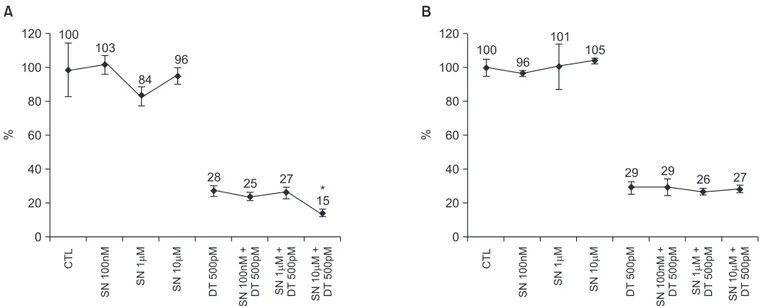
In the solitary selenium therapy model, there were no significant differences in cell growth among the control cells and cells treated with selenium at 100nM, 1μM, or 10μM in MDA-MB-231 cell lines (P = 0.824) and MCF-7 cell lines (P = 0.453) (Fig. 1). In the DT 500pM + SN combination therapy model, cell growth decreased significantly in the group with a DT 500pM + SN 10μM compared with the DT 500pM (15% vs.
28%, P = 0.004) in the MDA-MB 231 cell line (Fig. 1). However, cell growth did not significantly decrease in all DT 500pM + SN combination therapy groups compared with the solitary therapy group in MCF-7 cell lines (Fig. 1).
Combined effect of docetaxel and selenium in MDA-MB-231 and MCF-7 cells
Normalized isobolograms were constructed for MDA-MB-231 and MCF-7 cells using dose-response curves. In MDA-MB-231 cells, synergism was generally identified when the cells were
*
SN10M+ DT500pM
100
80
60
40
20
SN100nM
%
0 120
CTL SN1M SN10M DT500pM SN1M+ DT500pM
SN100M+ DT500pMn
100
80
60
40
20
%
0 100 120
103
84 96
28 25 27
15
100
101 105 96
29 29 27
26
A B
SN10M+ DT500pM
SN100nM
CTL SN1M SN10M DT500pM SN1M+ DT500pM
SN100M+ DT500pMn
Fig. 1. Cell growth after combined treatment of docetaxel at 500pM and selenium at 100nM, 1μM, or 10μM in the MDA- MB-231 cell line (A) and the MCF-7 cell line (B). Cells were stained with trypan blue to determine cell viability and were quantified using a hemocytometer. Relative cell growth rates are shown as percent survival versus indicated cells after treatment with the drug at different concentrations. The data represent the means of at least three independent experiments and the corresponding standard errors. *P-value less than 0.05.
exposed to 100μM of selenium with docetaxel (Fig. 2). And, an additive effect was generally identified when the cells were exposed to other doses of selenium with docetaxel. In MCF-7 cells, an additive effect was generally identified when the cells were exposed to selenium with docetaxel (Fig. 2).
Apoptosis and cell cycle analysis after combined treatment with docetaxel and selenium in the MDA-MB-231 and MCF-7 cell lines
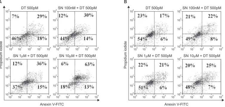
In the MDA-MB-231 cell lines, early apoptosis in all combined therapy groups was not significantly different from the DT 500pM group (13%–15% vs. 18%, P = 0.125). However, late apoptosis increased in the DT 500pM + SN 10μM compared with the DT 500pM and other combination therapy groups (63%
vs. 29%–36%) (Fig. 3). Cell cycle analysis indicated a dominant increase in cell cycle arrest in the G2/M phase in the DT 500pM + SN 10μM groups compared with the other treatment groups
(Fig. 4).
In MCF-7 cell lines, the amounts of early apoptosis (6%–8 % vs.
6%) and late apoptosis (21%–25% vs. 17%) were not significantly different in all the DT 500pM + SN combination therapy groups compared with the DT 500pM group (Fig. 3). Cell cycle analysis indicated a small increase in cell cycle arrest in the G2/M phase in the DT 500pM + SN combination therapy groups compared with the DT 500pM group, but no remarkable difference in cell cycle arrest in the G2/M phase was observed among the DT 500pM + SN combination therapy groups (Fig. 4).
DISCUSSION
In this experimental study, we investigated the combined effect of selenium and docetaxel-based chemotherapy on the growth suppression of two breast cancer cell lines (MDA- MB-231 and MCF-7). The results of our study showed that the Fig. 2. Normalized isobologram for selenium and docetaxel in the MDA-MB-231 (A) and MCF-7 (B) cell lines. Diagonal experimental combination data points, represented by dots located lower left, on the diagonal line, or upper right, indicate synergism, additivity, and antagonism, respectively. Fractional affected-combination index (Fa-CI) plot in combination index analysis in the MDA-MB-231 (C) and MCF-7 (D) cell lines. Combination index values <1, =1, or >1 indicate synergism, additivity, and antagonism, respectively.
0.5
0 0.5 1.0
Selenium
Docetaxel 0
1.0
0.5
0 0.5 1.0
Selenium
Docetaxel 0
1.0
Additive Synergistic
100 M Antagonistic
Additiv Synergistic e
Antagonistic
A B
0.5
0 0.5 1.0
Combinationindex
Fa 0
2.0
1.5
1.0
0.5
0 0.5 1.0
Combinationindex
Fa 0
2.0
1.5
1.0 Antagonistic
Synergistic Additive
Antagonistic
Synergistic Additive
C D
synergistic effect of selenium on chemotherapy with docetaxel for advanced breast cancer cells was cell line-specific and selenium dose-specific. Compared with solitary docetaxel therapy, only the MDA-MB-231 cell lines treated with DT 500pM + SN 10μM showed a higher suppression of cancer cell growth.
Additionally, we found increased percentages of apoptotic cells and increased DNA contents in the G2/M phase in the MDA- MB-231 cell lines treated with DT 500pM + SN 10μM, and this result was consistent with the cell growth results. However, in our study, we could not observe any synergistic effect of selenium on docetaxel therapy in the MCF-7 cell line.
Clinically, selenium may be an attractive natural additive for chemotherapy for advanced breast cancer, which is not easily curable. Selenium is involved in antioxidant reactions and redox regulation pathway as a component of seleno-proteins and as a source of seleno-metabolites [20]. Numerous experimental and clinical studies have demonstrated that selenium supple- mentation reduces the incidence of human cancers, including breast, prostate, lung, colon, and liver cancers [21-23]. Selenium has been established as a crucial cancer preventive element and is thought to reduce cancer progression. Although there have been no definite clinical studies, some laboratory studies have shown that selenium inhibits breast cancer cell growth in a dose-dependent manner.
Multiple chemotherapeutic agents have been developed to treat patients with advanced breast cancer. Docetaxel is generally accepted as the chemotherapy agent for patients with advanced
breast cancer in whom prior chemotherapy has failed [5], but its clinical efficacy can be compromised by serious docetaxel- related side effects such as serious neutropenia, fluid retention, hair loss, nail changes, and hypersensitivity reactions [8].
Controlling side effects associated with docetaxel therapy may improve quality of life and clinical outcomes in patients with chemotherapy. Adverse effects of chemotherapy are associated with an increase in free radicals and related oxidative damage in normal human cells [24]. Recently, selenium compounds have been suggested as toxicity antagonists that prevent side effects related to chemotherapy. Some animal studies have suggested that sodium selenite may reduce the side effects of chemotherapy, such as cisplatin or Adriamycin [15,17,18].
Patients often suffer from secondary lymphedema after surgical and radiological therapy because of fluid retention due to surgical- or radiation-related damage to the lymphatic system [13]. Selenium may improve redox balance in sparsely perfused edematous tissue and hence, may be a useful agent in controlling and alleviating lymphedema [25].
Considering that selenium can inhibit cancer cell growth in breast cancer patients, and can reduce the side effects of chemotherapy and lymphedema, consensus about the use of combination therapy using selenium may be a benefit treatment option in these patients. We first identified the independent effect of selenium on cancer cell growth in stepwise dose in- crements before evaluating the effects of selenium combination therapy. This experiment therefore precludes bias and reduces
A B
Propidiumiodide
104
103
102
101
100 100
DT 500pM
104 103 102 101
SN 100nM + DT 500pM
104 100 101 102 103
104
103
102
101
100
SN 1 M + DT 500pM
104 100 101 102 103
SN 10 M + DT 500pM
104 100 101 102 103 Anexin V-FITC
104
103
102
101
100
104
103
102
101
100
Propidiumiodide
104
103
102
101
100 100
DT 500pM
104 103 102 101
SN 100nM + DT 500pM
104 100 101 102 103
104
103
102
101
100
SN 1 M + DT 500pM
104 100 101 102 103
SN 10 M + DT 500pM
104 100 101 102 103 Anexin V-FITC
104
103
102
101
100
104
103
102
101
100
Fig. 3. Apoptosis after combined treatment with docetaxel at 500pM and selenium at 100nM, 1μM, or 10μM in the MDA- MB-231 cell line (A) and the MCF-7 cell line (B). All cells were stained with fluorescein isothiocyanate (FITC) conjugated Annexin V in a buffer containing propidium iodide and were analyzed using flow cytometry. For each treatment, the percentage of viable cells is shown in the lower left quadrant, for which both Annexin V and propidium iodide levels are low.
Data from a representative experiment (from a total of three) are shown.
confusion regarding efficacy of selenium plus docetaxel combination therapy. It clearly verifies the efficacy of selenium as an additive to the docetaxel chemotherapy in a dose- dependent manner. Considering that only 10μM of selenium can significantly reduce the cell growth in MDA-MB-231 cell lines, the selenium combined effect is considered dose- dependent and is targeted to specific cell lines, that is, MDA-MB cell lines. Apoptosis analysis using the Annexin V-FITC staining showed high concentrated apoptosis of cells in 10μM selenium combination therapy, which inhibited MDA-MB-231 cells significantly. These results are consistent with our cell analysis data that demonstrated increased arrest in the G2/M cycle in the combination therapy group.
We could not demonstrate the exact mechanism underlying the selenium combined with docetaxel chemotherapy in
this study. The mechanisms by which selenium might act on the cancer progression are not well known; however, ap- optosis and programmed cell death have been postulated as primary mechanism for inhibiting cancer growth by selenium compounds [26]. Few studies have shown that sele- nium inhibited the growth of human breast cancer cells by mitochondria-mediated apoptosis and arresting the G1 cell cycle [20,27]. Selenium-induced G1-phase arrest was associated with decreased expression levels of cyclin D1, cyclin D3, CDK4, and CKD6 and increased expression levels of p15 INK4B, p21 Waf1/
Cip1, and p53 [28].
An interesting finding of the study is that only estrogen receptor-negative MDA-MB-231 cells showed a significant sy- nergistic effect of selenium with the docetaxel combination therapy. Most breast cancer patients are female, and estrogen is Fig. 4. Cell cycle analysis after combined treatment with docetaxel at 500pM and selenium at 100nM, 1μM, or 10μM in the MDA-MB-231 cell line (A) and the MCF-7 cell line (B). The percentage of cells at each stage of the cell cycle was analyzed using flow cytometry after DNA staining with propidium iodide. Data from a representative experiment (from a total of three) are shown.
G0/G1: 3%
S: 2%
G2/M: 95%
A
DT 500pM SN 100nM + DT 500pM SN 1 M + DT 500pM SN 10 M + DT 500pM 200
Counts
0 160 120 80 40
1,000 0 200 400 600 800
200
Counts
0 160 120 80 40
1,000 0 200 400 600 800 200
Counts
0 160 120 80 40
1,000 0 200 400 600 800 200
Counts
0 160 120 80 40
1,000 0 200 400 600 800
DNA contents DNA contents DNA contents DNA contents
280
0 210 140 70
200 0 50 100 150
0 320 240 160 80
250 0 50 100 150 200 0
200 0 40 80 120 160 0
280 210 140 70
200 0 50 100 150
400 300 200 100
B
DT 500pM SN 100nM + DT 500pM SN 1 M + DT 500pM SN 10 M + DT 500pM
Counts
0
1,000 0 200 400 600 800
Counts
0
1,000 0 200 400 600 800
DNA contents DNA contents
300 240 180 120 60
Counts
0
1,000 0 200 400 600 800
DNA contents 300
240 180 120 60
Counts
0
1,000 0 200 400 600 800
DNA contents 300
240 180 120 60
300 240 180 120 60 G0/G1: 47%
S: 32%
G2/M: 21%
G0/G1: 49%
S: 30%
G2/M: 21%
G0/G1: 44%
S: 20%
G2/M: 36%
G0/G1: 17%
S: 15%
G2/M: 68%
800
0 600 400 200
200
0 50 100 150 250
G0/G1: 12%
S: 11%
G2/M: 77%
0 320 240 160 80
200
0 50 100 150 250
G0/G1: 6%
S: 4%
G2/M: 90%
0
250 0 50 100 150 200 800
600 400 200
0 800 600 400 200
250 0 50 100 150 200
G0/G1: 4%
S: 2%
G2/M: 94%
strongly associated with breast cancer cell growth. Estrogen is thought to be responsible for the regulation of the expression of selenium-binding protein, which induces the inhibition of cancer cell proliferation during selenium treatment [29]. In previous in vivo and in vitro studies, selenium induced greater cell death and apoptosis in estrogen receptor-positive MCF-7 cells but not in estrogen-negative MDA-MB-231 cells [29,30]. The mechanism of growth inhibition in estrogen receptor-positive MCF-7 cells is thought to occur through disruption of estrogen signaling [30]. However, selenium seemed to act differently on cancer cells when combined with docetaxel in the combined chemotherapy model. In the combined model of docetaxel chemotherapy with selenium, a synergistic effect of selenium on cell death was not observed in estrogen receptor-positive MCF-7 cells but was observed in estrogen-negative MDA-MB-231 cells. The mechanism responsible for cell death through disruption of estrogen signaling does not seem to explain our study results. The cytotoxic effect of docetaxel occurs mainly through blocking mitosis by inhibiting mitotic spindle assembly, and selenium may help induce this antimitosis reaction in MDA-MB-231 cells when given in combination with docetaxel instead of through the direct inhibition of cell growth by itself. However, it is unclear why the synergistic effect of selenium in combined docetaxel therapy differed between the
estrogen receptor-negative MDA-MB-231 and estrogen receptor- positive MCF-7 cells. The different hormonal characteristics of cancers may be closely associated with the synergistic effect of selenium on the actions of docetaxel, but we cannot offer any further explanation. Further studies are needed to explore and understand these differences.
In conclusion, we have identified that the combined therapy of selenium with docetaxel inhibits cell proliferation through apoptosis and cell arrest in the G2/M phase in MDA-MB-231 breast cancer cells. Chemotherapy combined with selenium may be beneficial for the treatment in some advanced breast cancer cases. Further studies are warranted to elucidate the molecular mechanism of the growth inhibition in combined treatment of breast cancer cells.
CONFLICTS OF INTEREST
No potential conflict of interest relevant to this article was reported.
ACKNOWLEDGEMENTS
This work was supported by Konkuk University.
1. Ko BS, Noh WC, Kang SS, Park BW, Kang EY, Paik NS, et al. Changing patterns in the clinical characteristics of korean brea- st cancer from 1996-2010 using an online nationwide breast cancer database. J Breast Cancer 2012;15:393-400.
2. Leclere B, Molinie F, Tretarre B, Stracci F, Daubisse-Marliac L, Colonna M, et al. Trends in incidence of breast cancer among women under 40 in seven European countries: a GRELL cooperative study. Cancer Epidemiol 2013;37:544-9.
3. Siegel R, Naishadham D, Jemal A. Can cer statistics, 2012. CA Cancer J Clin 2012;
62:10-29.
4. Yvon AM, Wadsworth P, Jordan MA. Ta xol suppresses dynamics of individual micro- tubules in living human tumor cells. Mol Biol Cell 1999;10:947-59.
5. Kleeberg UR, Fink M, Tessen HW, Nenne- cke A, Hentschel S, Bartels S. Adjuvant therapy reduces the benefit of palliative
treatment in disseminated breast cancer:
own findings and review of the literature.
Onkologie 2013;36:348-56.
6. Valero V, Holmes FA, Walters RS, Theriault RL, Esparza L, Fraschini G, et al. Phase II trial of docetaxel: a new, highly effective antineoplastic agent in the management of patients with anthracycline-resistant metastatic breast cancer. J Clin Oncol 1995;13:2886-94.
7. Martin M, Segui MA, Anton A, Ruiz A, Ramos M, Adrover E, et al. Adjuvant doce- taxel for high-risk, node-negative breast cancer. N Engl J Med 2010;363:2200-10.
8. Baker J, Ajani J, Scotte F, Winther D, Martin M, Aapro MS, et al. Docetaxel-related side effects and their management. Eur J Oncol Nurs 2009;13:49-59.
9. Hamilton SJ. Review of selenium toxicity in the aquatic food chain. Sci Total Envi- ron 2004;326:1-31.
10. Harris HR, Bergkvist L, Wolk A. Selenium
intake and breast cancer mortality in a cohort of Swedish women. Breast Cancer Res Treat 2012;134:1269-77.
11. Holmes MD, Stampfer MJ, Colditz GA, Rosner B, Hunter DJ, Willett WC. Dietary factors and the survival of women with breast carcinoma. Cancer 1999;86:826-35.
12. Watrach AM, Milner JA, Watrach MA, Poirier KA. Inhibition of human breast cancer cells by selenium. Cancer Lett 1984;25:41-7.
13. Erickson VS, Pearson ML, Ganz PA, Adams J, Kahn KL. Arm edema in breast cancer patients. J Natl Cancer Inst 2001;93:96- 111.
14. Letavayova L, Vlckova V, Brozmanova J.
Selenium: from cancer prevention to DNA damage. Toxicology 2006;227:1-14.
15. Boucher F, Coudray C, Tirard V, Barandier C, Tresallet N, Favier A, et al. Oral sele- nium supplementation in rats reduces cardiac toxicity of adriamycin during
REFERENCES
overexpression protect mammalian cells against UV-induced DNA damage. Biol Trace Elem Res 2007;115:227-42.
21. Kellen E, Zeegers M, Buntinx F. Selenium is inversely associated with bladder cancer risk: a report from the Belgian case- control study on bladder cancer. Int J Urol 2006;13:1180-4.
22. van den Brandt PA, Goldbohm RA, van 't Veer P, Bode P, Dorant E, Hermus RJ, et al.
A prospective cohort study on selenium status and the risk of lung cancer. Cancer Res 1993;53:4860-5.
23. Peters U, Chatterjee N, Church TR, Mayo C, Sturup S, Foster CB, et al. High serum selenium and reduced risk of advanced colorectal adenoma in a colorectal can- cer early detection program. Cancer Epi- demiol Biomarkers Prev 2006;15:315-20.
24. Weijl NI, Cleton FJ, Osanto S. Free radicals and antioxidants in chemotherapy-in- duced toxicity. Cancer Treat Rev 1997;
23:209-40.
25. Zimmermann T, Leonhardt H, Kersting S, Albrecht S, Range U, Eckelt U. Reduction of postoperative lymphedema after oral tumor surgery with sodium selenite. Biol ischemia and reperfusion. Nutrition 1995;
11(5 Suppl):708-11.
16. Fujieda M, Naruse K, Hamauzu T, Miya- zaki E, Hayashi Y, Enomoto R, et al. Eff ect of selenium on Cisplatin-induced neph- rotoxicity in rats. Nephron Exp Nephrol 2006;104:e112-22.
17. Francescato HD, Costa RS, Rodrigues Ca- margo SM, Zanetti MA, Lavrador MA, Bianchi MD. Effect of oral selenium ad- ministration on cisplatin-induced nephro- toxicity in rats. Pharmacol Res 2001;43:77- 82.
18. Ohkawa K, Tsukada Y, Dohzono H, Ko- ike K, Terashima Y. The effects of co- ad ministration of selenium and cis- platin (CDDP) on CDDP-induced toxicity and antitumour activity. Br J Cancer 1988;58:38-41.
19. Tan L, Jia X, Jiang X, Zhang Y, Tang H, Yao S, et al. In vitro study on the indivi- dual and synergistic cytotoxicity of adri amycin and selenium nanoparticles aga inst Bel7402 cells with a quartz cry- stal microbalance. Biosens Bioelectron 2009;24:2268-72.
20. Baliga MS, Wang H, Zhuo P, Schwartz JL, Diamond AM. Selenium and GPx-1
Trace Elem Res 2005;106:193-203.
26. Suzuki M, Endo M, Shinohara F, Echigo S, Rikiishi H. Differential apoptotic res- ponse of human cancer cells to organo- selenium compounds. Cancer Chemother Pharmacol 2010;66:475-84.
27. Luo H, Wang F, Bai Y, Chen T, Zheng W.
Selenium nanoparticles inhibit the growth of HeLa and MDA-MB-231 cells through induction of S phase arrest. Colloids Surf B Biointerfaces 2012;94:304-8.
28. Rao L, Puschner B, Prolla TA. Gene ex- pression profiling of low selenium status in the mouse intestine: transcriptional activation of genes linked to DNA damage, cell cycle control and oxidative stress. J Nutr 2001;131:3175-81.
29. Zhang S, Li F, Younes M, Liu H, Chen C, Yao Q. Reduced selenium-binding pro tein 1 in breast cancer correlates with poor survival and resistance to the anti-pro- liferative effects of selenium. PLoS One 2013;8:e63702.
30. Vekariya KK, Kaur J, Tikoo K. ERa signaling imparts chemotherapeutic selectivity to selenium nanoparticles in breast cancer.
Nanomedicine 2012;8:1125-32.