R E S E A R C H A R T I C L E
Open Access
Anti-cancer effect of Scutellaria baicalensis
in combination with cisplatin in human
ovarian cancer cell
Bo Yoon Choi
1, Jong Cheon Joo
2, Yeon Kyu Lee
1, Ik-Soon Jang
3, Soo Jung Park
4*and Yoon Jung Park
1*Abstract
Background: Ovarian cancer is one of the major causes of death among females in worldwide. Cisplatin is a primary anti-cancer drug against ovarian cancer, but the recurrent tumors after treatment frequently show acquired
chemoresistance. Extract of Scutellaria baicalensis (SbE) has been reported to have functional compounds including baicalin, which has anti-cancer effects. However, the anti-cancer effects of SbE in ovarian cancer and its underlying mechanisms are elusive.
Methods: We investigated that the effects of SbE and/or cisplatin on cell death in the cisplatin sensitive ovarian cancer cell line A2780 (CSC) and the counterpart cell line that has cisplatin resistance (CRC). Molecular mechanisms of the effects, focusing on apoptosis and autophagy, were examined.
Results: Treatment of cisplatin or SbE reduced cell viability significantly in CSC and too much lesser extent in CRC. Cisplatin-induced cell death in CSC was mediated by p53-induced apoptosis acompanied by expresson of damage-regulated autophagy modulator (DRAM). In CRC, decreased DRAM expression (p < 0.01) hindered p21-mediated cell death and contributed to cisplatin resistance. Treatment of SbE also induced cell death in CSC by p53-dependent apoptosis, not in CRC. Autophagy was not induced by neither cisplatin nor SbE. Intriguingly, the combinational treatment of SbE and cisplatin significantly decreased cell viability in CRC. The cell death was mediated by autophagy with increased expression of Atg5 and Atg12 (p < 0.05), rather than p53-dependent pathway with repressed expression of p21 (p < 0.001) through HDAC1 activation.
Conclusions: The combined treatment of SbE with cisplatin was effective in CRC, leading to cell death via Beclin1-independent autophagy, suggesting that SbE treatment in combination with cisplatin has a potential as a chemotherapeutic agent in cisplatin-resistant ovarian cancer.
Keywords: Cell death, Drug resistance, Epigenomics, Herbal medicine, Ovary Neoplasms Background
Ovarian cancer has remained one of common and lethal gynecological cancers for women in worldwide [1]. About the 70% of ovarian cancers are diagnosed at advanced stage [2] and a five-year survival rate was recorded about the 40% in all cancer staging of ovarian cancer [3]. Although thera-peutic methods against cancer such as chemotherapy and
surgery have rapidly developed in the past decades, identi-fication of successful treatments against the ovarian can-cer has been challenged due to its high rate for late-stage diagnosis and acquisition of drug resistance.
Cisplatin is a commonly used drug in the treatment of ovarian cancer, but it often faces the challenge of chemoresistance after repeated treatments, resulting in limiting drug effectiveness [4]. The resistance can be caused by multiple mechanisms, including inadequate cisplatin accumulation, cisplatin inactivation, enhanced
DNA repair, and activation of survival signaling
pathways [5]. Previous studies have found that expres-sion level of the genes, related to intracellular drug * Correspondence:[email protected];[email protected]
4Department of Sasang Constitutional Medicine, College of Korean Medicine,
Woosuk University, Jeonju, Republic of Korea
1Department of Nutritional Science and Food Management, College of
Science & Industry Convergence, Ewha Womans University, Seoul, Republic of Korea
Full list of author information is available at the end of the article
© The Author(s). 2017 Open Access This article is distributed under the terms of the Creative Commons Attribution 4.0 International License (http://creativecommons.org/licenses/by/4.0/), which permits unrestricted use, distribution, and reproduction in any medium, provided you give appropriate credit to the original author(s) and the source, provide a link to the Creative Commons license, and indicate if changes were made. The Creative Commons Public Domain Dedication waiver (http://creativecommons.org/publicdomain/zero/1.0/) applies to the data made available in this article, unless otherwise stated.
accumulation, drug inactivation, DNA repair system, and survival signaling pathway, was significantly differ-ent between CSC and CRC [6–9].
Drug resistant cancer cells show uncontrolled cell pro-liferation and reduced cell death such as apoptosis and autophagy not responding to the treatment [10]. Apop-tosis is a programmed process of unnecessary or dysfunctional cell death via DNA fragmentation, nuclear
fragmentation, chromatin condensation, membrane
blebbing and cell shrinkage [11]. p53 is a key modulator of cellular stress responses. p53 triggers apoptosis through up-regulating target genes in many cell types in-cluding cancer cells [12]. Target genes induced by p53 include the cyclin-dependent kinase inhibitor p21 gene and the pro-apoptotic Bax gene leading to apoptosis [13–15]. DRAM gene is another target gene, which is an important component of p53-induced apoptosis and triggers autophagy [16]. Autophagy is an intracellular self degradative process that dismantles unnecessary or dysfunctional cytoplasmic components and organelles in the lysosome. In cancer cells, some of anti-cancer thera-peutic agents promote autophagy-induced cell death [17]. Autophagic pathway occurs through the formation of double membrane vesicle called autophagosome that encloses cytoplasmic components and organelles and then autophagosome transfers to lysosome for degrad-ation [17]. Autophagosome formdegrad-ation involves multiple factors such as Beclin 1, autophagy-related protein (Atg)12-Atg5, and microtubule-associated protein light chain 3 (LC3) complexes [18]. The transfer to lysosome also requires DRAM in its membrane [19].
Extract of Scutellaria baicalensis (SbE) is an herbal medicine that have been used for oxidant and anti-inflammatory activities [20]. It is known to have multiple functional compounds including baicalin and baicalein. Baicalin is a flavone glycoside that has been reported to have anti-cancer effects in breast cancer and prostate cancer [21, 22]. Although baicalin as a single compound has been studied for its anti-cancer properties, few stud-ies are available for anti-cancer effects of the extract [23]. In this study, we investigated whether SbE contrib-uted to overcome cisplatin resistance using a cisplatin-resistant ovarian cancer cell model and its possible mechanisms.
Methods
Preparation of SbE
Lyophilized SbE was obtained from Hanpoong Pham & Foods Co., Ltd. (Jeonju, Korea). 300 g SbE was refluxed for 3 h in 3 L of 30% ethanol, passed through 1μm filter, evaporated, and dried in vacuum less than 60 °C and pul-verized. SbE, obtained with 115.3 g (38.43% yield), was dissolved in dimethyl sulfoxide (DMSO) to make stock solutions of 250 mg/mL and then was diluted with
serum-free RPMI 1640 for the working concentrations
(100 ~ 400μg/mL), resulting in the percentage of DMSO
to dissolve the extract was less than 0.16%, in final. Equal amounts of DMSO were included in controls.
Liquid chromatography-mass spectrometer (LC-MS) analysis
A liquid chromatograpy mass spectroscopy (LC-MS) analysis was achieved using an Agilent 6410B triple quadrupole (Agilent Technologies, Wilmington, DE, USA) equipped with electrospray ionization (ESI) (Agilent Technologies, Wilmington, DE, USA), accord-ing to a manufacturer’s protocol. Briefly, 100 mg sample dissolved in 1 mL of MeOH and centrifuged. Volume of sample injection into HPLC system (1200 Series LC,
Agilent Technologies, Wilmington, DE, USA) was 5μL.
150 cm × 2 mm2, 4μm Synergi Hydro-RP 80 Å column
(Phenomenex, Torrance, CA, USA) was used for LC sep-aration at 30 °C. ESI activated at 3 kV and 380 °C as a source temperature. LC-ESI-MS was measured under the following conditions: capillary voltage = 3 kV, cone voltage = 30 kV, source offset = 30 V, nebulizer pres-sure = 15 bar, desolvation gas flow-rate = 650 L/h, cone gas flow-rate = 150 L/h, fragmentor voltage = 90 V, col-lision voltage = 20 V. 0.1% formic acid in distilled water as mobile phase A and 0.1% formic acid in acetonitrile as mobile phase B separated the sample and went into the ESI chamber at a flow rate of 0.5 mL/min for 20 min. Sample was detected by multiple-reaction moni-toring mode (MRM) of monimoni-toring the transition pairs at m/z 252.1/136.1.
Cell culture
The cisplatin sensitive ovarian cancer cell lines (CSC) A2780 and the cisplatin resistant cell lines (CRC) A2780cis were obtained from Dr. Jung-Hyuck Ahn (Ewha Womans University school of medicine, Seoul, Korea). A2780 and A2780cis cells were cultured in RPMI 1640 (Welgene, Daegu, South Korea) supplemented with 10% fetal bovine serum (FBS) (Atlas, Fort Collins, CO, USA), 1% penicillin/streptomycine (Gibco, Gaithersberg, MD,
USA) in a humidified atmosphere of 5% CO2 at 37 °C.
A2780cis cells were supplemented 100 μM of cisplatin
(sigma, St. Louis, MO, USA) in medium every even cell passage. To investigate anti-cancer effects of SbE, cells were cultured in RPMI 1640 supplemented with 10% FBS and 1% penicillin/streptomycine. After 24 h,
100 ~ 400 μg/mL of SbE and/or 10 ~ 100 μM of
cis-platin or 28 ~ 56 μM of baicalin diluted in serum
free RPMI 1640 were treated the cells for 24 h.
MTT assays
Cell viability was measured by MTT assays [24]. 1X10⁴
incubated at 37 °C. After 24 h, a range of concentrations of SbE and/or cisplatin or baicalin were treated to wells and incubated at 37 °C for 24 h. After 22 h treatment of SbE and/or cisplatin or baicalin,
3-[4,5-dimethylthiazol-2-yl]-2,5-diphenyltetrazoliumbromide (MTT) (Sigma
Aldrich, St, Louis MO, USA) solution like one-tenth the original culture volume was added in the treated cells for 2 h. MTT solution was dissolved in phosphate buff-ered saline (PBS) to make stock solution 5 mg/mL. Then DMSO was added to convert MTT to purple formazan in mitochondria of viable cells. Microplate reader (Biochrom, Berlin, Germany) read viable cells using the absorbance of 562 nm.
RNA isolation and reverse transcription
A2780 and A2780cis cells were harvested after treated with various concentrations of SbE and/or cisplatin or baicalin for 24 h. Total RNA was extracted using the trizol reagent (Life Technologies, Gaithersburg, MD, USA) and isopropanol precipitation. The pellets were dissolved in Tris-EDTA (TE) buffer. 500 ng RNA was used for complementary DNA (cDNA) synthesis using RevertAid reverse transcriptase (Thermo Scientific, Waltham, MA, USA), according to the manufacturer’s protocol.
Quantitative reverse transcriptase (qRT)-PCR
cDNA was used for qRT-PCR to investigate gene expres-sion levels. qRT-PCR was performed with SYBR Green PCR Master mix (Qiagen, Hilden, Germany) using PCR machine, Rotor-Gene Q machine (Qiagen, Hilden, Germany). The primer pairs were the followings: for Atg5, 5′-TGGAGTAGGTTTGGCTTTGG-3′ and 5′- A TGGTTCTGCTTCCCTTTCA-3′, for Atg12, 5′-CCT TTGCTCCTTCCCCAGA-3′ and 5′-ATCCCCACGCC TGAGACTT-3′, for Bax, 5′-CGTGGACACAGACTC CCC-3′ and 5′-CCAATGTCCAGCCCATGATG-3′, for Beclin 1, 5′-ACCAACGTCTTTAATGCAACCT-3′ and CATGGAGCAGCAACACAGTC-3′, for DRAM, 5′-CATCCCCATGATTGTCTGTG-3′ and 5′-AAAGGCC ACTGTCCATTCAC-3′, for HDAC1, 5′- GGTCTC TACCGAAAAATGGAAA-3′ and 5′-TTGCTGTACTC CGACATGTTATC-3′, for p21, 5′-TGTCTTGTACCC TTGTGCCT-3′ and 5′- GGCGTTTGGAGTGGTA GAAA-3′, for p53, 5′-GCTGCTCAGATAGCGATGGT-3′ and 5′-CACGCACCTCAAAGCTGTTC-5′-GCTGCTCAGATAGCGATGGT-3′, and for TBP, AGCCAAGAGTGAAGAACAGTCC-3′ and 5′-CACAGCTCCCCACCATATTC-3′. Amplification was done at 95 °C for 5 min, followed by 40 cycles at 95 °C for 5 s and at 60 °C for 10 s. The relative expression of each gene of interest was calculated by normalization against TATA-box binding protein (TBP) expression levels in each sample.
Statistical analysis
Results of cell viability and mRNA expression levels were indicated as mean ± standard deviation. The results were analyzed using two tailed Student’s t-test using Microsoft Excel 2010 (Microsoft, Redmond, WA, USA) and one-way analysis of variance (ANOVA) followed by Duncan post hoc test using SAS 9.4 (SAS Inc., Cary, NC, USA). P < 0.05 was considered to indicate a statis-tical significance in all experiments.
Results
Ovarian cancer cell models differently respond to cisplatin and cisplatin treatment induces p53-dependent apoptosis in CSC, not in CRC
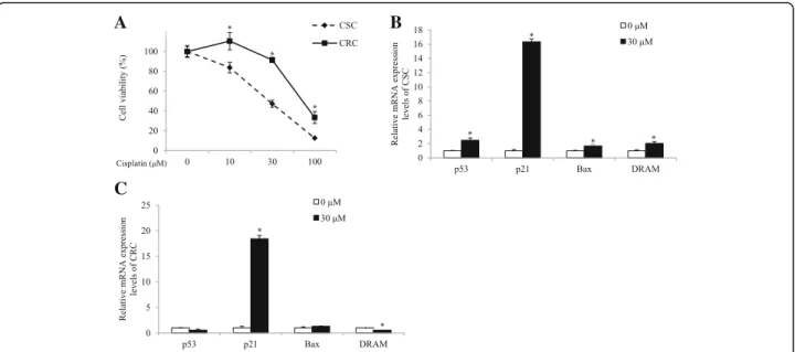
To investigate the mechanisms underlying cisplatin re-sistance, we used a pair of ovarian cancer cell lines; the cisplatin sensitive A2780 as CSC and its counterpart that acquires the resistance as CRC. We firstly confirmed that CRC was less sensitive to cisplatin, compared to CSC. The response to cisplatin was measured by cell via-bility using an MTT assay. CSC and CRC were treated
with from 10 μM to 100 μM of cisplatin for 24 h. As
shown in Fig. 1a, cisplatin treatment decreased cell viability and the response to cisplatin in CRC was sig-nificantly lower than that in CSC. Cell viability in CRC was significantly higher than it in CSC in 10μM, 30 μM, and 100μM of cisplatin-treated groups (p < 0.001). Cell viabilites in CSC decreased to 84%, 47%, and 13% by
10 μM, 30 μM, and 100 μM of cisplatin treatment,
re-spectively, compared with the non-treated group, while those in CRC decreased to 110%, 91%, and 33%. The difference of response to cisplatin between CSC and
CRC was greater in the 30μM of cisplatin-treated group
than in the 10μM or 100 μM of cisplatin-treated groups.
Therefore, we used 30 μM of cisplatin treatment for
further experiments.
We examined whether the difference in the induction of p53-mediated apoptotic pathway could explain the cisplatin resistance-involving mechanism in CRC. Ex-pression levels of p53, p21, Bax, and DRAM genes were measured in the cells treated with 30μM of cisplatin for 24 h. As shown in Fig. 1b, mRNA expression levels of pro-apoptotic p53 (p < 0.05) and its target genes such as p21 (p < 0.001), and Bax (p < 0.05), and DRAM (p < 0.05) significantly increased in the cisplatin-treated group, compared with the non-treated group in CSC, indicating that the cell death of CSC with cisplatin treat-ment invovled apoptosis through the p53-dependent pathway. On the other hand, CRC did not show signifi-cant changes in the expression of p53 and Bax (Fig. 1c). Although mRNA levels of p21 significantly increased, expression of DRAM rather decreased upon cisplatin treatment (p < 0.001; Fig. 1c). The results suggested that cisplitin treatment failed to induce apoptosis in the
CRC, unlike CSC, due to the decreased expression level of DRAM gene, which is required for activation of the p53-dependent pathway.
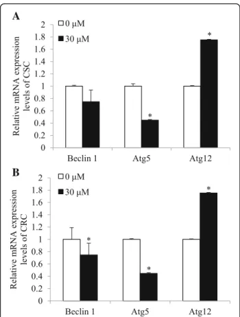
Cisplatin-induced cell death is not mediated by autophagy in ovarian cancer models
We investigated whether cisplatin-induced cell death is also mediated by autophagy. CSC and CRC were treated
with 30 μM of cisplatin for 24 h and mRNA levels of
autophay genes, Beclin 1, Atg12, and Atg5 genes were measured. In CSC, mRNA expression levels of Beclin 1 and Atg5 did not significantly alter or decreased, respect-ively, while those of Atg12 increased in cisplatin-treated group compared with the non-treated group (p < 0.01; Fig. 2a). Similarily, in CRC, mRNA expression levels of Beclin 1 and Atg5 decreased, while those of Atg12 in-creased upon cisplatin treatment (p < 0.01; Fig. 2b). The results suggest cisplatin-induced cell death in CSC is not mediated by autophagy, since Atg5 induction, which is esstial for the conjugation with Atg12 in autophagy, was not complete. In CRC, neither the p53-mediated apop-tosis nor autophagy did not taken place, indicating the resistance against cisplatin.
SbE is more effective to decrease cell viability than baicalin both in CSC and CRC
Next, we investigated whether SbE induced cell death in CSC and CRC to test a potential of SbE as an anti-cancer agent. We first analyzed quantitatively baicalin content in SbE using LC-MS analysis. The baicalin is a
major flavonoid in SbE and has been reported for its ef-fects on various cancer cells to inhibit cell proliferation and to induce cell death [21]. The quantity of baicalin in SbE was measured (Fig. 3a) and calculated according to a standard curve (Fig. 3b). As a result, we found that the content of baicalin in SbE was 74 mg/g (7.4%). Next, we investigated whether SbE and baicalin induced cell death in CSC and CRC at the various concentrations using an MTT assay. First, the cell lines were treated with from
200 μg/mL to 400 μg/mL of SbE and from 28 μM to
56μM of baicalin, i.e. equivalent to the amounts in the extract, for 24 h to compre the effect of the extract and the single compound. As shown in Fig. 3c and d, both SbE and baicalin treatment decreased cell viability, how-ever, the response of the SbE was significantly greater than that of baicalin. In CSC, cell viability in 200μg/mL
and 400 μg/mL of SbE-treated groups were significantly
lower than it in 28 μM and 56 μM of baicalin-treated
groups, respectively (p < 0.01 and p < 0.001,
respect-ively). Likewise, in CRC, 200 μg/mL and 400 μg/mL of
SbE treatment were significantly decreased compared to
28 μM and 56 μM of baicalin treatment, respectively
(p < 0.05 and p < 0.01, respectively). Cell viability in
CSC decreased to 53% and 33% by 200 μg/mL and
400 μg/mL of SbE treatment, respectively, compared
with the non-treated group (Fig. 3c), while thoes in CRC decreased to 71% and 55% (Fig. 3d). On the other hand, cell viability in CSC decreased to 77% and 67% by
28 μM and 56 μM of baicalin treatment, respectively
(Fig. 3c), while those in CRC decreased to 86% and 80%
C
A
B
Fig. 1 Effects of cisplatin on the cell viability and mRNA expression levels of p53, p21, Bax, and DRAM in CSC and CRC. Cells were exposed 10 ~ 100μM of cisplatin for 24 h. a Cell viability according to cisplatin treatment in CSC and CRC was measured by MTT assay. After cells were exposed 30μM of cisplatin for 24 h, mRNA expression levels of (b). p53, p21, Bax, and DRAM in CSC and (c). p53, p21, Bax, and DRAM in CRC were quantified by qPCR. All mRNA expression levels were normalized against TBP. Each value is the mean ± SD. p < 0.05 was taken to define statistical significance. Two tailed Student’s t-test and one-way ANOVA followed by Duncan post hoc test
(Fig. 3d). Because the effects on cell viability of the ex-tract is greater than that of single compound, baicalin, we focused on the effect of SbE for a subsequent experi-ments. We tested the effect of SbE on cell viability in more various concentrations. It effectively decreased cell viability in a dose-dependent manner in CSC and CRC and the response to SbE in CRC was significantly lower than in CSC (Fig. 3e).
SbE treatment induces p53-dependent apoptosis in CSC, not in CRC
We investigated by which mechanism SbE induced the cell death. Firstly, we tested whether SbE induced p53-mediated apoptotic pathway and its effect was similar in CSC and CRC. Expression level of p53-mediated apop-totic pathway such as p53, p21, Bax, and DRAM genes
were measured in the cells treated with 200 ~ 400 μg/
mL of SbE for 24 h. In CSC, mRNA expression levels of p53 (p < 0.01), p21 (p < 0.05), Bax (p < 0.05), and DRAM (p < 0.05) significantly increased in SbE-treated group, compared with the non-treated group (Fig. 4a).
On the contrary, CRC showed that mRNA expression levels of p53 and DRAM decreased (p < 0.001) and those of p21 and Bax did not significantly alter after SbE treat-ment (Fig. 4b). The data suggested that SbE treattreat-ment induced apoptosis in the CSC, at least in part, via p53 pathway, but not in the CSC.
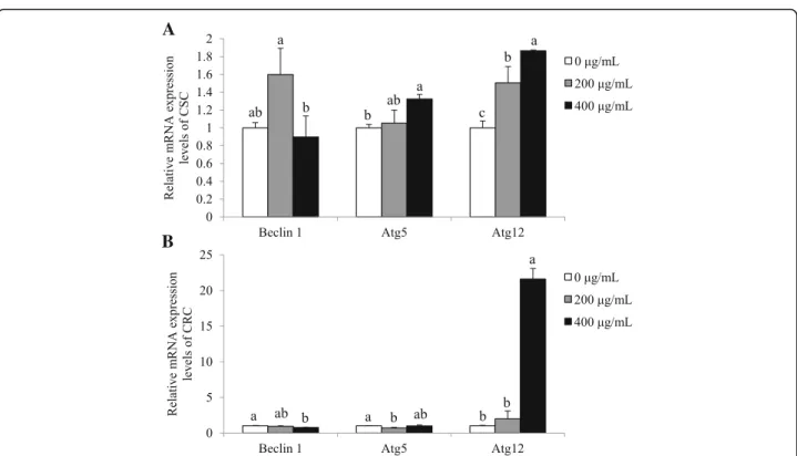
SbE treatment induces autophagy in CSC, not in CRC
We investigated whether SbE-induced cell death also in-volved the autophagy mechanism. Expression levels of autophagy genes, Beclin 1, Atg12, and Atg5 were
mea-sured in the cells treated with 200 ~ 400 μg/mL of SbE
for 24 h. In CSC, mRNA expression levels of Beclin 1 did not significantly alter, while those of Atg5 (p < 0.05) and Atg12 (p < 0.01) significantly increased in the SbE-treated group compared with the non-SbE-treated group (Fig. 5a). CRC, in contrast to CSC, showed that mRNA expression levels of Beclin 1 and Atg5 decreased (p < 0.05) after SbE treatment, while those of Atg12 in-creased (p < 0.001) in the SbE-treated group compared with the non-treated group (Fig. 5b). As similar to the response to cisplatin, the autophagy-mediated cell death was induced in CSC, but not in CRC. The results pro-posed that SbE could not induce cell death neither p53-mediated apoptotic pathway or autophagy and its mechanism is involved in the resistance in CRC.
CSC and CRC similarly respond to SbE combined with cisplatin
Next, we tested whether a combined treatment of SbE and cisplatin had additive or synergic effects in CSC and
CRC. CSC and CRC were treated with 200 ~ 400μg/mL
of SbE and 10μM ~ 100 μM of cisplatin for 24 h. The
response to the combined treatment in CRC was as sen-sitive as that in CSC by using MTT assays (Fig. 6). When the ovarian cancer cells were treated with the combined treatment, the cell viability between CSC and CRC grad-ually became closer depending on the concentration of the SbE at 30 uM of cisplatin, where the viability showed maximal difference between CSC and CRC upon only cisplatin treatment (Fig. 1a). The cell viability in CSC creased to 47%, 43%, and 14%, while those in CRC
de-creased to 91%, 64%, and 16% by 0 μg/mL, 200 μg/mL,
and 400 μg/mL of SbE with 30 μM of cisplatin
com-pared with the non-treated group (Fig. 6b). The data show that the combined treatment of SbE and cisplatin has a potential as a chemotherapeutic method to over-come chemoresistance.
The treatment of SbE, combined with cisplatin, induces not apoptotic but autophagic cell death in CRC
We further examined a possible mechanism underlying the combination effect. We tested whether the cell death upon the SbE treatment combined with cisplatin is
A
B
Fig. 2 Effects of cisplatin on mRNA expression levels of Beclin 1, Atg5, and Atg12 in CSC and CRC. After cells were exposed 30 μM of cisplatin for 24 h, mRNA expression levels of (a). Beclin 1, Atg5, and Atg12 in CSC and (b). Beclin 1, Atg5, and Atg12 in CRC were quantified by qPCR. All mRNA expression levels were normalized against TBP. Each value is the mean ± SD. p < 0.05 was taken to define statistical significance. One-way ANOVA followed by Duncan post hoc test
A
B
C
E
Fig. 3 Quantitative analysis of baicalin in SbE and effects of SbE and baicalin on the cell viability in CSC and CRC. a LC-MS analysis of baicalin in SbE. b Baicalin peak area in LC-MS as a function of baicalin concentration (correlation coefficient, R = 0.999) and the percent content of baicalin in SbE. Cells were exposed 200 ~ 400μg/mL of SbE or 28 ~ 56 μM of baicalin for 24 h. Cell viability according to SbE or baicalin treatment in (c) CSC and (d) CRC was determined by MTT assay. Cells were exposed 100 ~ 400μg/mL of SbE for 24 h. e Cell viability according to various concentrations of SbE treatment in CSC and CRC was determined by MTT assay. Each value is the mean ± SD. p < 0.05 was taken to define statistical significance. Two tailed Student’s t-test
A
B
Fig. 4 Effects of SbE on mRNA expression levels of p53, p21, Bax, and DRAM in CSC and CRC. After cells were exposed 200 ~ 400μg/mL of SbE for 24 h, mRNA expression levels of (a). p53, p21, Bax, and DRAM in CSC and (b) p53, p21, Bax, and DRAM in CRC were quantified by qPCR. All mRNA expression levels were normalized against TBP. Each value is the mean ± SD. p < 0.05 was taken to define statistical significance. One-way ANOVA followed by Duncan post hoc test
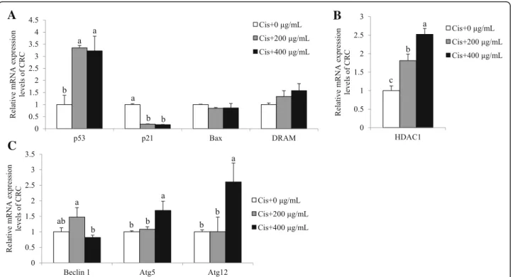
mediated by either p53 pathway and/or autophagic pathway
in the condition of 200 ~ 400μg/mL of SbE and 30 μM of
cisplatin treatment for 24 h. In CRC, mRNA expression level of p53 significantly increased in a SbE (p < 0.05), while that of p21 decreased (p < 0.001; Fig. 7a). In addition, the expression of Bax and DRAM did not significantly change
(Fig. 7a). Surprisingly, the dramatically decreased
expression of p21 was not consistent with the increased expression of p53, even though p21 is a well-known tran-scriptional target of p53. We investigated if the expression of p21 was regulated by additional factors such as epigen-etic modulators. HDAC1 and p53 have been demonstrated
as antagonistic regulators at the p21 locus. mRNA ex-pression levels of HDAC1 significantly increased upon
the combined treatment of SbE and cisplatin
(p < 0.05; Fig. 7b), restulting in the transcriptional re-pression of p21. Instead, we examined mRNA expres-sion levels of autophagy genes such as Beclin 1, Atg5, and Atg12. mRNA expression levels of Beclin 1 did not change (Fig. 7c) in the combination-treated group, compared with the non-treated group. How-ever, the expression of Atg5 and Atg12 increased upon the treatment (p < 0.05; Fig. 7c). Taken together, the results demonstrated that the combination therapy of
A
B
Fig. 5 Effects of of SbE on mRNA expression levels of Beclin 1, Atg5, and Atg12 in CSC and CRC. After cells were exposed 200 ~ 400μg/mL of SbE for 24 h, mRNA expression levels of (a) Beclin 1, Atg5, and Atg12 in CSC and (b) Beclin 1, Atg5, and Atg12 in CRC were quantified by qPCR. All mRNA expression levels were normalized against TBP. Each value is the mean ± SD. p < 0.05 was taken to define statistical significance. One-way ANOVA followed by Duncan post hoc test
A
B
C
Fig. 6 Combination effects of SbE and cisplatin on the cell viability in CSC and CRC. After cells were exposed 200 ~ 400μg/mL of SbE and 30 μM of cisplatin for 24 h. Cell viability according to the combination of SbE and (a) 10μM, (b) 30 μM, and (c) 100 μM of cisplatin treatment in CSC and CRC was measured by MTT assay. Each value is the mean ± SD. p < 0.05 was taken to define statistical significance. Two tailed Student’s t-test
SbE and cisplatin effectively induced cell death even in CRC via the autophagy pathway independent of Beclin 1.
Discussion
In this study, we investigated the effect and the possible molecular mechanism of SbE on cisplatin resistance of ovarian cancer by three steps. First of all, we validated the cell models whether CSC and CRC showed different sen-sitivity against cisplatin, as expected. Next, we examined whether SbE induced cell death in ovarian cancer and what molecular mechanism was involved. Lastly, we dem-onstrated the effects of combined treatment with SbE and cisplatin on cisplatin resistance in ovarian cancer.
We found that cisplatin induced cell death in CSC in a dose-dependent manner based on up-regulation of genes associating apoptosis pathway such as p53, p21, and Bax expression. As the cellular response to DNA damage, the tumor suppressor and transcription factor p53 and its targets, cyclin-dependent kinase inhibitor p21 and pro-apoptotic Bax, play important roles in apoptosis [25]. It is also critical to modulate the chemosensitivity of tumors by controlling cell death [26]. However, CRC showed cisplatin resistance even though the expression of pro-apoptotic protein p21 was up-regulated. One pos-sibility is due to decreased DRAM mRNA expression level. A previous study showed that DRAM is a major
component of p53-induced apoptosis [16]. Its knock-down decreased percentage of apoptosis even though mRNA expression levels of p53 and p21 increased in TetOn-p53 cells and RKO cells. Our results demonsta-rated that cisplatin treatment led to down-regulated ex-pression of the DRAM gene in CRC, while it did not in
CSC. Therefore, p53-mediated apoptotic pathway
seemed to be blocked due to the decreased expression of DRAM in the cisplatin resistant cells.
The cisplatin-mediated cell death was not dependent on autophagy. The mRNA expression of Atg5 levels was significantly decreased in both CSC and CRC. Because Atg12-Atg5 conjugate is an essential factor of autophagy, concordant expression of Atg12 and Atg5 expression is induced during autophagy. In a previous study, the Atg5 mutant was unable to generate the Atg12-Atg5 conju-gate, resulting in the decrease of autophagic activity rap-idly compared with the wild type [27].
Recently, the flavonoid baicalin enriched in herbal medicines including SbE [28] has reported to have anti-cancer properties in vivo and in vitro [21, 29]. We showed that an inhibitory effect of SbE as the extract on cell viability was greater than that of baicalin as a single compound in ovarian cancer cells. SbE induced cell death, depending on its concentration, by inducing ex-pression of apoptosis genes, such as p53, p21, and Bax, and autophagy genes such as Atg5 and Atg12 in CSC,
A
C
B
Fig. 7 Combination effects of SbE and cisplatin on mRNA expression levels of p53, p21, Bax, DRAM, Beclin 1, Atg5, and Atg12 in CRC. After cells were exposed 200 ~ 400μg/mL of SbE and 30 μM of cisplatin for 24 h, mRNA expression levels of (a) p53, p21, Bax, DRAM, (b) HDAC1, (c) Beclin 1, Atg5, and Atg12 in CRC were quantified by qPCR. All mRNA expression levels were normalized against TBP. Each value is the mean ± SD. p < 0.05 was taken to define statistical significance. One-way ANOVA followed by Duncan post hoc test. Cis; 30μM of cisplatin
but not in CRC. The results showed that CRC had lower efficiency against a single treatment of cisplatin or SbE, compared to CSC. However, combination treatment of SbE and cisplatin enhanced anti-cancer effects via
induc-tion of cell death. When 400μg/mL of SbE and 30 μM
of cisplatin were treated at the same time, cell viability was no difference between CSC and CRC. Intriguingly, the combination treatment did not fully induce p53-mediatic apoptotic pathway. Even though p53 expression increased, its target gene p21 expression decreased. One of the possibilities is the up-regulation of HDAC1, an epigenetic modulator. Recently, epigenetic mechanism has been highlighted in cancer field. Epigenetic modula-tion contributes to cancer development, progress, and treatment, since it regulates expression of oncogenes and tumor suppressor genes by altering chromatin struc-ture [30]. Chemical modifications on DNA and core histones, the octamer of proteins wrapping DNA, make chromatin condensate or unwind without altering DNA sequence [31]. Histone acetylation, an example of his-tone modifications, is a process that an acetyl group is bound to histone resulting in neutralizing DNA charge then forming euchromatin. Euchromatic structure allows a gene to be up-regulated by loosely unfolding nucleo-somes and making transcriptional factors easily access to DNA [32]. The acetylated histones are accomplished by histone acetyltransferases (HATs) and the acetyl group is detached by histone deacetylases (HDACs) from his-tones. A previous study showed that p53 and the epigen-etic regulator HDAC1 are antagonistic regulators of the p21 [33]. p53 transcriptionally activates p21 through binding to the transcription factor Sp1 in the activation of the p21 promoter. On the other hand, HDAC1 tran-scriptionally represses p21 gene expression by blocking the interaction between p53 and Sp1. In CRC, the com-bination treatment repressed p21 gene expression through HDAC1 activation, resulting in inactivation of apoptosis. However, further experiments are needed to verify whether up-regulation of HDAC1 expression re-sulted from the combined treatment alters histone acetylation levels, in particular at the p21 locus. Never-theless, the combination of SbE and cisplatin induced cell death via autophagy in CRC, showing effectiveness to overcome the resistance. It was mediated by non-canonical Beclin 1-independent autophagic cell death based on the increase of Atg5 and Atg12 expression, but no change in Beclin-1 expression. Beclin 1 is related to form a phagophore which is consist of autophagosome [18]. However, Beclin 1-independent autophagy has been reported [34, 35]. Resveratrol induced cell death through Beclin 1-independent Atg12-Atg5-dependent autophagy [34]. Arsenic trioxide also induced Beclin 1-independent autophagic pathway in ovarian cancer cells. In the arsenic trioxide-treated ovarian cancer cells, Atg5
knockdown reduced autophagy via altering the ratio of LC3-II/LC3-I, which is an indicator of the autophagic progress [36]. In contrast, Beclin 1 knockdown did not alter the ratio of LC3-II/LC3-I [35]. Our data suggest that the combination treatment of SbE and cisplatin pro-duced synergistic anti-cancer effect even in cisplatin re-sistant ovarian cells. Further analysis is needed to confirm the molecular alterations at protein levels and the extension of anti-cancer effects, i.e. the percentage of apoptotic and/or autophagic cell death.
Conclusions
Compared with SbE or cisplatin alone, the combination treatment of SbE and cisplatin had strengthened anti-cancer effects in ovarian anti-cancer cells. Although SbE in-duced cell death in ovarian cancer cells in a dose-dependent manner, the efficiency was significantly lower in CRC, compared to CSC. However, the combination treatment with cisplatin led to the effect on CRC, as similar as on CSC, suggesting the effectiveness of the combined treatment over chemoresistance. The combin-ation treatment in CRC induced autophagy by up-regulated expression of Atg5 and Atg12. It was different from the fact that SbE as a single treatment failed to in-duce apoptosis via p53 or autophagic pathways in CRC. Taken together, the results demonstrated that the com-bination treatment of SbE and cisplatin had a synergistic effect by inducing Beclin 1-independent autophagy in CRC. The findings suggest that the combination of SbE and cisplatin may be useful for a potential chemotherapy to treat ovarian cancer.
Abbreviations
3 MTT:3-[4,5-dimethylthiazol-2-yl]-2,5-diphenyltetrazoliumbromide; Atg: Autophagy-related protein; CRC: Cisplatin resistant human ovarian cancer cell line; CSC: Cisplatin sensitive human ovarian cancer cell line; DMSO: Dimethyl sulfoxide; DRAM: Damage-regulated autophagy modulator; FBS: Fetal bovine serum; HATs: Histone acetyltransferases; HDACs: Histone deacetylases; LC3: Microtubule-associated protein light chain; SbE: Extract of Scutellaria baicalensis
Acknowledgements
There is no contributor who does not meet the criteria for authorship.
Funding
This study was supported by the National Research Foundation of Korea (NRF 2014R1A1A2058942 and 2016R1D1A1A02937546). BYC and YKL were supported by Brain Korea 21 plus project (22A20130012143).
Availability of data and materials
The datasets supporting the conclusions of this article are included within the article.
Author’s contributions
SJP and YJP designed hypothesis and supervise experiments; JCJ and SJP provided the SbE; BYC, YKL and IJ performed the experiments; BYC and YJP analyzed the data, and wrote the manuscript. All authors read and approved the final manuscript.
Competing interests
Consent for publication Not applicable.
Ethics approval and consent to participate Not applicable.
Publisher’s Note
Springer Nature remains neutral with regard to jurisdictional claims in published maps and institutional affiliations.
Author details
1Department of Nutritional Science and Food Management, College of
Science & Industry Convergence, Ewha Womans University, Seoul, Republic of Korea.2Department of Sasang Constitutional Medicine, College of Korean Medicine, Wonkwang University, Iksan, Republic of Korea.3Department of
Bioconvergence, Korea basic science institute, Daejeon, Republic of Korea.
4Department of Sasang Constitutional Medicine, College of Korean Medicine,
Woosuk University, Jeonju, Republic of Korea.
Received: 22 December 2016 Accepted: 8 May 2017
References
1. Torre LA, et al. Global cancer statistics, 2012. CA Cancer J Clin. 2015;65(2):87–108. 2. McGuire WP, et al. Cyclophosphamide and cisplatin compared with
paclitaxel and cisplatin in patients with stage III and stage IV ovarian cancer. N Engl J Med. 1996;334(1):1–6.
3. Howlader N, Noone A, Krapcho M. SEER Cancer Statistics Review, 1975– 2013. Bethesda: National Cancer Institute; 2016.
4. Markman M, et al. Responses to second-line cisplatin-based intraperitoneal therapy in ovarian cancer: influence of a prior response to intravenous cisplatin. J Clin Oncol. 1991;9(10):1801–5.
5. Ohmichi M, et al. Mechanisms of platinum drug resistance. Trends Pharmacol Sci. 2005;26(3):113–6.
6. Xu X, et al. Genetic polymorphism of copper transporter protein 1 is related to platinum resistance in Chinese non-small cell lung carcinoma patients. Clin Exp Pharmacol Physiol. 2012;39(9):786–92.
7. Kelland LR. New platinum antitumor complexes. Crit Rev Oncol Hematol. 1993;15(3):191–219.
8. Dabholkar M, et al. ERCC1 and ERCC2 expression in malignant tissues from ovarian cancer patients. J Natl Cancer Inst. 1992;84(19):1512–7.
9. Sui L, et al. Survivin expression and its correlation with cell proliferation and prognosis in epithelial ovarian tumors. Int J Oncol. 2002;21(2):315–20. 10. Perego P, et al. Association between cisplatin resistance and mutation of
p53 gene and reduced bax expression in ovarian carcinoma cell systems. Cancer Res. 1996;56(3):556–62.
11. Green DR. Means to an end: apoptosis and other cell death mechanisms. Cold Spring Harbor: Cold Spring Harbor Laboratory Press; 2011. 12. Lowe SW, et al. p53-dependent apoptosis modulates the cytotoxicity of
anticancer agents. Cell. 1993;74(6):957–67.
13. Chiocca EA. Oncolytic viruses. Nat Rev Cancer. 2002;2(12):938–50. 14. El-Deiry WS, et al. WAF1, a potential mediator of p53 tumor suppression.
Cell. 1993;75(4):817–25.
15. Ahmad N, et al. Resveratrol causes WAF-1/p 21-mediated G1-phase arrest of cell cycle and induction of apoptosis in human epidermoid carcinoma A431 cells. Clin Cancer Res. 2001;7(5):1466–73.
16. Crighton D, et al. DRAM, a p53-induced modulator of autophagy, is critical for apoptosis. Cell. 2006;126(1):121–34.
17. Kondo Y, et al. The role of autophagy in cancer development and response to therapy. Nat Rev Cancer. 2005;5(9):726–34.
18. Kaur J, Debnath J. Autophagy at the crossroads of catabolism and anabolism. Nat Rev Mol Cell Biol. 2015;16(8):461–72.
19. Zhang X-D, et al. DRAM1 regulates autophagy flux through lysosomes. PLoS One. 2013;8(5):e63245.
20. Huang W-H, Lee A-R, Yang C-H. Antioxidative and anti-inflammatory activities of polyhydroxyflavonoids of Scutellaria Baicalensis GEORGI. Biosci Biotechnol Biochem. 2006;70(10):2371–80.
21. Zhou Q-M, et al. The combination of baicalin and baicalein enhances apoptosis via the ERK/p38 MAPK pathway in human breast cancer cells. Acta Pharmacol Sin. 2009;30(12):1648–58.
22. Ikezoe T, et al. Baicalin is a major component of PC-SPES which inhibits the proliferation of human cancer cells via apoptosis and cell cycle arrest. Prostate. 2001;49(4):285–92.
23. Kumagai T, et al. Scutellaria Baicalensis, a herbal medicine: anti-proliferative and apoptotic activity against acute lymphocytic leukemia, lymphoma and myeloma cell lines. Leuk Res. 2007;31(4):523–30.
24. Mosmann T. Rapid colorimetric assay for cellular growth and survival: application to proliferation and cytotoxicity assays. J Immunol Methods. 1983;65(1–2):55–63.
25. Culmsee C, Mattson MP. p53 in neuronal apoptosis. Biochem Biophys Res Commun. 2005;331(3):761–77.
26. Harris CC. Structure and function of the p53 tumor suppressor gene: clues for rational cancer therapeutic strategies. J Natl Cancer Inst. 1996;88(20): 1442–55.
27. Mizushima N, et al. A protein conjugation system essential for autophagy. Nature. 1998;395(6700):395–8.
28. Ye F, et al. Quality evaluation of commercial extracts of Scutellaria Baicalensis. Nutr Cancer. 2004;49(2):217–22.
29. Xu X-F, et al. Baicalin induces human mucoepidermoid carcinoma Mc3 cells apoptosis in vitro and in vivo. Investig New Drugs. 2011;29(4):637–45. 30. Egger G, et al. Epigenetics in human disease and prospects for epigenetic
therapy. Nature. 2004;429(6990):457–63.
31. Holliday R. Epigenetics: a historical overview. Epigenetics. 2006;1(2):76–80. 32. Jenuwein T, Allis CD. Translating the histone code. Science. 2001;293(5532):
1074–80.
33. Lagger G, et al. The tumor suppressor p53 and histone deacetylase 1 are antagonistic regulators of the cyclin-dependent kinase inhibitor p21/WAF1/ CIP1 gene. Mol Cell Biol. 2003;23(8):2669–79.
34. Scarlatti F, et al. Role of non-canonical Beclin 1-independent autophagy in cell death induced by resveratrol in human breast cancer cells. Cell Death & Differentiation. 2008;15(8):1318–29.
35. Smith D, et al. Arsenic trioxide induces a beclin-1-independent autophagic pathway via modulation of SnoN/SkiL expression in ovarian carcinoma cells. Cell Death & Differentiation. 2010;17(12):1867–81.
36. Wu J, et al. Molecular cloning and characterization of rat LC3A and LC3B—two novel markers of autophagosome. Biochem Biophys Res Commun. 2006;339(1):437–42.
• We accept pre-submission inquiries
• Our selector tool helps you to find the most relevant journal
• We provide round the clock customer support
• Convenient online submission
• Thorough peer review
• Inclusion in PubMed and all major indexing services
• Maximum visibility for your research Submit your manuscript at
www.biomedcentral.com/submit