Effects of allyl sulfur compounds and garlic extract on the
expressions of Bcl-2, Bax, and p53 in non small cell lung
cancer cell lines
Young-Sook Hong1,3, Yoon-Ae Ham1,
Ji-Hyung Choi1 and Jhingook Kim2
1
Department of Biochemistry, Medical Research Center, Division of Cancer Research, College of Medicine, Ewha Womans University, Seoul 158-056, Korea 2Department of Thoracic Surgery, College of Medicine,
Sung Kyun Kwan University, Seoul 135-230, Korea 3
Corresponding author: Tel, +82-2-650-5725; Fax, +82-2-653-8891; E-mail, [email protected]
Accepted 14 September 2000
Abbreviations: DADS, diallyl disulfide; DAS, diallyl sulfide; NSCLC, non small cell lung cancer
Abstract
Allyl sulfur compounds play a major role in the chemoprevention against carcinogenesis. The pre-sent study compared the antiproliferative effects of diallyl sulfide (DAS), diallyl disulfide (DADS) and garlic extract on p53-wild type H460 and p53-null type H1299 non small cell lung cancer cells (NSCLC). The DAS and DADS treatment of both H460 and H1299 cells resulted in the highest num-bers of cells in apoptotic state as measured by acri-dine orange staining, however, garlic extract treat-ment did not induce any significant apoptotic cells by MTT assay. DADS was found to be more effective in inducing apoptosis on NSCLC. The level of p53 protein in H460 cell was increased following DADS treatment. DAS and garlic extract treatment of H460 cells induced a rise in the level of Bax and a fall of Bcl-2 level. These results demonstrate that DAS, DADS and garlic extract are effective in re-duction of anti-proliperative gene in NSCLC and suggest that modulation of apoptosis-associated cellular proteins by DAS, DADS and garlic extract may be the mechanism for apoptosis which merit further investigation as potential chemoprevention agents.
Keywords: DAS, DADS, garlic extract, apoptosis,
NSCLC
Introduction
Epidemiological investigations provide strong evidence that environmental factors are modifiers in the occur-rence of some types of human cancer (Wynder and Gori, 1977; Parmer and Bakshi, 1983). Among these environ-mental factors, dietary habit is one of the important factors influencing cancer risk (Doll, 1992). Recent studies suggested that garlic and related allium foods might protect human from some types of cancer (Steinmetz et al., 1994), however, the protective effect by these foods was often inconsistent (Dorant et al., 1995). The reason for these inconsistencies remains to be clarified. Never-theless, laboratory investigations provided some convin-cing evidence that garlic and associated sulfur com-pounds reduced experimentally induced colon, esopha-geal, pulmonary, skin, forestomach and breast cancers (Wargovich et al., 1987; 1988; Sadhana et al., 1988; Hong et al., 1992; Liu et al., 1992; Dorant et al., 1993), suggesting that the protection provided by garlic and related compounds is not limited to a single tissue or to any particular type of carcinogen. Allyl sulfur compounds present in garlic probably account for much of its anticancer effect, and these allyl sulfur compounds have been shown to be effective both in the initiation and promotion phases of the carcinogenic process (Belman, 1983). However, in spite of numerous studies on the garlic oil component, a study on the whole water soluble garlic extract has never been reported.
p53 is involved in the activation of apoptosis by DNA damage induced by such agents as cisplatin. As a transcriptional activator, p53 increases the transcription of a number of genes, and the pattern of transcriptional regulation is critical in determining the cellular response to DNA damage. Indeed, it is known to activate the transcription of death agonist, Bax, but to repress the expression of the death antagonist, Bcl-2 (Miyashita et al., 1994; Perego et al., 1996).
The pharmacological role of allyl sulfide in prevention and treatment of cancer has received increasing atten-tion, but molecular mechanism of action of allyl sulfide compound are poorly defined. The cytotoxicity of most classical antitumor drugs thought to be mediated by their ability to induce apoptosis (Sen and D’Incalci, 1992). Several mechanism have been identified to modulation of apoptosis by induction of p53.
In this study, we examined a possible antiproliferative affect of DAS, DADS and garlic extract on human non
small cell lung cancer cell (NSCLC) and involvement of p53 in the induction of apoptosis of the treated cells.
Materials and Methods
Cell line and culture conditions
Non small cell lung cancer cell line (NCI-H460) with normal p53 gene and cell line (NCI-H1299) lacking p53 gene were purchased from ATCC (American Type Culture Collection). Each cells were cultured in RPMI-1640 (Biowhittaker, Wakersville, MD) medium contain-ing 10% FBS (Gibco-BRL, Grand Island, NY) and 1% penicillin-streptomycin (Gibco-BRL, Grand Island, NY) at 5% CO2 and 37oC.
MTT assay for cytotoxicity
To assess the cytotoxicity of organoallylsulfur compounds to the cells, MTT dye reduction assay was performed as described previously (Kim et al., 1997). Cells were plated out at a density of 3,000-4,000 cells per well into 96-well microtiter plates and allowed to attach overnight. After 24 h, the cells were treated with various con-centrations of organoallylsulfur compounds for 1 h, then replaced with new RPMI-1640 medium and incubated. After 48 h, all of cells were analyzed using MTT assay as follows: 50 µl of MTT (3,[4,4-dimethyl thiazol-2-yl]-2, 5-diphenyl-tetrazolium bromide, final concentration of 5 mg/ml; Sigma Chemical Co., St. Louis, MO) were added to each well, and further incubated at 37oC for 4 h. After
the incubation, the cells were centrifuged at 200×g for 5 min and supernatant was aspirated. Two hundred µl of dimethyl sulfoxide (DMSO) was added to each well to solubilize the formed formazine crystals. The absor-bance was recorded at 560 nm on a microculture plate reader (Becton-Dickinson Labware, Lincoln Park, NJ). Wells containing only RPM1-FBS and MTT were used as the controls. The IC50 (50% Inhibition Concentration)
values were defined as the concentration of an agent to induce 50% reduction in the absorbance. Each experi-ment was performed using 4 replicate wells for each drug concentration.
Western blot analysis of Bcl-2, Bax and p53
Cells were plated to 6-well plates and treated with DAS (purity 97%, Aldrich, Milwaukee, WI; 20 µM), DADS (purity 80%, Aldrich, Milwaukee, WI; 5 µM) or garlic extract (Bolak Co, Hwasung-Kun, Korea, 100 µg/ml), which was kindly provided by Bolak company. The garlic extract was prepared by homogenizing garlic in three times volume of water, and the homogenate was heated at 95oC and, after cooling, treated with protease to
eliminate protein. The above treated homogenate was concentrated, so that its solid component would be 60%.
At the end of treatment, the cells were collected and washed twice with cold PBS. The cells (1×10⁶) from each sample were solubilized in RIPA buffer [50 mM Tris-HCl (pH 8.0), 150 mM NaCl, 1% NP40, 0.1% SDS, 10 mM sodium deoxycholate, 1 mM phenylmethyl-sul-fonylfluoride] and kept on ice for 20 min. Cell lysates were centrifuged at 12,000 g at 4oC for 15 min and the
supernatants were stored at -70oC until assay. Protein
concentration was measured in each cell lysate by the Bradford method (Bradford, 1976). Equal amounts of protein lysates (50 µg) were resolved on 10% SDS-poly-acrylamide gels and transferred to PVDF membrane (Amersham, Buckinghamshire, UK) in glycine transfer buffer [192 mM glycine, 25 mM Tris-HCl (pH 8.8), 20% methanol(v/v)]. Membranes were blocked for 1 h in 2% skim milk solution with PBS containing 0.05% Tween 20, and then probed with monoclonal mouse anti-human Bcl-2 Ab (1 : 1000, Santa Cruz, Delaware, CA), polyclonal rabbit anti-human Bax Ab (1 : 1000, Santa Cruz, Delaware, CA), or monoclonal anti-human p53 Ab (1 : 500, Novocastra, Newcastle, UK) for 1 h at room temperature. Membranes were washed three times with 0.05% Tween 20 in PBS and incubated for 1 h with peroxidase-conjugated goat IgG fraction to mouse or rabbit (1 : 2000, Santa Cruz, Delaware, CA). After wash-ing, the immunoactive proteins were detected using the ECL Western blotting detection system (Amersham, Buckinghamshire, UK) according to the manufacturer's instructions. To measure the expression of each gene, the relative intensity was calculated by comparing with the intensity of β-actin using densitometry (Pharmacia LKB Biotechnology, Uppsala, Sweden).
RNA extraction and northern blot analysis
After treatment, cells were washed with PBS and total RNA was extracted from the cells using the TRIzol Kit (Gibco-BRL, Gland Island, NY). Twenty micrograms of RNA from each sample was loaded, electrophoresed on 1% agarose containing 2.2 M formaldeyde, and trans-ferred to a nylon membrane (Amershame, Bucking-hamshire, UK). Bcl-2 cDNA probe was labeled with [α
-32P]dCTP using Megaprime labelling system
(Amer-shame, Buckinghamshire, UK). The membrane was hybridized with [α-32P]dCTP labeled bcl-2 cDNA probe for 24 h at 42oC. After washing with 0.1% SDS in
2×SSC (15 min, two times) at 42oC, 0.1% SDS in
1×SSC (30 min, 1 time) and 0.1×SSC (30 min, 1 time) at 42oC, the membrane was exposed to X-ray film for 3 days. To control for variability in the loaded quantity of RNA, the membrane was reprobed with β-actin cDNA probe.
Acridine orange staining of apoptotic cells
Cells treated with DAS, DADS or garlic extract were washed in PBS, and fixed in 75% methanol and 25%
glacial acetic acid for 30 min in ice. Fixed cells were washed twice in PBS and stained with acridine orange (0.1% in PBS, Sigma Chemical Co., St. Louis, MO). Stained cells were washed in PBS and observed under a fluorescence microscope (×100, BX50, Olympus, Japan).
Statistics
Statistical analysis was performed using student’s t-test for differences between groups. Statistical significance
was defined as p < 0.05.
Results
Cytotoxicity of organoallylsulfur compounds and garlic extract
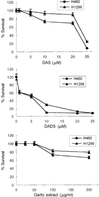
In order to examine the antiproliferative effects of the organoallylsulfur compounds and garlic extract on each cell line, cells were treated with various concentration of DAS, DADS and garlic extract for 1 h and then replaced with new medium. After 48 h, MTT analysis was carried out and was compared with the values obtained from the controls; these values are expressed as % survival.
As shown in Figure 1, Cells treated with DAS from 1 to 25 µM concentration exhibited viability 99-8% in H460 cells and 99-24% in H1299 cells, respectively. And Cells treated with DADS from 1 to 25 µM concentration exhibited viability 61-8% in both H460 cells and H1299 cells. Also Cells treated with garlic extract from 25 to 200 mg/ml concentration exhibited viability 99-66% in H460 cells and 99-79% in H1299 cells, respectively. The growth of H460 and H1299 cells was significantly
inhibit-Figure 1. Cytotoxic effect of DAS, DADS, and garlic extract on NSCLC. The cells were treated with various concentration of DAS, DADS and garlic extract for 1 h and then replaced with new RPMI-1640 medium supplemented with 10% FBS and incubated for 48 h. At 48 h, the cellular survival rate was measured using MTT assay. H460; p53 wild-type cell line and H1299; p53 deleted type cell line. Data are the means ± standard deviation of four times samples. Student's t-test value was less than 0.05.
Figure 2. Western blots showing expressions of Bcl-2, in NSCLC. The cells were treated with DAS (20 µM), DADS (5 µM) and garlic extract (100 µg/ml) for 1 h, then replaced with new RPMI-1640 medium and incubated. After 6 h, proteins were isolated as described in materials and methods. Two additional experiments performed in the same manner gave similar results. Relative intensity was calculated by comparing with the intensity of β-actin using densitometry. Student’s t-test value was less than 0.05. Lane 1; control, lane 2; DAS, lane 3; DADS, lane 4; garlic extract in H460 cells. lane 5; control, lane 6; DAS, lane 7; DADS, lane 8; garlic extract in H1299 cells.
ed by DAS and DADS, whereas was slightly inhibited by garlic extract.
Effects of the organoallylsulfur compounds and garlic extract on the expressions of Bcl-2, Bax and p53 Protein
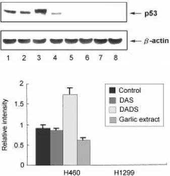
H460 and H1299 cells were treated with the organo-allylsulfur compounds and garlic extract, and Western blot was performed to examine the expressions of Bcl-2, Bax and p53 genes that participate in cell apoptosis (Figures 2-4). When treated with all of the allyl compounds, the expression of Bax increased slightly in H460 and H1299. The expression of Bcl-2 was slightly decreased when treated with all of the allyl compounds in H460 and H1299, and especially, the expression of Bcl-2 was significantly decreased when treated with DADS in H1299. In H460 (p53 wild), p53 did not show a significant increase after treated with DAS and garlic extract but increased when treated with DADS.
Effects of the organoallylsulfur compounds and garlic extract on the expressions of Bcl-2 mRNA
To determine whether the suppression of Bcl-2 protein is due to a drug-induced decrease in message, we next
measured the level of Bcl-2 mRNA by Northern blotting. As shown in Figure 5, the expression of Bcl-2 gene was decreased when H460 and H1299 cells were treated with organoallylsulfur compounds and garlic extract.
Morphological changes of apoptosis, visualized by acridine orange staining
When cells were treated with the organoallylsulfur compounds and garlic extract, morphological changes
Figure 3. Western blots showing expressions of Bax in NSCLC. The cells were treated with DAS (20 µM), DADS (5 µM) and garlic extract (100 µg/ ml) for 1 h, then replaced with new RPMI-1640 medium and incubated. After 6 h, proteins were isolated as described in materials and methods. Two additional experiments performed in the same manner gave similar results. Relative intensity was calculated by comparing with the intensity of β-actin using densitometry. Student’s t-test value was less than 0.05. Lane 1; control, lane 2; DAS, lane 3; DADS, lane 4; garlic extract in H460 cells. lane 5; control, lane 6; DAS, lane 7; DADS, lane 8; garlic extract in H1299 cells.
Figure 4. Western blots showing expressions of p53, in NSCLC. The cells were treated with DAS (20 µM), DADS (5 µM) and garlic extract (100 µg/ ml) for 1 h, then replaced with new RPMI-1640 medium and incubated. After 6 h, proteins were isolated as described in materials and methods. Two additional experiments performed in the same manner gave similar results. Relative intensity was calculated by comparing with the intensity of β-actin using densitometry. Student’s t-test value was less than 0.05. Lane 1; control, lane 2; DAS, lane 3; DADS, lane 4; garlic extract in H460 cells. lane 5; control, lane 6; DAS, lane 7; DADS, lane 8; garlic extract in H1299 cells.
Figure 5. Northern blot showing expressions of bcl-2. The cells were treated with DAS (20 µM), DADS (5 µM) and garlic extract (100 µg/ml) for 1 h, then replaced with new RPMI-1640 medium and incubated. After 6 h, total RNA were isolated as described in materials and methods. The autoradiogram are representative of similar results obtained in three replicate experiments. β-actin probe was used to normalize amounts of total RNA in each lane. Lane 1; control, lane 2; DAS, lane 3; DADS, lane 4; garlic extract in H460 cells. lane 5; control, lane 6; DAS, lane 7; DADS, lane 8; garlic extract in H1299 cells.
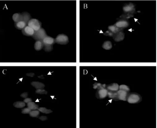
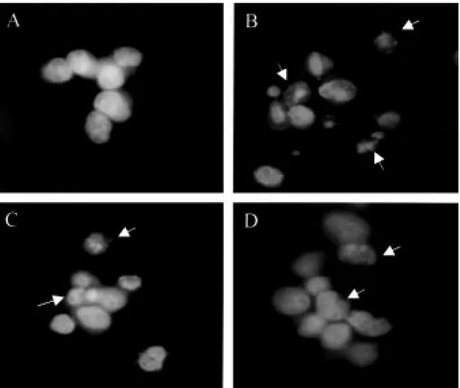
of the cells were detected by acridine orange staining. When treated with DAS, DNA segmentation of nucleus was observed in all the cells. Also, when treated with DADS and garlic extract, DNA segmentation was ob-served in H460 cells and H1299 cells (Table 1, Figure 6, 7). The results showed that apoptotic cells were signifi-cantly higher in cells treated with DAS and DADS among three agents used.
Discussion
The anticarcinogenic benefits of garlic and associated allyl sulfur compounds are not limited to carcinogen formation and bioactivation, but also appear to be relat-ed to changes in the rate of cellular proliferation and apoptosis (Sundaram and Milner, 1996a, 1996b). DADS
is one of the several compounds derived from garlic, and about 60% of the sulfur found in processed garlic oil has been reported to be DADS (Dausch and Nixon, 1990). Sundaram et al. reported that organoallylsulfur compounds in the processed garlic was able to sup-press growth of canine mammary tumor cell and that DADS also suppresses further growth in colon (HCT-15), lung (A549) and skin (SK MEL-2) cell lines (Sun-daram and Milner, 1993). In the present study, we confirmed that DAS, DADS and the water soluble whole garlic extract suppressed the growth of H460 and H1299 cells. The cytotoxic effects of organoallylsulfur compounds or garlic extract in H460 cells expressing wt-p53 were more sensitive than H1299 cells having null p53. These results revealed that the p53 protein plays an important role in the apoptotic response of a cell with DNA damage. However, DADS was able to induce strong
Table 1. Induction of apoptosis following exposure of NSCLC cells to DAS, DADS and garlic extract. Cells were treated with 5 µM DAS, 20 µM DADS and 100 µg/ml garlic extract for 1 h. After stained with 0.1% acridine orange, cells with nuclear fragmentation were counted under a fluorescence microscopy (×100). Each value represents the average of three independent determinations, and mean ± standard deviation
H460 H1299
Control DAS DADS Garlic extract Control DAS DADS Garlic extract
Cancer cells 217 142 137 155 214 159 151 181
Apoptotic cells 3 78 83 65 6 61 69 39
% of apoptosis 1.4 ± 0.7 35.5 ± 4.2 37.8 ± 3.8 29.5 ± 3.1 2.7 ± 1.4 27.7 ± 3.7 31.4 ± 4.2 17.7 ± 3.1
Figure 6. Morphological analysis of H460 cells treated with DAS, DADS and garlic extract. The cells were treated with 20 µM DAS, 5 µM DADS and 100 µg/ml garlic extracts for 1 h. After incubation, new RPMI-1640 medium supplemented with 10% FBS was added and incubated for 48 h. At 48 h, the cells were stained with acridine orange and analyzed under a fluorescence microscope. A, control; B, DAS; C, DADS; D, garlic extract.
cytotoxicity effect on the null p53 lung cancer cell line, H1299. The cytotoxic effects of anticancer drugs were known to be linked with the expression of oncogene or tumor suppressor genes such as p53 and Bcl-2.
Recent studies on the Bcl-2 oncogene have demon-strated its role in the prevention of apoptosis induced by a wide variety of stimuli and conditions (Chiou et al., 1994; Dole et al., 1994). The antiapoptotic activity of Bcl-2 correlates with its intracellular ratio to Bax. High levels of Bax have been shown to favor apoptosis in cells subjected to growth factor deprivation, whereas high levels of Bcl-2 prolong cell survival under the condi-tions (Baff et al., 1993). A number of studies have shown that constitutive expression of Bcl-2 proto-oncogene in a variety of cells results in a heightened resistance to chemotherapeutic agents that function by inducing ap-optosis (Hanada et al., 1993). In the present study, when H460 cells were treated with DAS or garlic extract, the ratio of Bcl-2/Bax decreased compared to that of the control. To examine the effects of DAS, DADS and garlic extract, we investigated to Northern and Western blot. The expression of genes related to both p53 dependent and independent apoptotic pathways. In Northern blot, the expression of bcl-2 gene was decreased treated with DAS, DADS and garlic extract in both H460 cells and H1299 cells (Figure 5).
The expression of Bcl-2 tended to decrease in H460 cell for all compounds, slightly decreased in H1299 when treated with DAS and garlic extract, significantly decreased when treated with DADS. When treated with all of the allyl sulfur compounds, the expression of Bax increased slightly in two NSCLC, showing that apopto-sis induced by the organoallylsulfur compounds and garlic extract is due to increased Bax expression and decreased Bcl-2 expression. The downstream of p53 is known to control and affect the Bax and Bcl-2 genes participating in apoptosis. It was reported that p53 indu-ces apoptosis by either increasing transcriptional activity of proapoptotic genes such as Bax or suppressing the activity of the antiapoptotic gene of the bcl-2 family (St Croix and Kerbel, 1997; Miyake et al., 1998). Thus, in order to determine whether the ability of organoallyl-sulfur compounds in inducing apoptosis and changes in the expressions of Bax/Bcl-2 are due to the effect of p53, changes in the expression of p53 was also ex-amined in p53-wild type H460 and p53-null type H1299. In p53-wild type H460, DAS and garlic extract did not show changes in the level of p53 expression. However, when treated with DADS, the level of p53 increased in H460. When treated with DAS and garlic extract, H460, the p53-wild type, did not increase p53 level but changes were observed in the expressions of Bax and Bcl-2 and
Figure 7. Morphological analysis of H1299 cells treated with DAS, DADS and garlic extract. The cells were treated with 20 µM DAS, 5 µM DADS and 100 µg/ml garlic extracts for 1 h. After incubation, new RPMI-1640 medium supplemented with 10% FBS was added and incubated for 48 h. At 48 h, the cells were stained with acridine orange and analyzed under a fluorescence microscope. A, control; B, DAS; C, DADS; D, garlic extract.
changes were observed in the expressions of Bax and Bcl-2 in H1299, the p53-null type, suggesting that ap-optosis due to DAS or garlic extract is through the interaction of Bax/Bcl-2 with the mechanism other than p53 downstream. In case of DADS, p53 level increased in H460, showing that DADS increased the level of p53, controlling Bax and Bcl-2, inducing apoptosis. Also, changes were seen in the expressions of Bax and Bcl-2 in H1Bcl-299, suggesting DADS inducing apoptosis through the p53-independent pathway.
In conclusion, the results of our study demonstrated that organoallylsulfur compounds and garlic extract have apoptotic potential on non small cell lung cancer cells. Although the exact mechanism involved in their protec-tive effects against carcinogenesis are not clearly under-stood at present, our results suggested that mechanism of apoptosis induced by organoallylsulfur compounds was regulated through p53-dependent or p53-indepen-dent related Bax/Bcl-2 dual pathway.
Acknowledgment
This work was supported by Ewha Womans University Research Fund.
References
Baff, G., Miyashita, T., Williamson, J. R. and Reed, J. C. (1993) Apoptosis induced by withdrawal of interleukin-3(IL-3) from an IL-3-dependent hematopoietic cell line is associated with repartitioning of intracellular calcium and is blocked by enforced Bcl-2 oncoprotein production. J. Biol. Chem. 268: 6511-6519
Belman, S. (1983) Onion and garlic oils inhibit tumor promotion. Carcinogenesis 4: 1063-1067
Bradford, M. M. (1976) A rapid and sensitive method for the quantitation of microgram quantities of protein utilizing the principle of protein-dye binding. Anal. Biochem. 72: 248-254
Chiou, S. K., Rao, L. and White, E. (1994) Bcl-2 blocks p53-dependent apoptosis. Mol. Cell. Biol. 14: 2556-2563 Dausch, J. G. and Nixon, D. W. (1990) Garlic: a review of its relationship to malignant disease. Prev. Med. 19: 346-361 Dole, M., Nunez, G., Merchant, A. K., Maybaum, J., Rode, C. K., Bloch, C. A. and Castle, V. P. (1994) Bcl-2 inhibits chemotherapy-induced apoptosis in neuroblastoma. Cancer Res. 54: 3253-3259
Doll, R. (1992) The lessons of life: keynote address to the nutrition and cancer conference. Cancer Res. 52: 2024S-2029S
Dorant, E., van den Brandt, P. A. and Goldbohm, R. A. (1995) Allium vegetable consumption, garlic supplement intake, and female breast carcinoma incidence. Breast Cancer Res. Treat.
33: 163-170
Dorant, E., van den Brandt, P. A., Goldbohm, R. A., Hermus, R. J. and Sturmans, F. (1993) Garlic and its significance for the prevention of cancer in humans: a critical view. Br. J. Cancer 6: 424-429
Hanada, M., Krajewski, S., Tanaka, S., Cazals-Hatem, D., Spengler, B. A., Ross, R. A., Biedler, J. and Reed, J. C. (1993) Regulation of Bcl-2 oncoprotein levels with differentiation of human neuroblastoma cells. Cancer Res. 53: 4978-4986 Hong, J. Y., Wang, Z. Y., Smith, T. J., Zhou, S., Shi, S., Pan, J. and Yang, C. S. (1992) Inhibitory effects of diallyl sulfide on the metabolism and tumorigenicity of the tobacco-specific carcinogen 4-(methylnitrosamino)-1-(3-pyridyl)-1-butanone (NNK) in A/J mouse lung. Carcinogenesis 13: 901-904 Kim, J., Lee, S. K., Hwang, E. S., Kim, J. S., Kim, K .and Lee, J. H. (1997) Limited cytotoxic effect of adenoviral-mediated p53 gene transfer in variable non-small cell lung cancer (NSCLC) cell lines. J. Korean Cancer 29: 565-575
Liu, J. Z., Lin, R. I. and Milner, J. A. (1992) Inhibition of 7,12-dimethylbenz[a] anthracene-induced mammary tumors and DNA adducts by garlic power. Carcinogenesis 13: 1847-1851 Miyake, H., Hanada, N., Nakamura, H., Kagawa, S., Fujiwara, T., Hara, I., Ito, H., Gohji, K., Arakawa, S., Kamidono, S., Saya, H. (1998) Overexpression of Bcl-2 in bladder cancer cells inhibits apoptosis induced by cisplatin and adenoviral-mediated p53 gene transfer. Oncogene 16: 933-943 Miyashita, T., Krajewski, S., Krajewska, M., Wang, H. G., Lin, H. K., Liebermann, D. A., Hoffman, B. and Reed, J. C. (1994) Tumor suppressor p53 is a regulator of bcl-2 and bax gene expression in vitro and in vivo. Oncogene 9: 1799-1805 Palmer, S. and Bakshi, K. (1983) Diet, nutrition, and cancer: interim dietary guidelines. J. Natl. Cancer Inst. 70: 1151-1170 Perego, P., Giarola, M., Righetti, S. C., Supino, R., Caserini, C., Delia, D., Pierotti, M. A., Miyashita, T., Reed, J. C. and Zunino, F. (1996) Association between cisplatin resistance and mutation of p53 gene and reduced bax expression in ovarian carcinoma cell systems. Cancer Res. 56: 556-562 Sadhana, A. S., Rao, A. R., Kucheria, K. and Bijani, V. (1988) Inhibitory action of garlic oil on the initiation of benzo[a]pyrene-induced skin carcinogenesis in mice. Cancer Lett. 40: 193-197 Sen, S. and D'Incalci, M. (1992) Apoptosis. Biochemical events and relevance to cancer chemotheraphy. FEBS Lett. 307: 122-127
St Croix, B. and Kerbel, R. S. (1997) Cell adhesion and drug resistance in cancer. Curr. Opinion Oncol. 9: 549-556 Steinmetz, K. A., Kushi, L. H., Bostick, R. M., Folsom, A. R. and Potter, J. D. (1994) Vegetables, fruit, and colon cancer in the Iowa Women's Health Study. Am. J. Epidemiol. 139: 1-15 Sundaram, S. G. and Milner J. A. (1993) Impact of organo-sulfur compounds in garlic on canine mammary tumor cells in culture. Cancer Lett. 74: 85-90
Sundaram, S. G. and Milner, J. A. (1996a) Diallyl sulfide inhibits the proliferation of human tumor cells in culture. Biochim. Biophys. Acta 1315: 15-20
Sundaram, S. G. and Milner, J. A. (1996b) Diallyl sulfide indu-ces apoptosis of human colon tumor cells. Carcinogenesis 17: 669-673
Wargovich, M. J. (1987) Diallyl sulfide, a favor component of garlic (Allium sativum), inhibits demethylhydrazine-induced colon cancer. Carcinogenesis 8: 487-489
Wargovich, M. J., Woods, C., Eng, V. W., Stephens, L. C. and
Gray, K. (1988) Chemoprevention of N-nitrosomethylbenzyl-amine-induced esophageal cancer in rats by the naturally occurring thioester, diallyl sulfide. Cancer Res. 48: 6872-6875 Wynder, E. L. and Gori, G. B. (1977) Contribution of the environment to cancer incidence: an epidermiologic exercise. J. Natl. Cancer Inst. 58: 825-832