https://doi.org/10.11626/KJEB.2020.38.1.021
Original article
규조류 Skeletonema pseudocostatum Medlin
(Thalassiosirales, Bacillariohyta)의 형태적 특징과 분자계통학적 위치
한경하1, 이 준2, 박준상3, 윤주연1, 김현정1,4, 곽경윤1, 오석진4, 신현호1,*
1한국해양과학기술원 해양시료도서관, 2한국생명공학연구원 생물자원센터
3한국해양과학기술원 생명공학연구센터, 4부경대학교 해양학과
Morpho-molecular characterization of diatom
Skeletonema pseudocostatum (Thalassiosirales, Bacillariophyta) from the Korean coast
Kyong Ha Han1, Zhun Li2, Joon Sang Park3, Joo Yeon Youn1, Hyun Jung Kim1,4, Kyeong Yoon Kwak1, Seok Jin Oh4 and Hyeon Ho Shin1,*
1
Library of Marine Samples, Korea Institute of Ocean Science and Technology, Geoje 53201, Republic of Korea
2
Biological Resource Center, Korean Collection for Type Cultures, Korea Research Institute of Bioscience and Biotechnology, Jeongeup 56212, Republic of Korea
3
Marine Biotechnology Research Center, Korea Institute of Ocean Science & Technology, Busan 49111, Republic of Korea
4
Department of Oceanography, Pukyong University, Busan 48513, Republic of Korea
Korean J. Environ. Biol.
38(1) : 21-29 (2020) ISSN 1226-9999 (print) ISSN 2287-7851 (online)
Korean Journal of Environmental Biology
* Corresponding author Hyeon Ho Shin Tel. 055-639-8440
E-mail. [email protected]
Received: 5 November 2019 Revised: 3 January 2020
Revision accepted: 15 January 2020
Abstract: Morphology of a strain of unspecified Skeletonema species established from Korean coast was examined by light, fluorescence and scanning electron microscopy, and SSU (small subunit) and LSU (large subunit) rDNA of the strain were also sequenced.
The specimen was characterized by solitary or short chains, and each cell contained 1-2 chloroplasts. The valve face was slightly convex, and the terminal fultoportula processes (TFPPs) were open and showed narrow distal ends that could be truncated or spiny with claw-like protrusions. The basal part of the TFPPs was tubular and oblique to the cell axis.
The intercalary fultoportula processes (IFPPs) were also narrow, completely open, and joined in a 1 : 1 junction. Occasionally, one IFPP was connected with two opposing IFPPs.
The morphological features of the specimen were identical to those of Skeletonema pseu
docostatum. Molecular phylogeny based on SSU rDNA revealed that the Korean strain is nested within a clade comprising S. pseudocostatum and S. tropicum. However, based on D1-D2 LSU rDNA sequences, a clade including S. pseudocostatum and a Korean strain was separated from the S. tropicum clade. This indicates that the Korean strain can be identified as S. pseudocostatum. This species represents the first record from Korean coastal waters.
Keywords: Skeletonema, morphology, SSU rDNA, LSU rDNA, phylogeny
서 론
Skeletonema
속은 바베이도스 주변의 에오세 후기 퇴적물로부터 관찰된
Skeletonema barbadense Grev.
을 기준 종(type species)으로 하여 명명되었다(Greville 1865).Greville(1866)은 홍콩 연안에서 발견된 돌말류 표본과 요크셔 연안의 해초류(ascidian)의 위장에서 발견된 표본 이 동일한 종임을 확인하고, 그 종을
Melosira costata Grev.
로 명명하였다. 이후 Cleve(1873)는 자바해로부터 관찰된
M. costata
와S. brabadense
의 형태적 유사성을 확인하고, M.costata
를S. costatum
(Grev.) Cleve으로 재명명 하였다.Skeletonema
속의 형태적 특징은 ‘받침돌기(fultoportula process)에 의해 세포 간에 연결되어 형성되는 사슬 군체’ 로서 분명하게 정의되지만(Hasle 1973; Round et al. 1990), 속 내 종을 구별할 수 있는 형태적 특징이 모호할 뿐 아니 라, 형태적 변이 때문에, 대부분의Skeletonema
종의 보고는S. costatum
으로서 기록되어 왔다(e.g. Jung et al. 2009). 하지 만, 최근에Skeletonema
종의 분자계통학적 분석을 통한 결 과는 속 내 다수의 숨은종(cryptic species)의 발견과 함께 다수의 신종 보고를 이끌었고(Sarno et al. 2005, 2007), 현 재 전 세계 보고된Skeletonema
속 내 종수는 21종으로 알 려져 있다(Guriy and Guiry 2019).S. pseudocostatum Medlin
은 받침돌기 외부형태, 입술돌 기(rimoportula process)의 위치와 유전적 차이에 의해S.
costatum
과 구별된다(Medlin et al. 1991). 그리고, Sarno etal.
(2005)은S. pseudocostatum
의 둘레띠(cingular band), 받 침돌기 등의 형태적 특징을 구체적으로 기재하여 다른 종 과의 형태적 차이를 보고하였다. 이후S. pseudocostatum
은 유럽(이탈리아, 네덜란드), 남아메리카(브라질), 아프리 카(이집트), 아시아(중국, 일본), 오스트리아, 뉴질랜드 등 전 세계 연안에서 다수 보고되어 왔으나(Sarno et al. 2005;Bergesch et al. 2009; Kaeriyama et al. 2011; McCarthy 2013;
Veen et al. 2015), 국내 연안에서의 출현은 보고된 바 없다. 국내에서
Skeletonema
속의 분류는 광학현미경하에 사 슬형태의 군체를 형성하는 것에 따라S. costatum
으로 동 정되어 왔다. Jung et al.(2009)은 국내 연안에 출현하는Skeletonema
종들을 대상으로 주사전자현미경을 이용하여구체적인 형태적 특징을 관찰하였다. 그 결과, S. dohrnii, S.
marioni, S. subsalsum, S. tropicum
등 4종을 확인하였지만 기 존에 빈번하게 보고되어 온S. costatum
의 출현은 확인하지못했다. 따라서, 국내 연안에서 출현하는
Skeletonema
종은 구체적인 형태 분석과 함께 분자계통학적 연구를 통해 동 정을 명확히 할 필요가 있다.본 연구는 한국해양과학기술원의 해양식물플랑크톤 기 탁등록보존기관에서 보존하고 있는
Skeletonema
속의 배양 주(strain)를 이용하여 종의 형태적 특징과 계통분류학적 특성을 분석하였다. 그 결과, 분석 종이 국내에서는 기록되 어 있지 않은S. pseudocostatum
으로 확인되어, 그 출현을 기 록한다.재료 및 방법
1. Skeletonema pseudocostatum의 배양주 (strain) 정보
S. pseudocostatum
은 1998년 5월 8일에 욕지도(34°38ʹ 30.2ʺN, 128°16ʹ22.7ʺE)에서 20μm 망목 크기의 식물플랑 크톤 네트를 수평 예인하여 채집되었다. S. pseudocostatum 의 단일 군체는 모세관 방법으로 분리하였고, 온도 20℃, 염분 33psu 및 광량 100μmol m-2 s-1하에 f/2 배지에서 배 양하였다. 단일 분리된 종주는 미세조류은행에서 보관, 관 리되어왔으나, 현재는 한국해양과학기술원 해양시료도서 관에 위치한 해양식물플랑크톤 기탁등록보존기관에 해당 종이 이관되어 배양주 LIMS-PS-0848로 등록하여 보존하 고 있다.2. 형태적 분석
S. pseudocostatum
의 군체(chain) 길이와 모양, 엽록체 구조 등의 형태적 특징은 광학현미경(Primo Vert, Zeiss, Germany)과 형광현미경(Axio Imager 2; Zeiss, Germany) 으로 관찰하였고, 돌기(process)의 구조와 위치 등의 미 세구조는 전계방출형 주사전자현미경( JSM7600, Jeol, Tokyo, Japan)으로 관찰하였다. 주사전자현미경 관찰을 위 해S. pseudocostatum
배양주는 최종농도 2%인 Lugol 용액 으로 고정한 후, 에탄올 시리즈(10, 30, 50, 70, 90, 99, 99%) 로 시료를 15분씩 탈수하였고, 임계점 건조법(critical point drying method)(Spi-DryTM Regular Critical Point Dryer, SPI Supplies, West Chester, PA, USA)을 통해 건조하였다. 건조 된 시료는 aluminum stub에 고정하고 백금코팅(platinum)하여 5~15Kv, 8.0mm working distance 하에서 관찰하였다.
3. 분자생물학적 분석
S. pseudocostatum
의 Genomic DNA(gDNA) 추출을 위 해 대수성장기의 배양주 1mL을 1.5mL tube에 분주하 여 원심분리기로 침전시킨 후 상등액을 제거하고 세 포만 농축하여 -20℃의 냉동실에 보관하였다. 농축 된 배양주의 gDNA은 DNeasy Plant Mini Kit(Qiagen, Valencia, CA)를 사용하여 매뉴얼에 따라 추출되었다. Small subunit(SSU) rDNA 유전자 부위의 염기서열 분석 을 위해 forward primer: SSU11+(5ʹ-TGATCCTGCCAG TAGTCATACGCT-3ʹ), reverse primer: SSU1147-(5ʹ-AG TTTCAGCCTTGCGACCATAC-3ʹ)을 사용하였고 (Alverson et al. 2007), large subunit(LSU) rDNA는 for- ward primer: LSU D1R(5ʹ-ACCCGCTGAATTTAAGCATA- 3ʹ), reverse primer: LSU R2(5ʹ-ATTCGGCAGGTGAGTTG TTAC-3ʹ)를 이용하여(Takano and Horiguchi 2006), PCR을 수행하였다. PCR 반응액의 경우 5μL 10X Ex Taq Buffer (Mg2+plus), 1.25U Ex Taq polymerase(Takara, Japan), 1 μM primer 및 1μL DNA을 주입하여 최종 50μL가 되도 록 하였고, PCR 수행조건은 pre-denature는 95℃에서 2 분, denature는 95℃에서 20초, annealing은 55℃에서 1 분, elongation은 72℃에서 1분씩 30회 반복 후, post- elongation은 72℃에서 5분간 실시하였다(T100TM Thermal Cycler; Bio-Red, Hercules, CA). PCR 산물은 1%
agarose gel에 전개하고 Midori Green Advance(NIPPON Genetics, Co., Ltd., Tokyo, Japan)로 염색하여 UV 하에서 DNA 밴드를 확인하였고, QIAquick PCR purification kit (Qiagen, Valencia, CA)를 사용하여 PCR 산물을 정제한 후 ABI PRISM® 3700 DNA Analyzer(Applied Biosystems, USA)을 사용하여 염기서열을 분석하였다.
4. 분자계통학적 분석
S. pseudocostatum
의 SSU rDNA 염기서열은 Alversonet
A B C D F
E
G
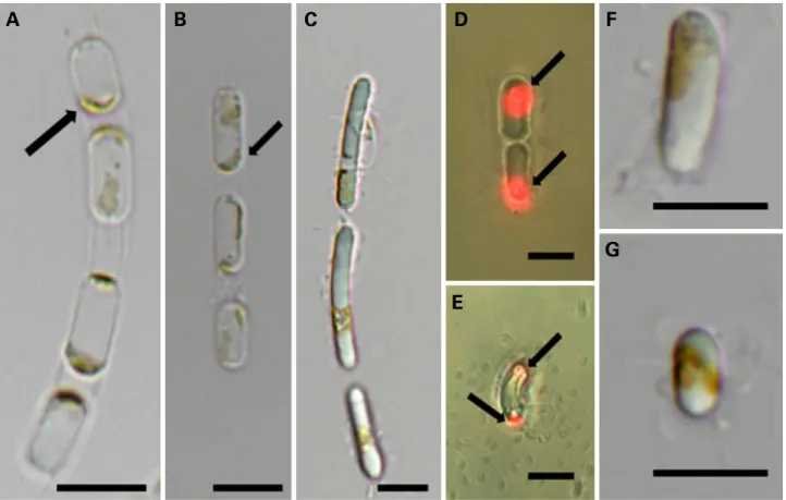
Fig. 1. Light and fluorescence micrographs of Skeletonema pseudocostatum (LIMS-PS-0848). (A-E) Colony with 1-2 chloroplasts in each
cell (arrows); (F, G) single cell. Scale bars: A-G
=10 μm.
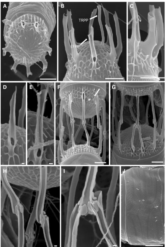
Fig. 2. Scanning electron micrographs of Skeletonema pseudocostatum (LIMS-PS-0848). (A) Terminal valve with subcentral TRPP; (B) terminal valve with TFPPs and TRPP; (C) detail of a terminal valve; (D, E) detail of TFPPs showing spiny ends; (F) intercalary valves with 1 : 1 IFPP junctions and IRPP (arrow); (G) intercalary valves with 1 : 2 IFPP junctions; (H) detail of 1 : 1 IFPP junctions; (I) detail of a 1 : 2 IFPP junction; (J) singular band. Scale bars: A-C, F-J
=1 μm; D, E
=0.1 μm.
A B C
D E F G
H I J
al.
(2005) 및 Sarno et al.(2005)을 참고하여 총 40개, LSU rDNA는 Sarno et al.(2005)을 참고하여 총 36개의 정렬 된 염기서열 정보를 추가한 후 MEGA v. 7.0.26 프로그램 을 사용하여 매뉴얼로 정렬하였다. 정렬된 염기서열의 적 합한 유전자 모델을 찾기 위해 MEGA v. 7.0.26 프로그램에 서 Modeltest를 수행하였고, 최적 모델은 SSU rDNA의 경 우 TIM3+G(A:C:G:T=0.2867:0.1822:0.2397:0.2914;p-inv=0; gamma shape=0.3090), LSU rDNA는 GTR+I(A :C:G:T=0.2573:0.1962:0.2932:0.2534; p-inv=0.6370;
gamma shape=0)을 선정하여 MEGA v. 7.0.26 프로그램 을 사용하여 최대유사분석(Maximun-likelihood analysis;
ML)을 분석하였다. 그리고, 계통수의 유연 관계를 평가하 기 위해 1,000회의 bootstrap을 수행하였다. 베이즈 추론 (Bayesian Inference; BI)은 MrBayes 3.1.2를 사용하였고, 분 석이 완료된 이후에는 분석결과를 바탕으로 하여 각 종 간 의 계통유연관계를 밝히는 분자계통도를 작성하였고, 분 자계통도의 확인은 MEGA 프로그램으로 수행하였다.
결과 및 고찰
1. Skeletonema pseudocostatum의 형태적 특징 Skeletonema pseudocostatum
의 둘레면 보기(girdle view) 에서 엽록체는 1~2개이고, 세포의 말단에 위치해 있다 (Fig. 1A~E). 각 세포는 길게 돌출한 가장자리 받침돌기에 의해 사슬 형태로 연결되었고 직선이나 곡선의 군체를 형성하였다(Fig. 1A~C). 군체는 2~6개의 세포로 구성되어 있고, 뚜껑(valve)의 직경은 6~17.3μm(n=50)로, 같은 배 양주 내에서도 세포마다 길이 차이를 보였으며, 단일 세포 로 존재할 때에도 길이 차이를 확인할 수 있었다(Fig. 1F, G).
주사전자현미경을 이용한
S. pseudocostatum
의 관찰 결 과, 뚜껑은 원형이고(Fig. 2A), 뚜껑면(valve face)은 평평하 거나 살짝 볼록했다(Fig. 2A, B). 말단세포의 가장자리 받 침돌기 끝(terminal fultoportula process; TFPP)은 갈라지 거나 갈고리 모양이고(Fig. 2B~D), 길이는 1.67±0.5μm (n=36)이며, 개수는 8.10±1.1개로 뚜껑의 크기에 따라 다양했다(Fig. 2B, C). 한 개의 말단세포 입술돌기(terminal rimoportula process; TRPP)는 뚜껑면 중앙으로부터 살 짝 벗어나 위치해 있었고(Fig. 2A, B), 두꺼운 원통형의 나팔관 모양이며, 길이가 1.1±0.6μm(n=6)이었다(Fig.2B). 그리고, 연결세포 받침돌기(intercalary fultoportula process; IFPP)는 일반적으로 1:1 결합으로 맞물려 있으나 (Fig. 2F~H), 1:2 결합도 종종 관찰되었다(Fig. 2I). 망목은 poroid이고, 방사상으로 배열하며 1 μm 당 13개(n=4)이 다(Fig. 2J).
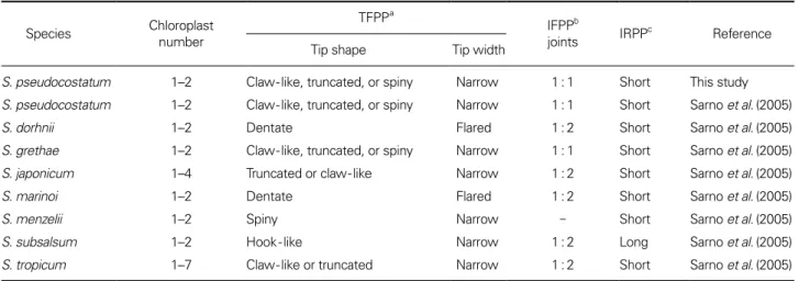
Sarno et al.(2005, 2007)의 분석 결과에 의하면,
Skele tonema
속은 종에 따라 TFPP와 TRPP의 개수 및 길이, IFPP 결합, 둘레띠 모양 등의 형태가 다르다. 이에 따라S.
pseudocostatum
을 포함한 8종의Skeletonema
의 미세구조 를 비교하였다(Table 1). S. pseudocostatum은 TFPP가 끝 이 갈라지거나, 갈고리 또는 가시 모양인 것이S. grethae, S.
Table 1. Morphological comparison of S. pseudocostatum to other Skeletonema species Species Chloroplast
number
TFPP
aIFPP
bjoints IRPP
cReference
Tip shape Tip width
S. pseudocostatum 1–2 Claw-like, truncated, or spiny Narrow 1 : 1 Short This study S. pseudocostatum 1–2 Claw-like, truncated, or spiny Narrow 1 : 1 Short Sarno et al. (2005)
S. dorhnii 1–2 Dentate Flared 1 : 2 Short Sarno et al. (2005)
S. grethae 1–2 Claw-like, truncated, or spiny Narrow 1 : 1 Short Sarno et al. (2005)
S. japonicum 1–4 Truncated or claw-like Narrow 1 : 2 Short Sarno et al. (2005)
S. marinoi 1–2 Dentate Flared 1 : 2 Short Sarno et al. (2005)
S. menzelii 1–2 Spiny Narrow - Short Sarno et al. (2005)
S. subsalsum 1–2 Hook-like Narrow 1 : 2 Long Sarno et al. (2005)
S. tropicum 1–7 Claw-like or truncated Narrow 1 : 2 Short Sarno et al. (2005)
a: Terminal fultoportula process, b: Intercalary fultoportula process, c: Process of the intercalary rimoportula
japonicum, S. menzelii, S. tropicum
과 유사한 형태적 특징을 보인다(Table 1). 하지만, 이 종들은 TFPPs의 형태, 규산화 정도, 군체모습, 엽록체수 등에 의해S. pseudocosatum
으로 부터 구별된다. S. grethae은 TFPPs의 형태적 차이에 의해S. pseudocostatum
과 구별되고, S. japonicum은 단단한 규산질각과 상대적으로 큰 기공에 의해 구별되며, S. menzelii은 단일세포로 관찰된다는 것에서 차이를 보이고, S. tropicum 은 엽록체의 수가 많고, 더 긴 군체를 형성한다는 것에서
S. pseudocostatum
과 다르다(Sarno et al. 2005). 이처럼 종 에 따라 일부 종은 서로 비슷한 형태적 특징을 가지기도Fig. 3. Maximum-likelihood (ML) tree showing the phylogenetic position of Skeletonema pseudocostatum based on partial nuclear-en-
coded SSU rDNA sequences. Pleurosira laevis (AF525670), Lithodesmium undulatum (Y10569), Helicotheca tamesis (X85385), Ditylum
brightwellii
(X8538) and Porosira pseudodenticulata (X85398) was selected as the outgroup. The numbers on each node are the bootstrap
values (%) and the Bayesian Posterior Probability (PP). Only bootstrap values above 50 and PP above 0.7 are shown. Scale bar
=number of
nucleotide substitutions per site.
하지만, 주사전자현미경을 통한 세부적 형태 특징 비교로
Skeletonema
속의 종들을 구분할 수 있을 것으로 판단된다.2. Skeletonema pseudocostatum의 계통 분류학적 위치
S. pseudocostatum
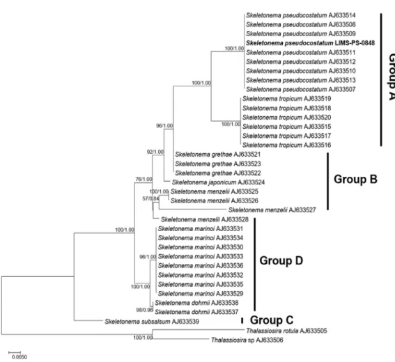
의 계통학적 위치를 파악하기 위해 정 렬된 SSU rDNA 염기서열의 길이는 1033bp였고, LSU rDNA 염기서열의 길이는 778bp였고, 각 종 간의 계통유 연관계를 밝히는 분자계통도는 Figs. 3과 4에 나타내었다.Skeletoenma
속의 진화적 유연관계는 4개의 그룹(Group) 으로 구분되었고(Figs. 3, 4), S. pseudocostatum는S. tropicum
과 함께 Group A에 포함되었다. Skeletonema 속은 형태적으로 유사성을 보였던 만큼, 종간 유전적 거리도 가까웠 는데, 특히
S. tropicum
은 SSU rDNA 결과에서S. pseudo costatum
과 유전적으로 동등한 위치에 있었다. 이는 SSU rDNA을 통한 계통학적 분석으로 두 종을 구분할 수 없 다는 것을 의미한다. 하지만 LSU rDNA 구간의 염기서 열 분석 결과에서S. tropicum
는S. pseudocostatum
과 함께Group A로 구분되었지만, 유전적 거리에서 차이를 나타
내었고, 유사도 분석(similarity analysis)결과 본 배양주와 97% 일치하는 결과를 보였다. 이처럼 SSU rDNA 결과에서 는 구분되지 않았던
S. tropicum
는S. pseudocostatum
이 LSU rDNA 결과에서 구분 가능하였다. 따라서Skeletonema
의 정 확한 종 동정을 위해 여러 구간의 분석이 요구된다.한편, LSU rDNA 염기서열의 유사도 분석(similarity
Fig. 4. Maximum-likelihood (ML) tree showing the phylogenetic position of Skeletonema pseudocostatum based on partial nuclear-encod-
ed LSU rDNA sequences. Thalassiosira rotula (AJ633505) and Thalassiosira sp. (AJ633506) were selected as the outgroup. The numbers on
each node are the bootstrap values (%) and the Bayesian Posterior Probability (PP). Only bootstrap values above 50 and PP above 0.7 are
shown. Scale bar
=number of nucleotide substitutions per site.
analysis)결과 본 배양주는 여러 지역에서 기록된 분리 주의 염기서열과 100% 일치하는 결과를 보였다. 이는
S.
pseudocostatum
은 지리적 위치와 상관없이 염기서열에서유전적 차이가 나타나지 않는 단일 계통(monophyly)이라 는 것을 의미한다.
적 요
Skeletonema pseudocostatum
의 세포는 규산질 성분의 돌 기에 의한 사슬 형태로, 길이는 6~17.3μm였고, 엽록체는 세포 당 1~2개를 포함하고 있었다. 주사전자현미경 관찰 결과Skeletonema
종을 구분할 수 있는 가장자리 받침돌기 끝(terminal fultoportula process)은 끝이 갈라지거나, 갈 고리 모양이었고, 길이가 1.67±0.5μm이고, 개각의 가장 자리에 위치해 있으며, 개수는 8.10±1.1개로 개각의 크기 에 따라 다양하게 나타났다. 말단세포 입술돌기(terminal rimoportula process)는 두꺼운 원통형의 나팔관 모양으 로, 개각의 중앙 근처에 위치하였고, 길이는 1.1±0.6μm 였으며, 개수는 1개였다. 연결세포 받침돌기(intercalary fultoportula process)는 대부분 1:1 결합으로 서로 맞물려 있는 형태였고, 1:2 결합도 종종 발견되었다. 계통분석 결 과는 형태적 특징이 유사한 종 간의 유전학적 거리가 가깝 다는 것을 나타냈고, 지리적 기원이 다른 동일 종의 경우, 유전적 차이를 보이지 않았다. 이는S. pseudocostatum
은 지 리적 위치와 상관없이 유전적 차이가 나타나지 않는 단일 계통(monophyly)이라는 것을 의미한다.사 사
본 연구는 해양수산과학기술진흥원 해양수산생명공학 기술개발사업(No. 20170431)과 한국해양과학기술원 연 구사업(PE99721)의 지원을 받아 수행되었음.