황련의 항염증효과
윤광로2, 김영진1, 이은2, 이준무1*1)
1: 상지대학교 한의과대학 2: 상지대학교 보건과학대학
Anti-inflammatory Effect of Coptidis Rhizoma
Yoon Kwang-Ro2, Kim Young-Jin1, Lee Eun2, Lee Joon-Moo1*
1: College of Oriental Medicine, Sangji University 2: College of Health Sciences, Sangji University
ABSTRACT
Objectives: The present study investigated Inflammatory effect of Coptidis Rhizoma in lipopolysaccharide- exposed rats and Raw 264.7 cells.
Methods: The plasma concentration of IL-1β, IL-6 and TNF-α peaked at 5 h after LPS injection, and the values of the Coptidis Rhizoma extract groups were lower than those of the control group.
In the increment of cytokines concentration at 2 h and 5 h after LPS injection, the Coptidis Rhizoma groups were lower than that of control group. The plasma concentration of IL-10 peaked at 5 h after LPS injection, and the values of the Coptidis Rhizoma extract groups were higher than those of the control group. In the increment of cytokines concentration at 2 h and 5 h after LPS injection, the Coptidis Rhizoma groups were higher than that of control group.
Liver cytokines measurement was done at 5 h after LPS injection. The concentration of liver IL-1β and IL-6 in the Coptidis Rhizoma groups was lower than that of the control group. The concentrations of liver TNF-α, and IL-10 showed no significant differences among all the treatment groups.
Results: In the studies of lipopolysaccharide-exposed Raw 264.7 cells, the concentration of IL-1β, IL-6 and TNF-α in the lipopolysaccharide-exposed cells groups was higher than that of control group (normal group), and in the lipopolysaccharide-exposed cells groups, these values showed a tendency to decrease in the Coptidis Rhizoma groups. The concentration of IL-10 in the lipopolysaccharide- exposed cells groups was higher than that of control group (normal group), and in the lipopolysaccharide- exposed cells groups, the values showed a tendency to increase in the Coptidis Rhizoma groups.
Conclusions: These results indicate that the Coptidis Rhizoma extracts have an functional material for Inflammatory activities.
Key words : Anti-inflammatory effect, Coptidis Rhizoma, LPS shock, cytokines, Raw 264.7 cells
서 론
염증반응은 생체 내에서 항원침입이나 조직손상과 같 은 응급상황이 발생하였을 때에 림프구를 포함한 면역세
포들에 의해 수행되는 일련의 생체보호반응이다. 그러나 염증반응이 과도하게 나타나거나 지속적으로 나타나게 되면 주변조직의 손상과 더불어 생체기능에 이상을 가져와 질병의 상태를 악화시키거나 새로운 질병을 유도한다1).
* 교신저자 : 이준무, 강원도 원주시 우산동 283 상지대학교 한의과대학 경혈학교실
․ Tel : 033-730-0662 ․ E-mail : [email protected]
․ 접수:2009년 8월 24일 ․ 수정:2009년 9월 16일 ․ 채택:2009년 9월 23일
따라서 염증제어는 질병 치료에서 가장 기본적으로 수행 되는 임상적 대응 중의 하나이며, 염증반응의 정도에 따 라 염증성 질환자 및 수술환자들의 사망률은 크게 달라 질 수 있다2).
최근 그 동안 치료에 사용했던 대부분의 항염증제들이 장기복용 할 경우 출혈성 위장관 궤양, 신장기능 저하, 혈압상승 등의 부작용과 함께 심근경색 및 혈전형성 등 의 순환계 질환도 유발할 수 있다3)고 보고되었다. 따라 서 효능과 안전성이 우수한 새로운 항염증제 개발에 관 심을 가지게 되었으며, 다양한 분야에서 새로운 항염증 제 개발을 위한 많은 연구들이 수행되었다4-6). 특히 마 황7), 감국8-10) 및 하고초11) 등을 비롯한 여러 천연물들을 이용한 연구결과들은 새로운 항염증제를 개발하기 위한 연구대상으로 한약재 및 천연물들의 응용 가능성을 한층 더 높여주었고, 보다 더 다양한 천연물들을 대상으로 한 기초연구의 필요성을 인식시켜 주었다.
황련(Coptidis Rhizoma, Coptis japonica Makino)은 미 나리아재비과(Ranunculaceae)에 속하는 다년생 초목으로 서 주요성분은 isoquinoline계열 alkaloid인 berberine이며, 이외에 coptisine, epiberberlin, feluric acid, magnoflorine, palmatine, worenine 등의 성분을 함유하고 있다. 특히 berberine은 항균작용, 항염증, 지혈, 혈압강하 및 항암작 용 등이 우수하며, 중추신경억제, 신장염 치료효과 및 기 관지 평활근 확장작용이 보고되었다12). 황련 추출물의 항 염증 효과에 대한 연구로는 Lipopolysaccharide (LPS)를 준 복강대식세포에서 iNOS, COX-2 및 TNF-α의 생성저 해12), 췌장세포의 세포사멸억제13), Keratinocytes에서 TNF-α 생성억제14) 및 기억장애 개선효과15) 등이 보고되었다. 따 라서 황련에 내재하는 기능성 물질들, 특히 항염증효과에 대한 연구자들의 관심이 한층 더 높아졌다.
LPS는 병원균의 내독소로서 그람음성세균의 막 구조 물로 다당류, 인지질 및 소량의 단백질로 구성되어 있으 며, 여러 종류의 염증세포 및 조직구성 세포들이 생산하 는 cytokine들의 생산을 촉진한다16). 따라서 염증반응을 연구하는 실험모델로 많이 응용된다17).
본 연구는 항염증 효과가 우수하고, 부작용이 없는 새 로운 항염증제를 개발하기 위한 기초연구의 일환으로, 황 련 추출물을 급여한 흰쥐에게 LPS 투여로 급성기 염증 반응을 유발시킨 후, 혈액 및 간장의 전염증성 cytokines 들의 농도를 시간의 경과에 따른 변동을 조사하였으며, 한편으로는 황련 추출물을 처리한 Raw 264.7 cell에 LPS 를 처리한 후, 처리군별 전염증성 cytokines들의 생산량 을 조사하여, in vivo 및 in vitro에서 황련 추출물의 항 염증효과를 검토하였다.
재료 및 방법
1. 시험동물 및 시험군
평균체중이 181.25±5.73 g의 Sprague-Dawley계 수컷
32마리를 1주일간 시험식이에 적응시킨 후, 평균체중이 유사하게 대조군(생리식염수 100 mg/kg, body weight (BW)), 처리1군(황련 추출액 100 mg/kg, BW), 처리2군 (황련 추출액 200 mg/kg, BW) 및 처리 3군(황련 추출액 300 mg/kg, BW)으로 나누어, 각 처리군당 8마리씩 임의 배치했다.
2. 식이 및 물
식이 및 물은 시험기간 6주 동안 자유 섭취하도록 하 였다(Table 1).
Table 1. Composition of Experimental Diet Ingredients (%) Basal diet
Casein 20.0
α-Corn starch 35.0
Sucrose 11.0
Lard 4.0
Corn oil 1.0
Mineral mix1) 3.5
Vitamin mix2) 1.0
Cellurose powder 23.5
DL-methione 0.3
1) Mineral mix. (g/kg diet) : CaCO3, 29.29; CaHPO4․2H2O, 0.43;
KH2PO4, 34.30; NaCl, 25.06; MgSO4․7H2O, 9.98; Feric citrate hexahydrate, 0.623; CuSO4․5H2O, 0.516; MnSO4․H2O, 0.121;
ZnCl2, 0.02; KI, 0.005; (NH4)6 MO7O24․4H2O, 0.0025.
2) Vitamin mix (mg/kg diet) : Thiamine-HCl, 12; Riboflavin, 40;
Pyrodoxin-HCl, 8; Vitamin-B12, 0.005; Ascorbic acid, 300;
D-biotin, 0.2; Menadione, 52; Folic acid, 2; D-calcium pantothenate, 50; P-aminobenzoic acid, 50; Nicotinic acid, 60;
Cholin choloride, 2000(IU/kg diet); Rethinyl acetae, 5000(IU/kg diet); Cholecalciferol, 250 (IU/kg diet).
3. 황련 추출물 및 투여
시중에서 구입한 양질의 황련 500 g (건조중량)을 적 량으로 나누어 수조상에서 냉각수 환류하에 EtOH로 5시 간씩 3회 추출하고, 여과, 감압 농축하여 EtOH extract 115 g을 얻었다. 투여는 매일 오후 5시경에 죤대를 이용 하여 경구 투여하였고 대조군은 동일한 방법으로 생리식 염수를 투여했다.
4. LPS 처리
LPS 처리는 6주간의 사양기간이 종료된 후, 5 mg/kg 의 수준으로 각 처리군 모두 동일하게 복강 주사하였다.
5. 혈액 및 간장 채취
혈액 채취는 시험 최종일에 LPS 처리 직전(0 h), LPS 처리 후 2시간(2 h) 및 5시간 (5 h)에 각 처리군별로 심 장 천자법에 의해 채혈했다. 간장 채취는 LPS 처리 후 5
시간에 혈액 채취가 끝난 후 적출하였다.
6. Raw 264.7 세포배양과 cytokines 정량용 시료 채취
마우스 대식세포인 Raw 264.7 cells은 한국세포주은행 (서울)에서 구입하였으며, Dulbecco’s modified Eagle’s medium (DMEM)에 10% fetal bovine serum (FBS), penicillin (100 U/ml) 및 streptomycin (100 μg/ml)이 첨 가된 배지를 사용하여 37℃, 5% CO2 incubator에서 배양 하였다. 시험과정의 모든 cells는 80-90%의 confluency에 서 실험하였고, 20 passages를 넘기지 않은 cell만 사용하 였다.
LPS 처리시 황련 추출물이 Raw 264.7 cells의 전염증 성 cytokines 생산에 미치는 영향을 검토하기 위하여 4 well dish에 cells를 분주하고(1×10⁶/ml), 황련 추출물을 농도별로 처치한 다음, 1시간 후에 각각 LPS 1 μg/ml를 처치하였으며, LPS 처치 후 6시간 후에 시료를 채취하여 Cytokines을 측정하였다.
7. Cytokines 정량
혈장 cytokine 정량용 시료는 채혈 직후, 혈장을 분리 하여 -80℃에 냉동 보관하였다. 간장 cytokine 정량용 시 료는 1 g의 간장을 채취하여 5 ml의 cold phosphate buffered saline (PBS, pH 7.4, containing a protease inhibitors cocktail)과 함께 혼합하여 얼음 위에서 분쇄 (homogenized)하였다. 분쇄혼합물을 4℃, 15,000 rpm, 15 분간 원심분리한 후, 상층부를 0.45 ㎛ 필터로 여과하고, 다시 원심분리해서 상층부를 -80℃에 냉동 보관하였다.
Raw 264.7 cells들이 생산한 cytokines 정량을 위해 배 양액을 4℃, 15,000 rpm, 15분간 원심분리한 후, 상층부를 0.45 ㎛ 필터로 여과하고, 다시 원심분리해서 상층부를 -80℃에 냉동 보관하였다.
Cytokine (IL-1β, TNF-α, IL-6 및 IL-10)정량은 시판 Kit (Biosource International, USA)를 이용했다. TNF-α 의 최저 측정농도는 0.7 pg/ml이며, 다른 cytokine들은 3-8 pg/ml이다. 간장 cytokines 정량은 5 ml의 PBS에 생 간장 1 g를 혼합한 조정액으로 측정하였으며, pg/mg 단 위로 나타내었다.
8. 통계처리
실험결과는 SPSS package를 이용하여 one-way ANOVA 검정을 수행하였으며, 각 처리군간의 유의성 검정은 Duncan’s multiple range test에 의해 p<0.05 수준에서 실시했다.
결과 및 고찰
염증성 질환 및 수술 후의 환자관리에서 염증반응의 적절한 제어는 질병치유뿐만 아니라 환자들의 생존율과 직결된다. 따라서 임상현장에서는 항염증 효과가 우수하 고 부작용이 없는 항염증제의 구비가 필수적이다. 그러나 현재 응용되고 있는 항염증제들은 부작용으로 인해 환자 에 따라 제한적으로 응용되고 있다. 따라서 항염증 효과 가 우수하고, 부작용이 없는 새로운 항염증제의 개발이 필요하다. 이러한 견지에서 본 연구는 항염증제의 개발 을 위한 기초연구로, 황련의 항염증 효과를 in vivo 및 in vitro에서 보다 더 구체적으로 검토해 보기 위하여 황련 추출물을 급여한 흰쥐에게 LPS 투여로 급성기 염증반응을 유발시킨 후, 혈액 및 간장의 전염증성 cytokines농도의 시간대별 변화를 조사하였다. 한편으 로는 Raw 264.7 cell에 LPS 처리 후, 황련 추출물이 각종 전염증성 cytokines 생산에 미치는 영향을 비교 검 토하였다.
본 연구에서 혈장 cytokine 측정은 LPS 처리 전(0 h), 처리 후 2시간(2 h) 및 5시간(5 h)에 실시하였으며, 간장 cytokine 측정은 LPS 처리 후 5시간(5 h)에 실시하였다.
LPS처리 후, 혈장 및 간장의 cytokine 측정시간의 채택 은 염증반응으로 나타나는 cytokines의 농도에 크게 영향 을 미친다. 따라서 본 연구에서 채택된 시간은 여러 연구 자들의 실험결과를 참고하여 rat의 내독소 쇼크를 검토하 기 위한 시료 채취 시간으로 적당하다고 생각되어 결정
하였다17,18). LPS 처리농도는 5 mg/kg으로 하였으며, 이
농도는 단시간에 rat와 마우스에 내독소의 쇼크를 주어 간장과 혈액내의 cytokine농도를 높인다는 다른 연구자의 실험결과를 참고했다19-22).
1. Plama cytokines
1) Plasma IL-1β
각 처리군별 Plasma IL-1β 농도의 시간대별 변동을 보면 LPS처리 후, 2시간부터 증가하여, 5시간에도 계속 적으로 증가하는 경향을 나타내었다. 이러한 결과는 LPS 처리 후 4-6시간에 혈장 IL-1β농도의 피크가 나타났다는 다른 연구자의 결과17)와 유사했다. IL-1β는 in vivo나 in vitro에서 LPS 독성의 mediator로서 알려진 전염증성 cytokine이다. IL-1β의 생물학적 기능은 TNF-α와 유사 하다. 또한 이들 두 cytokine들은 여러 형태의 실험에서 상호 상가효과를 나타낸다는 것이 밝혀졌으며, 급성기 염 증반응에서 급격히 증가한다17). 본 연구에서는 LPS 처리 후, 2시간에서는 처리군간에 유의한 차이를 나타내지 않 았으나, 5시간에서 황련 처리군들이 대조군보다 낮은 수 치를 보였으며, 시간대별 변동의 증가량도 황련처리군들 이 대조군보다 낮아 황련 처리군들의 염증반응이 대조군 보다 완화되었음을 시사해 준다(Table 2).
Table 2. Effect of Coptidis Rhizoma on Plasma IL-1β Concentration in Lipopolysaccharide-exposed Rats
Treatment IL-1β (pg/ml), Time (h)*
0 h 2 h 5 h
Control (saline, 100 mg/kg) 13.71±2.11NS 91.25±8.41NS 288.69±22.54b
Coptidis Rhizoma (100 mg/kg) 11.94±2.85NS 77.48±11.29NS 251.85±29.38b
Coptidis Rhizoma (200 mg/kg) 13.31±2.06NS 83.72±9.38NS 195.24±13.57a
Coptidis Rhizoma (300 mg/kg) 12.58±2.47NS 87.55±10.61NS 171.15±15.33a
* : 0 h, 2 h and 5 h after LPS injection.
a, b : Means in the same column with different superscripts are significantly different (p< 0.05). NS : Not significantly different (p> 0.05).
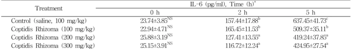
Table 3. Effect of Coptidis Rhizoma on Plasma IL-6 Concentration in Lipopolysaccharide-exposed Rats
Treatment IL-6 (pg/ml), Time (h)*
0 h 2 h 5 h
Control (saline, 100 mg/kg) 23.74±3.85NS 157.44±17.88b 637.45±41.73c
Coptidis Rhizoma (100 mg/kg) 22.94±4.71NS 165.45±11.53b 509.37±35.11b
Coptidis Rhizoma (200 mg/kg) 25.88±3.19NS 127.41±13.55a 419.24±37.85a
Coptidis Rhizoma (300 mg/kg) 25.15±3.91NS 116.72±12.24a 424.95±27.54a
* : 0 h, 2 h and 5 h after LPS injection.
a, b, c : Means in the same column with different superscripts are significantly different (p< 0.05). NS : Not significantly different (p> 0.05).
Table 4. Effect of Coptidis Rhizoma on Plasma TNF-α Concentration in Lipopolysaccharide-exposed Rats
Treatment TNF-α (pg/ml), Time (h)*
0 h 2 h 5 h
Control (saline, 100 mg/kg) 12.32±3.11NS 701.31±37.15b 768.27±43.72b
Coptidis Rhizoma (100 mg/kg) 15.02±2.98NS 693.58±51.26b 711.59±37.61b
Coptidis Rhizoma (200 mg/kg) 13.78±2.75NS 585.74±30.91a 605.92±30.55a
Coptidis Rhizoma (300 mg/kg) 12.66±3.51NS 611.28±35.59a 634.55±45.19a
* : 0 h, 2 h and 5 h after LPS injection.
a, b : Means in the same column with different superscripts are significantly different (p< 0.05). NS : Not significantly different (p> 0.05).
2) Plasma IL-6
Plasma IL-6의 농도는 LPS 처리 후 2시간에서부터 증가하여 5시간에도 높은 수치를 나타내었으며, 2시간의 각 처리군별 Plasma IL-6 농도는 황련 200 및 300 mg/kg 처리군들이 대조군보다 낮았으며, 5시간에서는 황 련 처리군들 모두가 대조군보다 낮았다. 각 처리군 별 시 간대별 증가량도 황련 추출물 처리군들이 대조군보다 낮 은 경향을 보였다. IL-6는 monocytes/macrophages 및 간장의 Kupper cell에서 생산되는 중요한 전염증성 cytokine 이다(Eduard et al., 2004). 본 실험의 결과에서 LPS 처리 후 5시간에 높은 수치를 보여 Mathiak 등(2000)이 LPS 유발 염증반응에서 혈장 IL-6 농도의 최고 피크 시간은 LPS처리 후 4-6시간이었다고 보고한 결과와 유사했으며, LPS 처리 후 5시간에 황련 추출물 첨가량이 증가함에 따라 Plasma IL-6의 농도가 하락하고, LPS 처리 후, 2시 간에서 5시간의 시간대별 증가량도 유사한 경향을 보여, 황련 추출물의 항염증효과를 시사해 주었다(Table 3).
3) Plasma TNF-α
Plasma TNF-α농도는 LPS 처리 후 2시간에 모든 처 리군 들이 급격하게 증가하여, 5시간까지 높은 수치를 유 지했다. 각 처리군별 Plasma TNF-α농도는 LPS 처리
후, 2시간 및 5시간 모두에서 황련 추출물 200 mg/kg 및 300 mg/kg 처리군들이 대조군보다 낮은 값을 나타내었 다. 시간대별 증가량도 LPS 처리 후, 2시간 및 5시간에 황련 추출물 200 mg/kg 및 300 mg/kg 처리군들이 대조 군보다 낮은 증가량을 나타내었다.
TNF-α는 LPS를 비롯한 여러 가지의 자극에 반응하 여 monocytes와 macrophage에 의해 방출되는 peptide mediator이다23). 이것은 endotoxin의 제거효과를 가지는 가장 중요한 mediator로 가정되었다24). 그러나 TNF-α는 LPS의 shock에 의해 Kuffer cell로부터 방출되며, 간장에 상처를 주고 간세포의 사멸을 일으키며25), TNF-α의 과 잉 생산은 광범위의 pathogenic 상태를 일으킨다. 따라서 최근의 연구에서 생체 내에서 TNF-α의 생산을 하락시키 는 방법을 연구하고 있다16). 본 실험의 결과에서 LPS 처 리 후, 2시간 및 5시간에서 TNF-α의 농도 및 증가량이 황련 추출물 200 mg/kg 및 300 mg/kg 처리군이 대조군 보다 낮은 경향을 보여 황련 추출물이 항염증기능에 관 여하고 있음을 시사했다(Table 4).
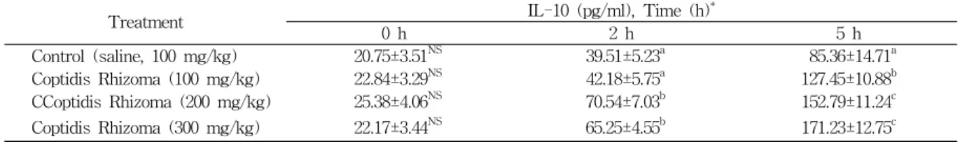
4) Plasma IL-10
Plasma IL-10 농도는 LPS 처리 후, 2시간에 증가하여 5시간에 최고치를 나타내었다. 각 처리군별 변동경향은
Table 5. Effect of Coptidis Rhizoma on Plasma IL-10 Concentration in Lipopolysaccharide-exposed Rats
Treatment IL-10 (pg/ml), Time (h)*
0 h 2 h 5 h
Control (saline, 100 mg/kg) 20.75±3.51NS 39.51±5.23a 85.36±14.71a
Coptidis Rhizoma (100 mg/kg) 22.84±3.29NS 42.18±5.75a 127.45±10.88b
CCoptidis Rhizoma (200 mg/kg) 25.38±4.06NS 70.54±7.03b 152.79±11.24c
Coptidis Rhizoma (300 mg/kg) 22.17±3.44NS 65.25±4.55b 171.23±12.75c
* : 0 h, 2 h and 5 h after LPS injection.
a, b, c : Means in the same column with different superscripts are significantly different (p< 0.05). NS : Not significantly different (p> 0.05).
Table 6. Effects of Coptidis Rhizoma on Liver Cytokines Concentration in Lipopolysaccharide-exposed Rats
Treatment IL-1β (pg/mg) IL-6 (pg/mg) TNF-α (pg/mg) IL-10 (pg/mg)
Control (saline, 100 mg/kg) 25.81±3.11b 10.11±1.45b 1.83±0.51NS 1.07±0.51NS Coptidis Rhizoma (100 mg/kg) 20.15±4.27ab 6.31±0.95a 1.91±0.85NS 1.48±0.55NS Coptidis Rhizoma (200 mg/kg) 16.38±3.92a 5.57±0.91a 1.49±0.57NS 1.74±0.43NS Coptidis Rhizoma (300 mg/kg) 17.21±3.75a 6.11±0.83a 1.11±0.62NS 1.91±0.71NS a, b : Means in the same column with different superscripts are significantly different (p< 0.05). NS : Not significantly different (p> 0.05).
LPS 처리 후, 2시간 및 5시간 모두에서 황련 추출물 첨 가군들이 대조군보다 높은 값을 나타내었으며, 시간대별 증가량도 황련 추출물 첨가군들이 대조군보다 높은 값을 나타내었다. IL-10은 lympocytes와 macrophages에 의해 생산되는 강력한 pleiotropic anti-inflammatory cytokine이 다26). 이것은 T helper type 1 cells, mono/ macrophages, polymorphonuclear cells에 의해서 IL-6 및 TNF-α 등의 전 염증성 cytokine들의 합성을 억제하며, in vitro와 in vivo에서 T-cell활성화를 감소시켰다21,27). 본 실험의 결과 에서 LPS처리 후 2시간과 5시간에서 황련 추출물 처리 군들의 IL-10농도 및 시간대별 증가량이 대조군보다 높 은 값을 나타내었는데, 이러한 결과가 IL-6 및 TNF-α 농도에 영향을 주었을 것으로 생각된다(Table 5).
2. Liver cytokines
1) IL-1β 및 IL-6
LPS 처리 5시간 후의 간장 IL-1β 및 IL-6의 농도는 황련 첨가군이 대조군보다 낮은 값을 나타내었다. LPS shock시에 간장의 활성화된 Kuffer cell들은 IL-1β를 지 속적으로 방출하며, LPS 처리 후 30분 이내에 간장 IL-1 β 농도는 급격하게 증가한다고 보고되었다28). 또한 Eduard 등(2004)도 LPS shock는 간장 IL-1β농도를 증가시켰다 고 보고하였으며, Sang 등(1999)은 LPS 처리 후 30분 이 내에 IL-6 mRNA가 급격하게 증가하였으며, 3시간 후에 도 이러한 현상은 지속되었다고 보고하였다. 본 연구에서 는 LPS 처리 후 5시간에 간장 cytokines를 측정하였다.
다른 연구자들의 연구 결과들을 참고해 볼 때 간장 cytokines 측정시간이 다소 차이는 있으나, LPS 처리 후 3시간 이후에도 증가된 cytokines 농도가 지속되었음을 고려해 보면, 본 연구의 결과도 LPS shock가 지속된 상 태에서 측정된 것으로 생각된다. 따라서 간장 IL-1β 및
IL-6 농도(Table 6)가 황련 추출물 처리군들 모두에서 대조군보다 낮은 값을 나타내어, 황련 추출물이 간장 IL-1β 및 IL-6 생산을 저해하였음을 시사했다(Table 6).
2) TNF-α
TNF-α는 LPS shock에 의해 간장 Kuffer cell로부터 방출되며, 간장세포의 사멸을 일으키고25), LPS 처리 후 2 시간에 급격하게 증가하는 것으로 보고되었다29). 본 실험 의 결과에서는 LPS 처리 후 간장 TNF-α 농도가 대조군 과 황련 추출물 처리군들 간에 유의한 차이를 나타내지 않았다. 이와 같은 결과는 간장에서 생산된 TNF-α가 혈 액으로 유입되어 소량으로 잔류하였는데 기인한 것으로 생각된다(Table 6).
3) IL-10
IL-10은 간장이 주요 공급원이며, 간장에서 IL-10의 강력한 cellular source는 macrophages, Kupffer cells, T 와 B lymphocytes 및 hepatocytes이다30). 특히 설치류에 서 IL-10은 LPS 처리 시에 Kufer cell에서 TNF-α 및 IL-6를 하향 조절한다26,28). 본 연구의 결과에서는 IL-10 의 농도는 대조군과 황련 처리군 간에 유의한 차이를 나 타내지 않았으며 혈액 내 cytokines들의 변동 경향과 잘 부합되지 않는다. 이와 같은 결과는 TNF-α와 마찬가지 로 간장에서 생산된 IL-10이 혈류로 유출되고 난 후의 휴지기 상태로, 소량의 cytokines들만이 잔류하였기 때문 인 것으로 생각된다(Table 6).
3. Raw 264.7 macrophages cytokines
황련 추출물이 LPS 처리를 한 Raw 264.7 macrophages 의 전염증성 cytokines 생산에 미치는 영향을 Fig. 1, 2, 3 및 4에 나타내었다. 3개 전염증성 cytokines, 즉 IL-1β,
0 5 10 15 20 25 30
IL-1β Concentration(pg/ml)
a
b c d e
Con Ⅰ Ⅱ Ⅲ Ⅳ Fig. 1. Effect of Coptidis Rhizoma on IL-1β concentration in lipopolysaccharide induced Raw 264.7 macrophages
a, b, c, d, e : Means with different superscripts are significantly different (p< 0.05).
Con : Control.
Ⅰ : LPS (1 μg/ml). Ⅱ : LPS (1 μg/ml)+10 μg/ml Coptidis Rhizoma.
Ⅲ : LPS (1 μg/ml)+30 μg/ml Coptidis Rhizoma.
Ⅳ : LPS (1 μg/ml)+100 μg/ml Coptidis Rhizoma.
0 5 10 15 20 25 30 35 40 45
IL-6 Concentration(ng/ml)
a c
b
b b
Con Ⅰ Ⅱ Ⅲ Ⅳ
Fig. 2. Effect of Coptidis Rhizoma on IL-6 concentration in lipopolysaccharide induced Raw 264.7 macrophages
a, b, c : Means with different superscripts are significantly different (p< 0.05).
Con : Control.
Ⅰ : LPS (1 μg/ml). Ⅱ : LPS (1 μg/ml)+10 μg/ml Coptidis Rhizoma.
Ⅲ : LPS (1 μg/ml)+30 μg/ml Coptidis Rhizoma.
Ⅳ : LPS (1 μg/ml)+100 μg/ml Coptidis Rhizoma.
0 1 2 3 4 5 6 7 8
TNF-α Concentration(ng/ml)
a c
bc
b b
Con Ⅰ Ⅱ Ⅲ Ⅳ
Fig. 3. Effect of Coptidis Rhizoma on TNF-α concentration in lipopolysaccharide induced Raw 264.7 macrophages a, b, c : Means with different superscripts are significantly different (p< 0.05).
Con : Control.
Ⅰ : LPS (1 μg/ml). Ⅱ : LPS (1 μg/ml)+10 μg/ml Coptidis Rhizoma.
Ⅲ : LPS (1 μg/ml)+30 μg/ml Coptidis Rhizoma.
Ⅳ : LPS (1 μg/ml)+100 μg/ml Coptidis Rhizoma.
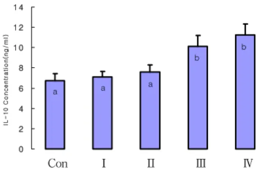
0 2 4 6 8 10 12 14
IL-10 Concentration(ng/ml)
a a a
b b
Con Ⅰ Ⅱ Ⅲ Ⅳ
Fig. 4. Effect of Coptidis Rhizoma on IL-10 concentration in lipopolysaccharide induced Raw 264.7 macrophages
a, b, c : Means with different superscripts are significantly different (p< 0.05).
Con : Control.
Ⅰ : LPS (1 μg/ml). Ⅱ : LPS (1 μg/ml)+10 μg/ml Coptidis Rhizoma.
Ⅲ : LPS (1 μg/ml)+30 μg/ml Coptidis Rhizoma.
Ⅳ : LPS (1 μg/ml)+100 μg/ml Coptidis Rhizoma.
IL-6 및 TNF-α의 농도들은 대조군인 정상군보다 LPS 처리군들 모두가 높은 수치를 나타내었다. 그러나 LPS 처리군들 간에서는 황련 처리군들 모두가 낮은 값을 나 타내었으며, 황련 첨가량이 증가함에 따라 감소하는 경향 을 보였다. IL-10의 농도는 정상군보다 LPS 처리군 모두 가 높은 경향을 보였으나, LPS 단일처리군 및 10 μg/ml 황련 추출물 처리군은 정상군과 유의한 차이를 나타내지 는 않았다. 그러나 30 μg/ml 및 100 μg/ml 황련 처리군 들은 이들 3개 처리군보다 유의하게 높은 수치를 나타내 었다. 이와 같은 결과들은 in vivo 실험에서 나타난 결과 들과 잘 부합된다. 즉 황련 추출물 처리군들에서 IL-β, IL-6 및 TNF-α의 농도가 하락하고, IL-10농도가 증가하 여 세포배양 실험에서도 황련이 염증반응을 완화시켰음 을 시사하고 있다(Fig 1~4).
결 론
본 연구는 새로운 항염증제를 개발하기 위한 기초연구 의 일환으로, 황련 추출물을 투여한 흰쥐에게 LPS 투여 로 급성기 염증반응을 유발시킨 후, 혈액 및 간장의 전염 증성 cytokines 농도의 시간대별 변동을 조사하였으며, 다른 한편으로는 황련 추출물을 처리한 Raw 264.7 cell에 LPS를 처리한 후, 처리군별 전염증성 cytokines들의 생 산량을 조사하여, in vivo 및 in vitro에서 황련 추출물 의 항염증 효과를 검토하였다.
그 결과 Plasma IL-1β, IL-6 및 TNF-α 농도는 LPS 처리 후, 2시간에서 급격히 상승하여, 5시간에서도 계속 적으로 증가하거나 그 상태를 유지하는 경향을 나타내었 다. LPS 처리 5시간 후 이들 3개 cytokines의 농도는 황 련 처리군들이 대조군보다 낮은 경향을 보였으며, 시간의 경과에 따른 증가량도 황련 처리군들이 대조군보다 낮았 다. Plasma IL-10 농도는 LPS 처리 후, 2시간에 증가하
여 5시간에서도 증가하는 경향을 보였고, 각 처리군 별 변동경향은 LPS 처리 후, 2시간 및 5시간에서 모두 황련 추출물 첨가군들이 대조군보다 높은 경향을 나타내었으 며, 시간의 경과에 따른 증가량도 황련추출물 첨가군들이 대조군보다 높은 값을 나타내었다.
LPS 처리 5시간 후, 간장에서의 IL-1β 및 IL-6의 농 도는 황련추출물 처리군들이 대조군보다 낮은 값을 나타 내었으며, 황련 처리군들 간에서는 유의한 차이를 나타내 지 않았으며, 간장 TNF-α농도 및 IL-10의 농도는 대조 군과 황련추출물 처리군들 간에 유의한 차이를 나타내지 않았다.
Raw 264.7 cell의 세포배양시험에서 IL-1β, IL-6 및 TNF-α의 농도는 대조군인 정상군보다 LPS 처리군들 모 두가 높은 수치를 나타내었다. 그러나 LPS 처리군들 간 에서는 황련처리군들 모두가 낮은 값을 나타내었으며, 황 련 첨가량이 증가함에 따라 감소하는 경향을 보였다. IL-10 의 농도는 정상군보다 LPS 처리군 모두가 높은 경향을 보였으며, LPS 처리군들 간에서는 LPS 단일처리군보다 황련 처리군들 모두가 높은 경향을 나타내었다.
이상의 결과들을 종합해보면, 황련에는 항염증에 관여 하는 기능성 물질들이 내재하고 있음을 시사해 준다.
참고문헌
1. Rabson A, IM Roitt, PJ Delves. Really Essential Medical Immunology. Oxford : Blackwell publishing Ltd. 2005 ; 1-14.
2. Chao CY, SL Yeh, MT Lin, WJ Chen. Effects of parenteral infusion with fish-oil or safflower-oil emulsion on hepatic lipids, plasma amino acids, and inflammatory mediators in septic rats. Nutrition.
2000 ; 16 : 284-8.
3. Boumpas DT, GP Chrousos, RL Wilder. Glucocorticoid therapy for immune mediated disease: Casic and clinical correlates. Ann Intern Med. 1993 ; 119 : 1198-208.
4. Barton CC, EX barton, PE Ganey, SL Kunkel, RA Roth. Bacterial lipopolysaccharide enhances aflatoxin B1 hepatotoxicity in rats by a mechanism that depends on tumor necrosis factor-α. Hepatology.
2001 ; 33 : 66-73.
5. Surh YJ. Anti-tumor promoting potential of selected spice ingredients with antioxidative and anti-inflammatory activities: a short review. Food Chem Toxicol. 2002 : 40(8) : 1091-7.
6. Seo WG, Pae HO, Oh GS, KY Chai, Kwon TO, Yun YG, Kim NY, Chung HT. Inhibitory rotundus rhizomes on nitric oxide and superoxide production by murine macrophage cell line, Raw 264.7 cells. J
Ethnopharmacol. 2001 ; 76 : 59-64.
7. 김태희, 양기숙, 황은진, 박성배. 마황의 면역작용에 미치는 영향. 생약학회지. 1991 ; 22(3) : 183-91.
8. Lee E. Anti-inflammatory effect of Scutellariae Radix. Korean J. Plant Res. 2007 ; 20(6) : 548-22.
9. 남정연, 이현선, 이승웅, 정미연, 최정호, 유은숙, 노문철, 김영국. 감국에서 분리한 Kikkanol F Monoacetate와 5-Hydroxy-6,7,3’,4’-tetramethoxyflavone의 IL-6생성억 제활성. 생약학회지. 2005 ; 36(3) : 186-90.
10. 김선재, 박윤미, 정순택. 감국추출물의 항충치효과와 Glucosyltransferase 저해활성탐색. Korean food culture.
2005 ; 20(3) : 341-5.
11. 고인자, 유승조, 이은방. 한국산 하고초류의 약물학 적 연구(1). 생약학회지. 1986 ; 17(3) : 232-41.
12. Lee DU, Kang YJ, Park MK, Lee YS, Seo HG, Kim TS, Kim CH, Chang KC. Effects of 13-alkyl- substituted berberine alkaloids on the expression of COX-II, TNF-alpha, iNOS, and IL-12 production in LPS-stimulated macrophages. Life Science. 2003 ; 73 : 1401-12.
13. Kim EK, Kwon KB, Han MJ, Song MY, Lee JH, Lv N, Ka SO, Yeom SR, Kwon YD, Ryu DG, Kim KS, Park JW, Park R, Park BH. Coptidis rhizoma extract protects against cytokine-induced death of pancreatic beta-cells through suppression of NK- kappaB activation. Exp Mol Med. 2007 ; 39 : 149-59.
14. Enk R, Ehehalt R, Graham JE, Bierhaus A, Remppis A, Greten HJ. Differential effect of Rhizoma coptidis and its main alkaloid compound berberine on TNF-alpha induced NFkappaB translocation in human keratinocytes. J Ethnopharmacol. 2007 ; 109 : 170-5.
15. Wang X, Xing D, Wang W, Su H, Tao J, Du L.
Pharmacokinetics of berberine in rat thalamus after intravenous administration of Coptidis rhizoma extract. Am J Chin Med. 2005 ; 33 : 935-43.
16. Marriot JB, M Westby, S Cookson, M Guckian, S Goodbourn, G Muller. CC-3052: a water-soluble analog of thalidomide and potent inhibitor of activation-induced TNF-α production. J Immunol.
1998 ; 161 : 4236-43.
17. Mathiak G, G Grass, T Herzmann, T Luebke, C Cu-Zetina, SA Boehm. Capase-1-inhibitor ac-YVAD- cmk reduces LPS-lethality in rats without affecting haematology or cytokine responses. Br J Pharmacol.
2000 ; 131 : 383-6.
18. Eduard FM, Martha SMR, Victor PA, Pablo M.
Immunomodulatory effects of thalidomide analogs on LPS-induced plasma and hepatic cytokines in the rat. Biochemical pharmacology. 2004 ; 68 :
1321-9.
19. Aono K, K Isobe, K Kuichi, Z Fan, M Ito, A Takeuchi. In vitro and in vivo expression of inducible nitric oxide synthase during experimental endotoxemia: involvement of other cytokines. J cell Biochem. 1997 ; 65 : 349-58.
20. Corral LG, GW Muller, AL Moreira, X Chen, M Wu, D Stirling. Selection of novel analogs of thalidomide with enhanced tumor necrosis factor-α inhibitory activity. Mol med. 1996 ; 25 : 964-9.
21. Sang H, GL Wallis, CA Stewart, K Yashige.
Expression of cytokines and activation of transcription factors in lipopolysaccharide-administered rats and their inhibition by phenyl N-tert-butylnitrone (PBN).
Arch Biochem Biophys. 1999 ; 363 : 341-8.
22. Harry D, R Anand, S Holt, S Davies, R Marley, B Fernando. Increased sensitivity to endotoxemia in the bile duct-ligated cirrhotic rat. Hepatology. 1999 ; 30 : 1198-205.
23. Chamulitrat W, ME Blazka, SJ Jordan, MI Luster, RP Mason. Tumor necrosis factor-α and nitric oxide production in endotoxin-primed rats administered carbon tetrachloride. Life Sci. 1995 ; 24 : 2273-80.
24. Harbrecht BG, M DiSilvio, AJ Demetris, RL Simmons, TR Billiar. Tumor necrosis factor-α regulates in vivo nitric oxide synthesis and induces liver injury during endotoxemia. Hepatology.
1994 ; 20 : 1055-60.
25. Hamada E, T Nishida, Y Uchiyama, J Nakamura, K Isihara, H Kazuo. Activation of Kupffer cells and caspases-3 involved in rat hepatocyte apoptosis induced by endotoxin. J Hepatol. 1999 ; 30 : 807-18.
26. Thompson KC, A Trowern, A Fowell, M Marathe, C Haycock, MJP Arthur et al. Primary rat and mouse hepatic stellate cells express the macrophage inhibitor cytokine interleukin-10 during the course of activation in vitro. Hepatology. 1998 ; 28 : 1518-24.
27. Moreira AL, EP Sampaio, A Zmuidzinas, P Frindt, KA Smith, G Kaplan. Thalidomide exerts its inhibitory action on tumor necrosis factor alpha by enhancing mRNA degradation. J Exp Med. 1993 ; 177 : 1675-80.
28. Simpson KJ, Lukacs NW, Colletti L, Strieter RM, Kunkel SL. Cytokines and the liver. J Hepatol.
1997 ; 27 : 1120-32.
29. Klapproth J, Geiger T, Andus T, Heirich PC. Fate and biological action of human recombinant interleukin 1β in the rat in vivo. Eur J Immunol.
1980 ; 19 : 1485-90.
30. Louis H, LeMoine O, Peny MO, Quertinmont E, Fokan D, Goldman M et al. Production and role of interleukin-10 in concanavalin A-induced hepatitis in mice. Hepatology. 1997 ; 25 : 1382-9.