Isolated Activation of the Upper Trapezius in Three Manual Muscle Testing Position : Convergence Study
Sungmin Ha
Professor, Department of Physical Therapy, Sangji University
세 가지 도수근력 검사 자세에서 상부 승모근의 독립적 수축 : 융합적 연구
상지대학교 물리치료학과 교수하성민
Abstract The purpose of this study was to investigate the optimal position among three manual muscle tested positions for upper trapezius in which to obtain an isolated upper trapezius EMG signal for the normalization of upper trapezius muscle EMG activity. A total of 28 healthy adult men participated in the experiment. The UT (upper trapezius) and LS (levator scapulae) muscle activities were measured using the TeleMyo 2400T and analyzed using MyoResearch software. The muscle activity of the US and LS was measured by performing three manual muscle test positions for the upper trapezius. The UT/LS ratio during the S-MVIC was 80.25 and was significantly higher than that during the T-MVIC (76.50; p
= 0.011) and the C-MVIC (60.95; p < 0.001). And, the UT/LS ratio during the T-MVIC and was significantly higher than that during the C-MVIC (p < 0.001). Based on the results of present study, we suggest a switch from T-MVIC to S-MVIC for the independent normalization reliability of upper trapezius EMG activity. The UT muscle strength or normalization test using S-MVIC will be able to measure muscle strength or activity of UT compared to previous measurement methods.
Key Words : EMG, Levator scapulae, Manual muscle test, Normalization, Upper trapezius
요 약 본 연구의 목적은 상부 승모근 근활성도에 대한 정규화를 하는 동안, 어떠한 도수 근력 검사 자세가 상부 승모근 의 선택적 활성화에 최적화된 자세인지를 알아보고자 하였다. 28명의 성인 남.녀가 본 연구에 참여하였다. 상부 승모근 과 견갑거근의 근활성도를 측정하기 위해 근전도를 이용하였다. 세 가지 도수근력 검사 자세에서 견갑거근에 대한 상부 승모근의 활성비를 측정하였다. 다른 측정 자세와 비교하여 S-MVIC 자세에서 유의하게 높은 상부 승모근/견갑거근 근활성비(S-MVIC: 80.25, T-MVIC: 76.50, C-MVIC: 60.95)를 보였다. 그리고 C-MVIC와 비교하여 T-MVIC가 유 의하게 높은 상부 승모근/견갑거근 근활성비를 보였다. 실험 결과를 토대로 기존의 T-MVIC 검사 자세에서 S-MVIC 로 상부 승모근 근전도 정규화 검사 자세를 바꾸는 것이 상부 승모근의 선택적 근활성도 수집의 정규화 신뢰도를 높이 는데 도움이 될 것이다. 이전 측정방법과 비교하여 S-MVIC를 이용한 상부 승모근 근력 또는 근활성도 측정방법은 상부 승모근의 근력 또는 근활성도의 독립적 측정이 가능하다.
주제어 : 근전도, 견갑거근, 도수근력측정 검사, 상부 승모근, 정규화
*This work was supported by Sangji University Research fund, 2018
*Corresponding Author : Sungmin Ha([email protected]) Received October 28, 2019
Accepted January 20, 2020 Revised December 5, 2019
Published January 28, 2020
1. INTRODUCTION
In order to compare the results obtained from the study using EMG with the difference in muscle usage between individuals and exercise methods, the normalization process is necessary[1]. Quantitative methods including maximum voluntary isometric contraction (MVIC), sub-maximal voluntary contraction (SVIC), and reference voluntary isometric contraction (RVC) is commonly used[2].
According to the characteristics of the studies, many literatures use specific position or methods for the characteristics of muscle to compare muscle usage patterns and exercise methods using EMG have been developed[3-5].
Which normalization method to choose is a very important factor and the muscles to be measured must be selectively or independently activated about the surrounding muscles[6,7]. It is very important to find a measurement posture that minimizes the effect of the surrounding muscle contraction[8,9]. For upper trapezius (UT) traditional normalization using MVIC, it is mainly measured through shoulder shrugging in sitting or standing[2,10]. However, this methods can lead over-activation of the levator scapulae (LS). If the UT is weak, excessive or compensatory activity of the LS makes accurate measurements difficult for measurement of traditional UT MVIC[11].
In this study, it study was to determine the optimal position among three tested positions in which to obtain an isolated UT EMG signal for the normalization of UT muscle EMG activity. In addition, this study suggests a quantitative measurement posture that can be a new alternative (selective or independent activation of the UT muscle) by comparing with the traditional method of measuring the UT muscle activities quantification.
2. METHODS
2.1 Participants
A total of 28 healthy adult men participated in the experiment. All 28 subjects were right-handed. The general characteristics of the subjects are presented in Table 1. Participants in the study had healthy shoulders and completed self-report to determine if there was an orthopedic or neurological disorder in the shoulder. And, participants were informed of the purpose of our study and its possible hazards. All subjects confirmed their consent to participate by completing a research consent form.
Participants were excluded from this study if 1) a dislocated or traumatic history of the shoulder complex, or 2) there was a history of shoulder surgery within the previous 6 months.
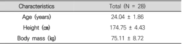
Table 1. Subject characteristics
Characteristics Total (N = 28)
Age (years) 24.04 ± 1.86
Height (㎝) 174.75 ± 4.43
Body mass (㎏) 75.11 ± 8.72
2.2 Instrumentation
The UT and LS muscle activities were measured using the TeleMyo 2400T (Noraxon USA, Inc., Scottsdale, AZ) and analyzed using MyoResearch software (XP Master Edition 1.07; Noraxon USA, Inc.). Before attaching EMG electrodes, the skin was shaved and gently rubbed with sandpaper to reduce skin impedance. To measure UT muscle activity, one electrode was placed at the upper crest of the shoulder, halfway between the C7 spinous process and the acromion[12]. Another surface electrode for LS muscle activity was placed between the posterior margin of the sternocleidomastoid muscle and the anterior margin of the UT[13,14]. The average value of the middle 3 seconds of the 5-second period was used for data analysis. For data analysis, the EMG signals were amplified and the sampling rate was 1000 Hz. A bandpass filter between 20 and 450
Hz was used and a notch filter at 60 Hz was applied. EMG data were processed into the root-mean-square (RMS) value, which was calculated from 50-ms data points of windows.
To calculate the UT/LS ratio, the UT amplitude was divided by the LS amplitude.
2.3 Experimental procedure
All subjects performed three test positions. The muscle activity of the US and LS was measured by performing three motions. The US and LS muscle activities of all the motions were measured in a sitting position so that rotation and bending of the trunk or lower extremity did not occur compensatively. During the three motions, we used a metronome to represent the sound signal. All subjects were instructed to practice for 5 minutes for each motion to familiarize themselves with the motions. Each motion was repeated three times. Subjects maintained each trial for 5 seconds. One minute of rest was given between repetitions, and 5 minutes of rest was given between three motions to prevent muscle fatigue. The three motions were performed randomly for each subject, so that the order effect did not occur.
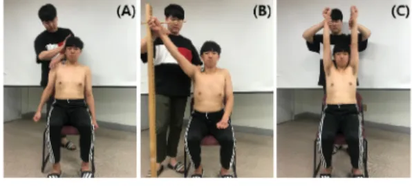
In the traditional MVIC (T-MVIC), subjects were sitting in a chair with the shoulder neutral position. The examiner placed the hand on the acromion of the subject’s shoulder and prepared to resist. The subjects performed the movement of elevating the acromion and the ends of the clavicle with the maximum force, and the examiner applied resistance inferiorly. At this time, the examiner fixed the subject's head by using the opposite hand to prevent excessive bending of the subject's neck in Fig. 1[10].
As an alternative method performed on the scaption plane MVIC (S-MVIC), subjects were sitting in chair with shoulder neutral position and the palms facing the body and thumbs up.
The subjects were instructed to raise their arms in the scapular plane about 30 degrees forward
from the frontal plane. The arm of the subject was able to follow the target bar to keep the scapular plane constant while raising the arm. An additional target bar was used to maintain the arm raising angle at 140 degrees, and the scaption motion was performed with maximum force at this point in Fig. 1.
Another alternative method performed on the coronal plane MVIC (C-MVIC), subjects were sitting in chair with shoulder neutral position and the palms facing the forward. The subjects were instructed to raise their arms in the frontal plane up to 180 degrees. Also, the opposite arm was raised to 180 degrees so that the body or neck do not bending in the opposite direction.
The subject contracted with maximum force to gather both hands, and the examiner fixed the opposite arm to prevent movement and gave resistance to the wrist region of the arm to be measured in Fig. 1.
Fig. 1. Test positions
(A) T-MVIC, (B) S-MVIC, (C) C-MVIC
2.4 Statistical analysis
Variables were normally distributed, as determined by Kolmogorov-Smirnov tests; thus, parametric statistics were used. Data of this study were expressed as mean ± standard deviation.
Statistical analyses were performed using a SPSS 25.0 (SPSS Inc., Chicago, IL, USA). The EMG differences among three test positions were tested for statistical significance (p = .05) using a one-way repeated-measures analysis of variance (ANOVA) for each muscle. When we found a significant difference, we used post-hoc paired
Table 2. Ratio of upper trapezius to levator scapulae muscle activity for each test position Maximal Voluntary Isometric Contraction (Mean±SD)
Ratio T-MVIC S-MVIC C-MVIC F P
UT/LS 76.50 11.88 80.25 9.67 60.95 16.96 24.27 <0.001
Abbreviations: UT, upper trapezius; LS, levator scapulae
Table 3. Post hoc paired t-test analysis between test position
Ratio Test position comparison Mean difference P
UT/LS
T-MVIC versus S-MVIC 3.75 - 7.30 = 0.011
S-MVIC versus C-MVIC 19.30 - 17.70 < 0.001
C-MVIC versus T-MVIC 15.55 - 18.92 < 0.001
Abbreviations: UT, upper trapezius; LS, levator scapulae
t-tests to determine which values were significantly different. And, a Bonferroni adjustment with the α level set at 0.016 (.05/3) was used for post-hoc t tests.
3. RESULTS
The ratios of UT to LS muscle activity (UT/LS ratio) during three positions are shown in Table 1. The UT/LS ratio during the S-MVIC was 80.25 and was significantly higher than that during the T-MVIC (76.50; p = 0.011) and the C-MVIC (60.95; p < 0.001). And, the UT/LS ratio during the T-MVIC and was significantly higher than that during the C-MVIC (p < 0.001) in Table 2.
4. DISCUSSION and CONCLUSION
Previous reported that compensatory movement or muscle activation in the surrounding muscles leads to decreasing reliability of the normalization of the UT muscle during EMG normalization or measuring UT muscle strength. In particular, it is necessary to find out how to independently measure the contraction of the UT muscle while minimizing the compensatory activity of surrounding muscles. The purpose of this stud was to investigate the optimal position among three
tested positions in which to obtain an independent UT muscle activity
Our study showed that the UT/LS EMG ratio was significantly higher in the S-MVIC position than in all other test positions, following the order S-MVIC > T-MVIC > C-MVIC. There are several possible explanations for our results. For the independent UT/LS EMG ratio, high UT activity and low LT muscle activity are required.
First, compared to other methods, the S-MVIC method can maximize the activation of UT muscles[15]. Active force length tension curve is described by a racheting filament model and has its maximum near the muscle's normal rest or mid-range length. At this length is the most interaction between the actin and myosin filaments, accounting for the largest active force production. At shorter or longer lengths the filaments have less overlap to form cross-bridges;
they therefore produce less force[16]. T-MVIC and C-MVIC are measured at the maximum and minimum muscle length of the UT, respectively.
Therefore, UT muscle activity is lower than that of S-MVIC measured in the middle range.
Second, the LS muscle as a scapular retractor, elevator, and downward rotator based on anatomy[10]. Therefore, the greater the angle of scapular upward rotation or scapular abduction, the lower the muscle activity of the LS. Previous study reported that maximum range of LS EMG is in the range of shoulder abduction 0-90 degrees.
In the case of S-MVIC and C-MVIC, the UT / LS ratio was high as the muscle activity of LS was lowered because it was performed in the range of 140-180 shoulder abduction[17]. Minimizing excessive activity of surrounding muscles is very important for independent UT muscle activity[7,18]. Based on the results of our study, we suggest a switch from T-MVIC to S-MVIC for the independent normalization reliability of UT EMG activity.
This study has some limitations. First, the generalization of the results of this study is limited because subjects were young; further studies should determine whether our findings can be generalized to subjects beyond this age range. We also did not measure force or torque for UT muscle during normalization procedure.
Further study is need to examine force or torque for UT muscle.
5. CONCLUSION
The purpose of this study was to investigate the optimal position among three manual muscle tested positions for upper trapezius in which to obtain an isolated upper trapezius EMG signal for the normalization of upper trapezius muscle EMG activity. Based on the results of present study, we suggest a switch from T-MVIC to S-MVIC for the independent normalization reliability of UT EMG activity.
REFERENCES
[1] G. A. Mirka. (1991). The quantification of EMG normalization error. Ergonomics, 34, 343-352.
DOI:10.1080/00140139108967318
[2] S. Bao, S. E. Mathiassen & J. Winkel. (1995).
Normalizing upper trapezius EMG amplitude:
Comparison of different procedures. Journal of Electromyogrphy & Kinesiology, 5(4), 251-257.
[3] S. M. Ha, H. S. Cynn, O. Y. Kwon, K. N. Park & G. M.
Kim. (2013). A reliability of electromyographic normalization methods for the infraspinatus muscle in healthy subjects. Journal of Human Kinetics. 28(36), 69-76.
DOI: 10.2478/hukin-2013-0007
[4] J. Sinclair, P. J. Taylor, J. Hebron, D. Brooks, H. T.
Hurst & S. Atkins. (2015). The Reliability of Electromyographic Normalization Methods for Cycling Analyses. Journal of Human Kinetics, 10(46), 19-27.
DOI: 10.1515/hukin-2015-0030
[5] K. J. Netto & A. F. Burnett. (2006). Reliability of normalisation methods for EMG analysis of neck muscles. Work, 26(2), 123-130.
[6] I. Jeon, S. M. Ha & S. H. Jung. (2018). Isolated Activation of the Infraspinatus Muscle in Four Manual Muscle Testing Positions. Journal of Musculoskeletal Science Technology, 2(2), 38-42.
DOI: 10.29273/jmst.2018.2.2.38
[7] T. Alenabi, R. Whittaker, S. Y. Kim & C. R. Dickerson.
(2018). Maximal voluntary isometric contraction tests for normalizing electromyographic data from different regions of supraspinatus and infraspinatus muscles: Identifying reliable combinations. Journal of Electromyogrphy & Kinesiology, 41, 19-26.
DOI: 10.1016/j.jelekin.2018.04.007
[8] M. H. Kang, J. S. Oh & J. H. Jang. (2014). Differences in muscle activities of the infraspinatus and posterior deltoid during shoulder external rotation in open kinetic chain and closed kinetic chain exercises.
Journal of Physical Therapy Science, 26(6), 895-897.
[9] J. W. Kim, J. Y. Yoon, M. H. Kang & J. S. Oh. (2012).
Selective activation of the infraspinatus during various shoulder external rotation exercises. Journal of Physical Therapy Science, 24(7), 581-584.
[10] F. P. Kendall & E. K. McCreary. (2005). Muscles:
Testing and Function. 5th ed. Baltimore, MD : Williams & Wilkins.
[11] S. A. Sharmann. (2002). Diagnosis and treatment of movement impairment syndromes. St. Louis : Mosby.
[12] J. R. Cram, S. G. S. Kasman & J. Holtz. (1998).
Introduction to surface electromyography. Maryland:
Aspen Publishers.
[13] K. Schüldt. (1988). On neck muscle activity and load reduction in sitting postures. An electromyographic and biomechanical study with applications in ergonomics and rehabilitation. Scandinavian journal of rehabilitation medicine. 19, 1-49.
[14] G. Sundelin & M. Hagberg. (1989). The effects of different pause types on neck and shoulder EMG activity during VDU work. Ergonomics, 32(5), 527-537.
DOI: 10.1080/00140138908966123
[15] D. H. Hardwick, J. A. Beebe, M. K. McDonnell & C. E.
Lang. (2006). A comparison of serratus anterior muscle activation during a wall slide exercise and other traditional exercises. Journal of Orthopaedic &
Sports Physical Therapy, 36(12), 903-910.
DOI: 10.2519/jospt.2006.2306
[16] K. T. Patton & G. A. Thibodeau. (2016). Anatomy &
Physiology (9th ed.), Elsevier.
[17] D. J. Eliot. (1996). Electromyography of levator scapulae: new findings allow tests of a head stabilization model. Journal of Manipulative Physiol Ther, 19(1), 19-25.
[18] H. A. Kim, U. J. Hwang, S. H. Jung, S. H. Ahn, J. H. Kim
& O. Y. Kwon. (2017). Effect of horizontal adduction force on infraspinatus and deltoid activities during the side-lying wiper exercise using pressure biofeedback. Journal of Human kinetics, 24(4), 77-83.
DOI: 10.2478/hukin-2014-0113
하 성 민(Sungmin Ha) [정회원]
․ 2004년 2월 : 연세대학교 재활학과(보 건학사)
․ 2009년 2월 : 연세대학교 일반대학원 재활학과 (이학석사)
․ 2012년 8월 : 연세대학교 일반대학원 물리치료학과 (이학박사)
․ 2014년 4월 ~ 현재 : 상지대학교 물 리치료학과 교수
․ 관심분야 : 근골격계 물리치료학
․ E-Mail : [email protected]