Article Info
Received October 8, 2020 Revised October 26, 2020 Accepted October 27, 2020 Corresponding Author Oh-yun Kwon
E-mail: [email protected] https://orcid.org/0000-0002-9699-768X
Key Words Head Neck Posture
Background: Forward head posture (FHP) is common postural malalignment. FHP is de- scribed relatively extension to upper cervical and lower cervical is relatively flexion. Although several researchers mentioned the lower cervical flexion posture in FHP, most of the studies related to FHP is focused on the deep cervical flexor function.
Objects: The purposes of present study is to compare the cervical strength (upper cervical extension [UCE], lower cervical extension [LCE], upper cervical flexion [UCF], lower cervical flexion [LCF]) between individuals with and without FHP.
Methods: Fifty-one participants are recruited. Participants who have the craniovertebral angle (CVA) less than 48 degree were classified to the FHP group (n = 24) and the others were included in without FHP group (n = 27). The cervical strength (UCE, LCE, UCF, LCF) were measured using Smart KEMA strength sensor and the strength data was normalized by body weight. All strength measurement conducted at head and neck neutral position in sitting.
Independent t-test was used to compare the cervical strength between individuals with and without FHP.
Results: The mean value of CVA was greater in without FHP group than with FHP group (p
< 0.000). The strength value of UCF (p < 0.002) and LCE (p < 0.001) was significant less in FHP group than without FHP group. But no significant differences were seen in the LCF and UCE strength between two groups.
Conclusion: UCF and LCE weakness in FHP group should be considered to evaluate and manage the individuals with FHP.
Copyright ⓒ Korean Research Society of Physical Therapy
This is an Open Access article distributed under the terms of the Creative Commons Attribution Non-Commercial License (http://creativecommons.org/licenses/by-nc/4.0) which permits unrestricted non-commercial use, distribution, and reproduction in any medium, provided the original work is properly cited.
INTRODUCTION
Forward head posture (FHP) is caused by improper work- ing posture with a computer, potentially leading to changes in head and neck alignment [1]. In the sagittal plane, it has been defined when the craniovertebral angle (CVA) is less than 50° [2]. FHP is associated with neck pain, temporomandibular disorder, and headache and imposes an abnormal load on the cervical muscles [1,3].
FHP causes muscle imbalance in the cervical spine. Increase in FHP is associated with weakness and lengthening of the up- per cervical flexor (UCF) muscles, such as the longus colli and longus capitis, and lower cervical extensor (LCE) muscles, such as the semispinalis cervicis, multifidus, and splenius cerivicis
[4]. In addition, FHP is associated with shortening of the up- per cervical extensor (UCE) muscles, including the semispinalis capitis, rectus capitis, and lower cervical flexor (LCF) muscle, such as the sternocleidomastoid [5-7].
It is important to achieve a balance between the strength of the cervical flexors and extensors for managing cervical disorders with FHP [1]. Cagnie et al. [8] reported that cervical extensor strength was significantly decreased in individuals with neck pain. Bokaee et al. [1] suggested that the ratio of isometric strength between the UCF and UCE may be a good indicator for evaluating individuals with FHP. Although several researchers have reported flexed posture of lower cervical in FHP [7,9], the research on LCE strength in individuals with and without FHP, is insufficient.
Physical Therapy Korea
PTK https://doi.org/10.12674/ptk.2020.27.4.272 pISSN: 1225-8962 eISSN: 2287-982X Phys Ther Korea. 2020;27(4):272-277
Original Article
Comparison of Upper and Lower Cervical Muscle Strengths Between Subjects With and Without Forward Head Posture
Ji-yeon Eun
1, PT, BPT, Oh-yun Kwon
2, PT, PhD, Ui-jae Hwang
1,2, PT, PhD, Sung-hoon Jung
1,2, PT, PhD, Sun-hee Ahn
1,2, PT, PhD
1
Department of Physical Therapy, Graduate School, Yonsei University,
2Department of Physical Therapy, College of Health Science,
Laboratory of Kinetic Ergocise Based on Movement Analysis, Yonsei University, Wonju, Korea
Nojiri et al. [10] found that the lower cervical region be- comes kyphotic as the lordosis of the upper cervical region in- creases. Therefore, the lower cervical region might be consid- ered as an element that determines the upright posture of the head with the upper cervical region. However, no study has in- vestigated the strength of the flexors and extensors of the up- per and lower cervical region in individuals with and without FHP. Therefore, the purpose of this study was to compare the strength of the flexor and extensor muscles of the upper and lower cervical regions between individuals with and without FHP. We hypothesized that the UCF and LCE strength would be lower in the FHP group than in the group without FHP.
MATERIALS AND METHODS
1. Subjects
The participants were recruited using a social network sys- tem. The healthy adults aged between 20 and 45 years were included, and were divided into two groups based on the CVA:
those with CVA less than 48° were included in the FHP group, while those with CVA greater than 48° were included in the without FHP group [11]. Individuals with any pathology in the neck region and those who were unable to perform the strength test in the present study, were excluded [12]. Fifty- one subjects (age = 32.6 ± 4.8 years, height = 168.8 ± 7.8 cm, weight = 68.9 ± 15.5 kg) were recruited in this study. Twenty- four subjects (male: 15, female: 9) were included in the FHP group and 27 subjects (male: 10, female: 17) were included in the without FHP group. All participants signed an informed consent form approved by the Yonsei University Mirae Institu- tional Review Board (approval number: 1041849-202002-BM- 022-02).
2. Procedure 1) CVA
A mobile phone camera (iPhone; Apple Inc., Cupertino, CA, USA) was used to capture a photograph to measure the CVA.
The camera was installed on the right side of the subjects at a distance of 2 meters [13]. Reflex markers were attached to the C7 spinous process and tragus of right ear, respectively.
The subjects were asked to stand comfortably, with their right side toward the camera. The height of the camera was set to the subject's shoulder height [14]. After capturing a photo, a line was drawn parallel to the floor at the C7 level and another
one through the tragus and C7 spinous process (Figure 1) [15].
Image J imaging software (US National Institutes of Health, Bethesda, MD, USA) was used to analyze the CVA [16].
2) Strength measurement
Isometric strength of the UCE, LCE, UCF, and LCF was mea- sured using a Smart KEMA strength sensor (KOREATECH Co., Ltd., Seoul, Korea). One side of the sensor was fixed to an im- mobile bar with a nonelastic belt, and the opposite side was hung on the subject. The force signals were transmitted to an Android tablet for analysis with the Smart KEMA software, at a sampling frequency of 10 Hz, via a Bluetooth connection. The intra-rater reliability of the strength sensor has been presented in previous studies [7,17].
The subjects sat upright in a chair with a backrest [5,18]. The pelvis and upper thorax were strapped to the back of the chair to stabilize the upper body while measuring the neck strength.
During all strength measurements, the virtual line through the forehead and chin was perpendicular to the floor. To avoid compensatory movement, the examiner gave a verbal cue to maintain the head and trunk in a neutral position. The adjust- able axis of upper cervical flexion and extension is the con- cha of the ear, and lower cervical flexion and extension is the transverse process of C7 [5,18]. The subjects were instructed to isometrically contract the muscles twice for 5 seconds at the maximum force and a 30-second rest was provided to avoid muscle fatigue. The application calculated the average value at middle 3 seconds, and the average of the two values was used
Figure 1.
Figure 1. Craniovertebral angle.
for data analysis. All strength data were normalized by body weight and presented in terms of percentage (% body weight).
The procedure is shown in Table 1.
3. Statistical Analysis
The Kolmogorov-Smirnov test was used to confirm the nor- mal distribution. Independent t-tests were used to analyze the differences in the CVA, UCE, LCE, UCF, and LCF between groups with and without FHP. The level of statistical signifi- cance was set at 0.05. To confirm the intra-rater test-retest reliability of the method of strength measurement, intra-class correlation coefficient (ICC) [3, 1] model and 95% confidence intervals (CIs) were used. Statistical analyses were conducted
using PASW statistics 18 (IBM Co., Armonk, NY, USA).
RESULTS
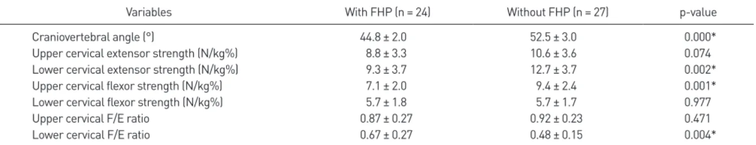
There were significant differences in the CVA between groups (p < 0.000). The strength values of UCE (p = 0.074) and LCF (p = 0.977) were not significantly different between the two groups. The strength values of LCE (p < 0.002) and UCF (p
< 0.001) were significantly lower in the FHP group than those in the without FHP group (Table 2). The ratio of flexor/exten- sor strength of upper cervical and lower cervical was shown in a Table 2.
In the present study, the intra-rater test-retest reliability of
Table 1.
Table 1. Measurement of strength in the UCE, LCE, UCF, and LCF
Measurement Figure Instruction
Upper cervical extensor The strap is placed on the subject’s chin, and the opposite end of the strap is fixed at the immobile bar located behind the subjects’ head. The strength sensor is placed between the subject and the immobile bar. The subject is indicated to rotate his/her chin superiorly against the resistance of the strap.
Lower cervical extensor The strap is placed behind the subject’s occipital bone and the opposite end of the strap is fixed on the immobile bar in front of the subject. The subject is indicated to move his/her head in a posterior translatory movement while maintaining the forehead and chin in neutral position.
Upper cervical flexor The strap is placed below the subject’s chin and the opposite end of the strap is fixed at the immobile bar located over the subject’s head at a height 1 m 50 cm. The strength sensor is placed between the subject and the immobile bar. The subject is indicated to rotate his/her chin inferiorly against the resistance of the strap.
Lower cervical flexor The strap is placed on the forehead of the subject and the opposite end of the strap is fixed at the immobile bar located behind the subject’s head. The subject is indicated to rotate his/her forehead in an antero-inferior direction.
UCE, upper cervical extensor; LCE, lower cervical extensor; UCF, upper cervical flexor; LCF, lower cervical flexor.
the Smart KEMA strength sensor was high (ICC > 0.85). The ICC values of the methods of strength measurement ranged from 0.97 to 0.99. The values (95% CI) were 0.98 (0.97 –0.99) for UCE, 0.99 (0.98–0.99) for LCE, 0.96 (0.93–0.98) for UCF, and 0.97 (0.95 –0.98) for LCF.
DISCUSSION
Abnormality of postures is considered to exist with muscle imbalance [19]. FHP is typically observed in patients with ab- normal posture of the head and neck [20]. The present study compared the difference in the strength of the flexors and ex- tensors of upper and lower cervical region between individuals with and without FHP. The results of the present study dem- onstrated weakness of the LCE and UCF in the group with FHP, suggesting that extensor muscles should be considered in the management of FHP.
FHP usually involves lengthened cervical flexors, including the longus colli and longus capitis, and is related to deep cervi- cal flexor weakness [7]. The longus capitis and longus colli are the most important muscles involved in the deep cervical flex- ion movement, and the deep cervical flexors are a part of the UCF anatomically [21]. Bokaee et al. [1] showed that the group with FHP had lower UCF strength than the group without FHP.
In addition, change in length of the posterior neck muscles may have an effect on UCF strength. FHP is usually associated with shortening of the suboccipital muscles [3]. Prolonged FHP displays hyperextension of the craniocervical junction [22].
Hyperextension of the craniocervical junction could limit the range of motion during craniocervical flexion, and the limited range of motion could prevent sufficient contraction of the UCF. Thus, the UCF may be weaker in the FHP group than in the without FHP group.
FHP has been described as the flexed position of the lower
cervical region [9]. Anatomically, the semispinalis cervicis and cervical multifidus are the LCE muscles that produce an exten- sor torque in the lower cervical region [4,5]. These muscles have a lengthened position in FHP. The load on the cervical muscles caused by head protrusion posture is larger in FHP than in the neutral head position [23], and the LCE produces an eccentric force during flexed posture of lower cervical.
Prolonged eccentric contraction could lead to loss of strength [24,25]. This strength loss could be the evidence of our result indicating LCE weakness in the FHP group.
There was no significant difference in the strength of LCF and UCE between the two groups. It is possible that the UCF influenced the measurement of the LCF strength while main- taining the neutral head position. Another possibility is that the flexion movement of the thorax was limited because the upper chest was fixed, and the LCF was not sufficiently mobi- lized for maximum effort. In the case of UCE, cervical protru- sion may have occurred as a compensatory movement. The force of the UCE and cervical protrusion may have combined, such that there was no difference in force between the two groups.
Bokaee et al. [1] compared the ratio of the upper cervi- cal flexor to extensor strength between individuals with and without FHP (1.26 vs. 1.55), and there was significant differ- ence (p < 0.008) between groups. In contrast, there was not significantly difference in the ratio of the upper cervical flexor to extensor strength in the present study. In our knowledge, there was little study for comparison of ratio of the upper and lower cervical flexor to extensor strength between individuals with and without FHP. The ratio of the upper cervical flexor to extensor strength could be affected by both the upper cervi- cal flexor and extensor strength. Although significantly differ- ence the ratio of the upper cervical flexor to extensor strength was not found between groups, the upper cervical flexor was Table 2.
Table 2. The mean values ± standard deviation of the craniovertebral angle and normalized isometric strength of the cervical muscles in the with FHP