A bucket-handle tear is a longitudinal, vertical or oblique tear of the meniscus with an attached fragment displaced away from the donor meniscus (1,2). In order
that surgical intervention is appropriate, it is important to visualize the displaced meniscal fragment as well as determine the type of meniscal injury(1,3).
Several magnetic resonance imaging (MRI) findings have been used for the diagnosis of bucket-handle tears:
(1) the double posterior cruciate ligament (PCL) sign, which indicates a meniscal fragment flipped anterior to the PCL, simulating two ligaments (1,2,4); (2) the flipped meniscus sign, which indicates a fragment flipped ante-
MRI of Bucket-Handle Te a rs of the Meniscus of the Knee
1Joon Yong Park, M.D., Young-uk Lee M.D., Eun-Chul Chung M.D., Hae-Won Park M.D., E u n - Kyung Youn M.D., Shin Ho Kook, M.D., Young Rae Lee, M.D.
Purpose: To determine the frequency of the MRI signs of meniscal bucke t - h a n d l e tears already known as the double PCL sign, the flipped meniscus sign, the absent b ow-tie sign, and the fragment-in-notch sign, and to compare the sagittal with the coronal images.
Materials and Methods : We retrospectively rev i ewed the MR findings of 37 patients in whom an initial interpretation of MR images had suggested meniscal bucke t - h a n d l e t e a r s. All underwent subsequent arthroscopic evaluation and in 28, bucke t - h a n d l e tears were confirmed. Sagittal double-echo and coronal fat-suppressed double-echo T 2 - weighted images were obtained. Sagittal images were evaluated to determine whether or not signs of bucket-handle tear were evident, and coronal images we r e c h e c ked for a torn meniscus with displaced fragment. We also evaluated the MR find- ings of the nine false positive cases.
Results : The prevalence rate of absent bow - t i e, double PCL, fragment-in-notch, and flipped meniscus signs was 96.4%, 53.6%, 17.9%, and 10.7%, respective l y. The detec- tion rate for displaced fragment was higher with coronal images (92.9%) than with sagittal images (78.6%). Among the nine false positive cases, a longitudinal tear in the discoid meniscus was most common. A false-positive diagnosis was much more fre- quent on sagittal than on coronal images.
Conclusion: The prevalence rate of absent bow-tie sign was very high, but was ac- companied by a relatively high rate of misinterpretation. Coronal fat-suppressed T2- weighted images provided more reliable clues for the diagnosis of bucket-handle tears, with a high detection rate of displaced fragment.
Index words :K n e e, MR
K n e e, ligaments, menisci, and cartilage
1Department of Radiology, Kangbuk Samsung Hospital, Sungkyunkwan University School of Medicine
Received May 3, 1999 ; Accepted July 23, 1999
Address reprint requests to : Young-Uk Lee, M.D., Department of Radiology, Kangbuk Samsung Hospital, Sungkyunkwan University School of Medicine,
#108 Pyeong-dong, Chongro-gu, Seoul 100-638, Korea
Tel. 82-2-2001-2342 Fax. 82-2-2001-2797 E-mail. [email protected]
riorly so the anterior horn appears large (5); (3) the ab- sent bow-tie sign, which means that only one slice or none of the body segment is seen on sagittal images (6);
and (4) the fragment-in-notch sign, representing the fragment which is located in the intercondylar notch and does not lie in the same sagittal plane as the PCL (1).
In the detection of bucket-handle tears, the sensitivity of MRI has varied. The purpose of this study was to de- termine the prevalence of the above MRI signs in the di- agnosis of bucket-handle meniscal tears and to compare their prevalence between sagittal and coronal images.
Materials and Methods
We retrospectively reviewed the MR findings of 37 patients, in whom an initial interpretation of MR find- ings had suggested bucket-handle tears. All underwent subsequent arthroscopic evaluation. In 28, bucket-han- dle tears were confirmed by arthroscopy. The remain- ing nine patients had tears which were not the bucket- handle type. Among the 28 patients with arthroscopical- ly-proven bucket-handle tear, 25 were male and three were female patients, and their ages ranged from 14 to 64 (average 33) years. Fifteen right and 13 left knees were involved and medial menisci (n=19) were torn more commonly than lateral menisci (n=9).
MR examination was performed using a 1.5T scanner (GE Medical Systems, Milwaukee, U.S.A.), and images were obtained in both the sagittal and coronal planes.
Sagittal dual-echo (TR/TE=2500/15,60) images and
coronal fat-suppressed dual-echo (TR/TE=2500/17,60) images were obtained with a 16cm FOV, 3mm slice thickness with a 1mm gap, 1NEX, and a 256X192 ma- t r i x .
For retrospective review, settings were different for each sagittal and coronal plane interpretation. The au- thors determined whether or not the following MRI findings were visible on sagittal images: the double PCL sign, the flipped meniscus sign, the absent bow-tie sign, and the fragment-in-notch sign (Fig. 1-3). Coronal im- ages were evaluated to determine whether they provid- ed information about bucket-handle tears, including a displaced meniscal fragment as well as a deficient or truncated torn meniscus. In nine cases in which find- ings of bucket-handle tear were false-positive, images obtained in both the sagittal and coronal planes were retrospectively evaluated.
R e s u l t s
The prevalence of each MRI sign is shown in Table 1.
The most sensitive finding was an absent bow-tie sign,
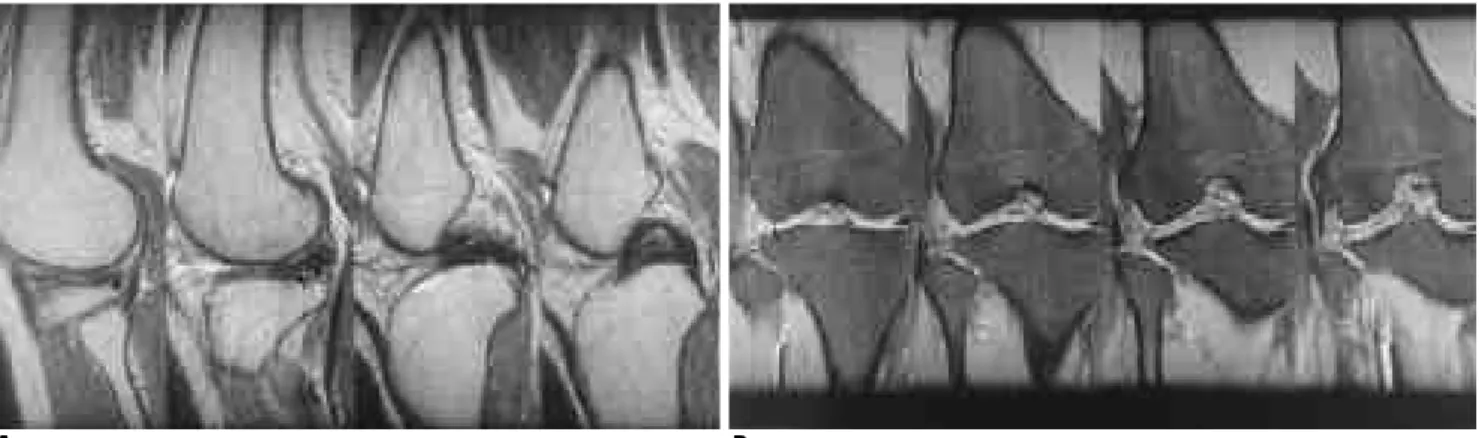
A B
Fig. 1. A bucket-handle tear of medial meniscus.
A . Far medial sagittal image (left) reveals absent bow-tie sign at body segment and more internal image (right) shows double PCL sign.
B . On serial coronal images, the torn donor meniscus (short arrow) is truncated and the displaced fragment (long arrow) is located in same orientation as PCL.
Table 1.Prevalence of MR Signs of Meniscal Bucket-Handle Tears on Sagittal Images (n=28)
MR Sign C a s e s Prevalence (%)
Absent bow-tie sign 2 7 9 6 . 4
Double PCL sign 1 5 5 3 . 6
Fragment in notch sign 5 1 7 . 9
Flipped meniscus sign 3 1 0 . 7
but this was also seen in other types of tear, including three which were longitudinal and one which was pe- ripheral (Fig. 4). The double PCL sign was found only in medial meniscal tears. Supposing that a bucket-handle tear with any sign can be diagnosed on sagittal images re- gardless of number or kind, the detection rate of bucket- handle tears as seen on sagittal images was higher (96.4%) than when visualised on coronal images (92.8%).
With regard to the detection of displaced fragments, these were clearly visualized on coronal images in 26 of 28 cases, and on sagittal images in 22 cases (Fig. 5).
The nine misinterpreted cases involved three longitu- dinal tears in discoid menisci, three radial tears, two longitudinal tears in non-discoid menisci, and one pe-
ripheral tear (Fig. 4). This misinterpretation was more common on sagittal images (9/9) than on coronal images ( 2 / 9 ) .
D i s c u s s i o n
A bucket-handle tear is the most frequent pattern of displaced meniscal injury, and results from a longitudi- nal tear (1,2). The medial meniscus is more frequently involved. In the detection of a bucket-handle tear by M- RI, sensitivity varied between 64% and 97% (1-6). The MRI findings of bucket-handle tears have been de- scribed. The double PCL sign (1,2,4), the flipped menis- cus sign (5), and the absent bow-tie sign (6) are mostly
A B
Fig. 2. A medial meniscal bucket-handle tear with flipped meniscus sign. A. On sagittal image, the posterior horn is hardly visible while anterior horn looks larger due to torn and anterioly flipped posterior horn. B. The anterior and posterior coronal images re- veal torn donor segment (short arrows) and anteriorly displaced fragment (long arrow).
A B
Fig. 3. Fragment-in-notch sign in bucket-handle tear of lateral meniscus.
A . The lateral meniscus is macerated with the displaced fragment (arrow) located in the intercondylar fossa on serial sagittal im- ages.
B. The torn fragment (arrows) of lateral meniscus is located in the notch, displacing the ACL medially and lying in different orien- tation from PCL.
seen on sagittal images, while the fragment-in-notch sign (1) and a truncated or small remnant meniscus can be seen in both the sagittal and coronal planes.
The diagnosis of bucket-handle tears through the i- dentification of displaced fragments is difficult. In spite of the involvement of a relatively long segment, tears
A B
Fig. 4. False positive case: Peripheral tear in lateral discoid meniscus.
A. The serial sagittal images show absent bow-tie sign, obliquely oriented tear line (arrows) and double PCL sign-like appearance.
B . On all serial coronal images, the lateral meniscus keeps the appearance of bow-tie, suggesting discoid meniscus, and shows pe- ripheral tear (arrows).
A B
C
Fig. 5. The far lateral sagittal images (A) reveal absent bow-tie sign suggesting tear of body segment. More medial sagittal im- ages (B), however, don’t disclose the fragment clearly. On coro- nal images (C), the donor segment (short arrow) of bucket-han- dle tear looks small and truncated. Note the displaced fragment (long arrow) just lateral to the midsubstance of the PCL.
may be overlooked because they run parallel to the sagittal plane (1,7.8).
In this study, the bow-tie sign was absent in 27 of 28 cases of bucket-handle tears. The application of the ab- sent bow-tie sign to the diagnosis of bucket-handle tears must be based on a knowledge of meniscal anatomy.
The average width of a normal meniscal body is 9-1 2 mm, and 4- or 5-mm thick sagittal images should reveal the body of the meniscus on two successive images (6.9). In most cases of bucket-handle tears with free edge deficit of the meniscal body, no body segment, or one, will be seen. This highly sensitive sign does not in- dicate a torn, displaced fragment, but implies a torn body segment, and this makes it non-specific for buck- et-handle tears.
As opposed to previous reports of high sensitivity of the double PCL sign for bucket-handle tears (2,4), our s- tudy revealed a sensitivity of 53.6%. This sign was sel- dom present in bucket-handle tears of the lateral menis- cus or with the fragment displaced more posteriorly than anterointernally and oriented differently from the PCL. Moreover, a non-bucket-handle tear developed in the discoid meniscus showed a positive result. The frag- ment-in-notch sign may also be positive in cases other than bucket-handle tears.
Many authors have emphasized the importance of coronal images in diagnosing bucket-handle tears (9,10).
Coronal imaging is to some extent superior because it can reveal both the bucket and the handle of the torn meniscus quite well. The torn donor segment is truncat- ed or smaller. In this study, the displaced fragment could in most cases be easily detected using double-e- cho, fat-suppressed T2-weighted coronal imaging.
In summary, the absence of a bow-tie sign on sagittal images was most prevalent in bucket-handle tears, though this was offset by a high rate of false-positive in- terpretation. The prevalence of other MR signs sugges- tive of a displaced fragment were low. Coronal fat-sup- pressed T2-weighted imaging provided a higher detec- tion rate of displaced meniscal fragment and a lower rate of misinterpretation. We therefore suggest that for the accurate diagnosis of bucket-handle tears, both coro- nal and sagittal images are evaluated.
R e f e r e n c e s
1. Wright DH, De Smet AA, Norris M. Bucket-handle tears of the medial and lateral menisci of the knee: value of MR imaging in de- tecting displaced fragments. A J R 1 9 9 5 ; 1 6 5 : 6 2 1 - 6 2 5
2 . Singson R, Feldman F, Staron R, Kiernan H. MR imaging of dis- placed bucket-handle tear of the medial meniscus. A J R 1 9 9 1 ; 1 5 6 : 1 2 1 - 1 2 4
3. Magee TH, Hinson GW. MRI of meniscal bucket-handle tears.
Skeletal Radiol1 9 9 8 ; 2 7 : 4 9 5 - 4 9 9
4. Weiss KL, Morehouse HT, Levy IM. Sagittal MR images of the knee: a low signal band parallel to the posterior cruciate ligament caused by a displaced bucket-handle tear. A J R 1 9 9 1 ; 1 5 6 : 1 1 7 - 1 1 9 5. Haramati N, Staron RB, Rubin S, Shreck EH, Feldman F, Kierman
H. The flipped meniscus sign. Skeletal Radiol 1 9 9 3 ; 2 2 : 2 7 3 - 2 7 7 6. Helms CA, Laorr A, Cannon WD Jr. The absent bow tie sign in buck-
e t -handle tears of the menisci in the knee. A J R 1 9 9 8 ; 1 7 0 : 5 7 - 6 1 7. De Smet A, Graf B. Meniscal tears missed on MR imaging: relation-
ship to meniscal tear patterns and anterior cruciate ligament tears.
A J R1 9 9 4 ; 1 6 2 : 9 0 5 - 9 1 1
8. Herman L, Beltran J. Pitfalls in MR imaging of the knee. R a d i o l o g y 1 9 8 8 ; 1 6 7 : 7 7 5 - 7 8 1
9. Silverman J, Mink , Deutsch A. Discoid menisci of the knee: MR imaging appearance. R a d i o l o g y 1 9 8 9 ; 1 7 3 : 3 5 1 - 3 5 4
1 0 . Mesgarzadeh M, Moyer R, Leder DS, et al. MR imaging of the knee:
expanded classification and pitfalls to interpretation of meniscal tears. R a d i o G r a p h i c s 1 9 9 3 ; 1 3 : 4 8 9 - 5 0 0
무릎관절에서 양동이 손잡이형 반월판 파열의 자기공명영상 소견1
1성균관대학교 의과대학 강북삼성병원 방사선과
박준용・이영욱・정은철・박해원・윤은경・국신호・이영래
목적 : 반월판의 양동이 손잡이형 열상을 시사하는 자기공명영상 소견인 이중 후십자인대 징후, 반월판 전치 징
후, 반월판 체부 결손 징후, 및 과와내 연골편 징후 등의 발현빈도를 알아보고, 시상면과 관상면 영상의 신뢰도 를 검증하고자 하였다.
대상 및 방법 : 자기공명영상에서 양동이 손잡이형 열상으로 추정되고 이어서 관절경 검사를 시행한 3 7예의 자기
공명영상을 후향적으로 관찰하였으며, 이 중 2 8예가 관절경 검사에서 양동이 손잡이형 파열로 진단되었다. 시상 면 이중에코 T 2강조영상과 관상면 이중에코 지방신호감쇄 T 2강조영상을 얻었다. 시상면 자기공명영상에서 상기 한 네 개 징후들의 존재유무를 판독하고 관상면 영상에서 반월판 파열과 전치된 조각이 보이는지를 관찰하였다.
또한 아홉개의 위양성 예들의 자기공명영상 소견도 관찰하였다.
결과 : 반월판 체부 결손, 이중 후십자인대, 과와내 연골편 및 반월판 전치 징후 등의 발현빈도는 각각 9 6 . 4 % ,
53.6%, 17.9%, 10.7% 이었고, 전위된 반월판 조각의 발견율은 관상면과 시상면이 각각 92.9%, 78.6%이었다. 위양 성 9예의 실제 파열 형태 중 가장 흔한 것은 원판형 반월판에 발생한 종열상의 파열이었고, 위양성의 진단적 오 류는 시상면에서 더 흔히 일어났다.
결론 : 양동이 손잡이형 파열의 진단에 있어 반월판 체부 결손의 발현빈도는 9 6 . 4 %로 매우 높게 나타났으나 위
양성 결과를 상대적으로 많이 초래하였다. 관상면의 지방신호감쇄 T 2강조영상은 전위된 반월판 파편을 찾는데 상당히 높은 발견율을 보여 양동이 손잡이형 반월판 열상 진단에 있어 신뢰성이 높은 검사방법으로 생각되었다.
대한방사선의학회지 1 9 99;41: 8 01- 8 0 6