갑상선암의 치료에서 초음파의 유용성
전북대학교 의과대학 외과학교실 유방ㆍ갑상선외과
윤현조, 강상율, 정성후
Usefulness of Ultrasound in the Management of Thyroid Carcinoma
Hyun Jo Youn, Sang Yull Kang, Sung Hoo Jung
Division of BreastㆍThyroid Surgery, Department of Surgery, Chonbuk National University Medical School, Jeonju, Korea
Received March 26, 2018 Revised April 30, 2018 Accepted May 4, 2018
Since the late 1960s, ultrasonography (US) has been used increasingly in the diagnosis and treatment of patients with thyroid carcinoma. As a noninvasive, rapid, and easily reprodu- cible high-resolution imaging study, the use of US has expanded from the detection of non- palpable thyroid carcinoma to an examination of the lymph node basins for staging purposes and treatment planning, fine needle aspiration guidance, intraoperative localization of thy- roid lesions and lymph nodes, and postoperative surveillance for recurrent thyroid carcinoma. The recent literature has shown that compared to radiologist-performed US, sur- geon-performed US is more accurate and suitable for thyroid carcinoma patients with lower local recurrence rates. This article reviews the clinical significance and usefulness of US in the management of patient with thyroid carcinoma.
Keywords: Lymph node, Thyroid carcinoma, Ultrasonography Correspondence to:
Sung Hoo Jung
Division of BreastㆍThyroid Surgery, Department of Surgery, Chonbuk National University Medical School, 567 Baekje-daero, Deokjin-gu, Jeonju 54896, Korea Tel: +82-63-250-2133 Fax: +82-63-271-6197 E-mail: [email protected]
서 론
갑상선암은 국내 여성 암 발생률 1위를 차지하는 암으 로 2015년 한 해 약 25,000명의 새로운 환자가 발생했 다.(1) 갑상선암의 발생율이 높은 이유로는 건강에 대한 관심 증가로 인한 검진의 활성화와 함께 갑상선 초음파를 포함한 영상학적 진단 기술의 발전을 여러 원인 중의 하나 로 꼽을 수 있다. 모든 갑상선암의 90%는 분화 갑상선암 (differentiated thyroid cancer)이며 갑상선 유두암 (papillary thyroid cancer)이 대부분을 차지하고 있 다.(2) 림프절 전이는 갑상선암 환자의 30-80%에서 발견 되며 재발의 가장 흔한 위험인자로 알려져 있다.(3) 신체 진찰은 작은 크기의 갑상선암이나 림프절 전이를 발견하 기에는 민감도가 낮아 갑상선암의 확실한 수술 전 진단이
나 수술 후 추적 관찰을 위해서는 좀 더 정확한 영상학적 검사가 필요하다.
갑상선 초음파는 갑상선 병변을 확인하는데 가장 흔히 사용되는 유용하고 안전하며 효율적인 방법으로 Ameri- can Thyroid Association과 National Comprehensive Cancer Network 등 여러 권위있는 단체에서 갑상선 질환 을 진단하고 치료하는 가이드라인으로 권고하고 있다.(4) 과거에는 갑상선을 영상화하기 위해 방사성 요오드를 축 적하여 촬영하는 갑상선 스캔(scan)이 필요하였으나 초음 파의 출현 이후 고해상도의 이미지, 실시간 촬영, 간단하 고 비침습적임, 조직 검사의 동시 시행 가능, 방사선 노출 이 없다는 점 등의 장점으로 많은 임상의들이 대다수의 갑 상선 환자들에게 스캔을 대체하여 시행하고 있다.(5)
전통적으로 갑상선암 환자의 수술 전, 후 병기 설정을 J Surg Ultrasound 2018;5:11-17
https://doi.org/10.46268/jsu.2018.5.1.11 JSUJournal of Surgical Ultrasound
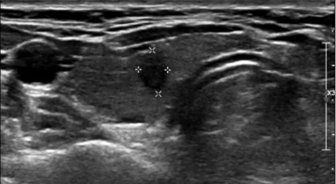
Fig. 1. Right papillary carcinoma in a 48-year-old woman.
Transverse gray-scale ultrasonographic image demonstrates that the nodule is 0.6 × 0.4 cm sized, hypoechoic, irregular margin and taller than wide.
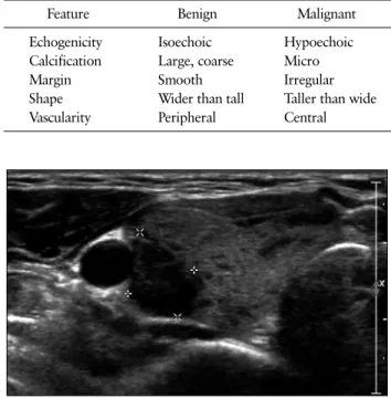
Fig. 2. Right papillary carcinoma in a 35-year-old man. Sagittal gray-scale ultrasonographic image shows microcalcifications in a 1.4
× 1.3 cm sized, hypoechoic and solid nodule.
위한 초음파는 영상의학과 전문의에 의해 시행되어 왔다.
하지만 최근 보고에 따르면 외과 의사에 의해 시행되는 초 음파가 영상의학과 의사에 의해 시행되는 초음파에 비해 좀 더 정확하고 수술 전 계획을 설계하는데 유용하여 국소 재발률을 낮추는 것으로 알려져 있다.(6-8)
갑상선암의 적절한 치료를 위해 진단 당시 병변의 범위 를 정확히 파악하는 것은 필수적이다. 즉, 수술 전에 이미 존재했던 병변을 발견하지 못하면 부적절한 제거로 인해 수술 후 잔존 암이 발생하여 재발의 위험을 높이고 결국 재수술을 필요로 하게 된다.(4) 갑상선암의 수술 전 진단 에 있어 갑상선 초음파는 가장 민감도가 높은 진단 방법으 로 알려져 있으며 세침흡인세포검사(fine needle aspi- ration cytology, FNAC)를 동시에 시행할 수 있고 수술 중 병변을 실시간으로 확인할 수 있으며 수술 후 추적 관찰 에도 매우 유용한 것으로 보고된다.(9-11)
이에 저자들은 문헌 고찰을 통해 갑상선암 환자의 적절 한 진단과 치료에 있어 초음파의 임상적 중요성과 유용성 에 관하여 살펴보고자 한다.
본 론
1. 수술 전 초음파
갑상선암 환자의 진료에 있어 갑상선 초음파는 가장 기 본적인 영상학적 검사 방법으로 갑상선의 원발 종양과 함 께 다른 종양들의 존재 여부와 경부 림프절 전이 여부 등을 평가해야 한다. 악성이 의심되는 갑상선 종양이나 림프절 이 관찰되는 경우에는 초음파 유도 하 FNAC를 통해 조직
학적으로 확인 후 수술 범위를 결정한다.(12,13) 1) 갑상선 결절
갑상선 결절은 중년 여성의 50% 이상에서 발견되는 흔 한 질환으로 이 중 약 5%만이 악성 결절이다.(14) 초음파 검사 단독으로는 갑상선 결절의 악성 여부를 정확하게 판 명할 수 없지만 임상의들이 악성 여부를 판단하는데 강력 한 단서를 제공한다. 악성을 시사하는 갑상선 결절의 특징 적인 초음파 소견으로는 단단하고(solid), 저에코성이며 (hypoechoic), 미세석회화(microcalcification), 가장자 리의 왜곡(distortion of the rim), 키 큰 형태(taller than wide), 중심부 혈관 신생(central vascularity), 갑상선외 침범(extrathyroidal extension) 등이 있다.
경계가 명확하고 낭성 소견을 보이는 갑상선 결절은 일 반적으로 양성인 경우가 많은 반면에 악성 결절은 경계가 불명확하고 단단하며 주변 정상 갑상선 실질보다 저에코 성을 보이는 경우가 흔하다(Fig. 1). 미세석회화는 갑상선 암 환자에서 85%의 민감도, 95%의 특이도와 94%의 정확 도를 가진다고 보고된다(Fig. 2).(15) 그러나 크고 거친 석 회화나 결절의 표면을 따라 존재하는 석회화(rim calcifi- cation)는 모든 종류의 결절에서 흔히 볼 수 있는 소견으 로 이전의 출혈이나 퇴행성 변화를 반영하기도 한다. 최근 에 보고된 체계적 고찰에서 키 큰 형태를 보이는 결절의 악성 민감도와 특이도는 각각 53%와 93%였으며, 특히 크 기가 큰 결절에서는 민감도가 낮은 것으로 알려져 있 다.(16,17) 혈류 분포의 양상도 악성 갑상선 결절을 예측 하는 인자로 알려져 있으며 결절 중앙 부분에 혈류가 증가 하는 소견이 결절 주변부에서 보이는 혈관 생성보다 악성 일 가능성이 높다.(18) 갑상선 초음파 상 갑상선암을 시사
Table 1. Ultrasonographic Features Associated with Benign and Malignant Thyroid Nodules
Feature Benign Malignant
Echogenicity Isoechoic Hypoechoic
Calcification Large, coarse Micro
Margin Smooth Irregular
Shape Wider than tall Taller than wide
Vascularity Peripheral Central
Fig. 3. Extrathyroidal extension of a right papillary carcinoma in a 47-year-old woman. Transverse gray-scale ultrasonographic image demonstrates a 1.2 cm sized, hypoechoic and solid nodule with indistinct lateral margins and invasion of the adjacent tissue which was confirmed intraoperatively.
Fig. 4. Metastatic left level II lymph node in a 58-year-old man with left papillary thyroid carcinoma. Transverse gray-scale ultraso- nographic image demonstrates an enlarged (1.0 cm sized) left level II lymph node with an abnormally thickened, irregular margin and loss of the fatty hilum.
하는 특징적인 단일 소견은 없지만 앞 서 살펴본 여러 특징 들을 함께 보이는 갑상선 결절인 경우에 악성의 가능성이 매우 높아진다.(19,20) 갑상선 결절의 양성과 악성을 시 사하는 특징적인 소견을 Table 1에 정리하였다.
갑상선암의 대부분을 차지하는 갑상성 유두암은 다병 소성(multifocality)의 특징이 있기 때문에 갑상선암 환자 에서 다발성 결절은 적절한 수술 계획을 수립하기 위해 주 의 깊게 살펴야 한다. 크기가 1 cm 이상인 결절은 더욱 세 밀하게 평가해야 하며 악성을 시사하는 초음파 소견을 보 이는 경우 FNAC를 적극적으로 시행해 악성 여부를 판단 해야 한다. 특히 의심스러운 결절이 양측에 있다면 향후 갑상선 전절제술을 시행해야 할 수 있으므로 원발 병소 반 대측 엽에 결절이 있는 경우 확실히 평가해야 한다. 136명 의 갑상선암 환자를 대상으로 연구한 보고(7)에 따르면 영 상의학과 의사가 시행한 갑상선 초음파의 22%에서 반대 측 결절을 간과하였으며, 또 다른 연구에서도 영상의학과 의사가 놓친 반대측 결절을 외과 의사가 발견하여 적절한 치료를 시행할 수 있었다고 보고하였다.(6) 갑상선암 환
자의 수술 전 초음파에서 반대측 결절의 악성 여부 판단은 갑상선 전절제술 시행 여부를 결정하는 매우 중요한 인자 이므로 항상 세밀하게 관찰해야 한다.
갑상선외 침범은 갑상선암 환자의 국소 재발과 전이의 위험 인자로 알려져 있다.(21) 수술 전 갑상선외 침범(특 히 뒤쪽 침범으로 인한 신경, 기도, 식도 등의 주요 기관 침습)을 정확하게 예측하지 못하면 좀 더 복잡한 수술 술 기를 시행하는데 어려움을 겪을 수 있으며, 수술 후 재발 의 위험을 증가시킬 수 있다. 따라서 수술을 직접 집도하 는 외과 의사는 수술 전 초음파에서 갑상선암의 갑상선외 침범 여부를 반드시 주의 깊게 살펴야 한다(Fig. 3).
2) 림프절
분화성 갑상선암 환자의 약 20-50%에서 경부 림프절 전이가 보고되므로 수술 전 명확한 병기 설정을 위해 경부 림프절의 전이 여부 평가가 매우 중요하다.(22)
전이된 림프절을 시사하는 초음파의 특징적인 소견으 로는 낭성 변화(cystic degeneration), 미세석회화, 주변 부 혈류 증가, 주변 근육과 비교해 고에코성, 지방문의 소 실(loss of fatty hilum), 둥근 형태 등이 있다(Fig. 4).
(23,24) 이 중 낭성 변화와 미세석회화는 가장 높은 특이 도를 보이며 림프절 전이 소견이 있는 경우 즉각적인 FNAC를 시행해야 한다.(25) 초음파의 악성 경부 림프절 의 민감도와 특이도는 각각 94.4%와 85.2%로 보고 된 다.(5)
갑상선암 환자의 수술 후 남아있는 전이 림프절은 재발
Fig. 5. Injection of vital dye during intraoperative ultrasonography in a 51-year-old woman with recurrent papillary thyroid carcinoma. (A) Intraoperative ultrasonography-guided vital dye injection to recurrent cervical lymph node. (B) The needle (arrow) was inserted to the 0.7 cm sized lymph node located in right level III.
의 가장 흔한 원인이며 불완전한 수술 전 검사를 의미하기 도 한다. 예방적 중앙 경부 림프절 절제술의 유용성에 대 하여 아직 이견이 많은 상황에 수술 전 중앙 경부 림프절의 확인은 매우 중요하며, 초음파 판독지에 이에 관한 기술도 필수적이다. 그럼에도 불구하고 갑상선암 환자를 대상으 로 영상의학과 의사에 의해 시행된 초음파에서 경부 림프 절에 대한 평가와 기술이 많이 부족했다는 보고가 있 어,(8) 수술 전 초음파에서 경부 림프절의 전이 여부를 명 확히 판단함으로써 적절한 수술과 이에 따른 예후 증진에 기여할 수 있을 것으로 생각한다.
3) 초음파 유도 하 세침흡인세포검사
초음파 상 악성을 시사하는 소견이 많은 갑상선 결절이 나 림프절의 정확한 진단을 위해서는 FNAC가 필수적이 다.(23) 과거에 FNAC는 주로 촉지되는 결절에서 시행되 었으나 초음파 유도 하 FNAC를 통해 촉지되지 않는 작은 결절에서도 정확한 조직검사가 가능하게 되었다. 초음파 유도 하 FNAC는 바늘의 위치를 확인하며 조직 검사를 시 행할 수 있어 부적절한 검체 획득을 줄이고 민감도와 특이 도를 증가시키는 장점이 있다.(26,27)
초음파 유도 하 FNAC의 정확도는 91-93%로 보고되고 있으며, 흡인된 검체에서 티로글로불린(thyroglobulin) 을 측정하거나 유전자 돌연변이(gene mutation) 또는 mRNA 발현 등을 살펴봄으로써 진단의 정확도를 향상시 킬 수 있다.(28-30)
2. 수술 중 초음파
갑상선암 환자에서 수술 중 초음파(intraoperative ul-
trasonography, IOUS)는 매우 유용하여 수술의 완결성 을 향상 시키고 특히 경부 림프절 재발 병변의 위치 결정에 탁월한 장점이 있다.
갑상선암 환자의 수술 시 IOUS를 시행한 군에서 시행 하지 않은 군에 비해 통계학적으로 의미있게 낮은 재발률 을 보였으며(1.9% vs. 12.5%, P<0.05),(31) Agcaoglu 등 (32)은 갑상선암 환자 중 변형근치경부절제술을 시행하는 25명의 환자를 대상으로 IOUS를 적용하였을 때 4예(16%) 에서 IOUS를 통해 II, IV와 V 구역에서 잔존하는 림프절 을 찾아 절제함으로써 수술의 완결성을 높일 수 있었다고 보고 하였다.
갑상선암의 경부 림프절 전이 시 수술적 절제가 표준 치 료인데 이전 수술에 의한 유착 및 반흔(scar) 조직에 의해 병변의 정확한 제거가 어려운 경우가 있다. 따라서 재수술 시에는 첫 수술보다 후두 신경과 부갑상선의 손상 등의 합 병증 발생률이 높으며 수술 시간도 더 오래 걸리는 것으로 알려져 있다.(33) 재발 갑상선암 환자에서 IOUS는 매우 유용하며 중앙경부림프절 전이와 병변의 크기가 2 cm 미 만인 경우 그리고 이전에 외부 방사선 조사를 받은 경우에 특히 효과적이다.(34) 재발 갑상선암 환자에서 병변의 IOUS 유도 하 생체 염료 주입은 수술실에서 외과 의사가 직접 시행하는 경우 수술 시 목표 병변의 위치를 가늠하는 데 매우 효과적이며, 또한 절제된 병변의 동결절편검사를 시행하지 않아도 되기 때문에 수술 시간 및 의료 비용을 절약할 수 있는 장점도 있다(Fig. 5).(35)
3. 수술 후 초음파
역사적으로 분화성 갑상선암 환자의 수술 후 재발 여부 감시는 혈중 티로글로불린과 전신 스캔으로 이루어졌다.
하지만 전신 스캔의 민감도가 낮은 것이 알려지면서 실제 임상에서는 혈중 티로글로불린과 초음파를 주로 사용하 고 있으며 재발 병변에 대해 96.3%의 민감도와 99.5%의 음성 예측도를 보이고 있다.(36)
갑상선암 환자의 수술 후 재발은 경부와 종격동 림프절 (74%), 갑상선 수술 부위(bed) (20%), 기도 또는 인접 근 육(6%)의 순으로 발생한다.(37) 이러한 재발들은 일반적 으로 촉지되지 않는 작은 병변으로 나타나기 때문에 수술 후 추적 관찰에서 초음파는 매우 중요한 역할을 담당한 다.(38,39)
정상적으로 갑상선 수술 부위는 섬유지방 증식성 조직 (fibrofatty proliferative tissue)으로 채워지기 때문에 잔존하는 정상 갑상선 조직과 재발 병변과의 주의 깊은 감 별을 요한다. 갑상선 수술 부위에서 재발한 결절의 초음파 소견은 수술 전 결절과 비슷하여 불규칙한 경계, 저에코 성, 미세석회화, 키 큰 형태 등의 특징이 있다.(40) 그러나 이러한 병변은 수술 후 육아종, 신경종, 반응성 림프절 또 는 부갑상선 선종 등과 감별을 요한다.(24)
경부 림프절(특히 II-VI 구역)은 갑상선암 환자의 가장 흔한 재발 부위이므로 수술 후 초음파로 주의 깊게 살펴야 한다. 전이 림프절을 의심할 수 있는 크기가 보고되고 있 지만(II 구역은 0.8 cm 이상, III-VI 구역은 0.5 cm 이상) 림프절의 크기만으로는 악성 림프절을 예측할 수 있는 정 확도가 낮아 수술 전과 유사한 전이 림프절을 시사하는 초 음파 소견을 보이는 림프절은 FNAC를 통해 재발 여부를 판단해야 한다.(37)
갑상선 엽절제술을 시행 받은 환자의 경우 수술 후 혈중 티로글로불린을 재발 예측 인자로 활용할 수 없기 때문에 수술 후 추적 관찰 시 갑상선 초음파의 유용성은 더욱 높아 진다.(41)
갑상선암 환자의 수술 또는 방사선 요오드 치료 후 초음 파 추적 관찰은 6-12개월 째에 시작하며 그 이후 환자의 재발 위험도에 따라 정기적인 간격으로 시행한다.(41) 향 후 갑상선암 환자의 수술 후 재발 감시를 위한 초음파의 적절한 시행 시기를 결정하기 위한 추가적인 연구가 필요 하다.
결 론
갑상선암 환자의 진료에서 초음파는 필수적인 영상학 적 진단 방법으로 진단, 병기 설정, 수술 범위 결정, 수술 의 완결성, 수술 후 추적 검사 등에 다양하게 적용된다. 갑 상선 수술을 집도하는 외과의는 보다 나은 양질의 진료를 위해 초음파를 능숙하게 사용하는 능력을 배양해야 한다.
향후 발전된 초음파의 적용을 통해 갑상선암 환자의 진단 및 치료 정확도를 높임으로써 궁극적으로 갑상선암 환자 의 삶의 질을 향상시킬 수 있을 것으로 생각한다.
REFERENCES
1. National Cancer Information Center. Cancer statistics [Internet]. Goyang: Ministry of Health and Welfare [cited 2018 Mar 14]. Available from http://www.
cancer.go.kr.
2. Siegel R, Ma J, Zou Z, Jemal A. Cancer statistics, 2014. CA Cancer J Clin 2014;64:9-29.
3. Stack BC Jr, Ferris RL, Goldenberg D, Haymart M, Shaha A, Sheth S, et al. American Thyroid Association consensus review and statement regarding the anat- omy, terminology, and rationale for lateral neck dis- section in differentiated thyroid cancer. Thyroid 2012;22:501-8.
4. Yeh MW, Bauer AJ, Bernet VA, Ferris RL, Loevner LA, Mandel SJ, et al. American Thyroid Association statement on preoperative imaging for thyroid cancer surgery. Thyroid 2015;25:3-14.
5. Sipos JA. Advances in ultrasound for the diagnosis and management of thyroid cancer. Thyroid 2009;19:
1363-72.
6. Mazzaglia PJ. Surgeon-performed ultrasound in pa- tients referred for thyroid disease improves patient care by minimizing performance of unnecessary pro- cedures and optimizing surgical treatment. World J Surg 2010;34:1164-70.
7. Carneiro-Pla D, Amin S. Comparison between pre- consultation ultrasonography and office surgeon- performed ultrasound in patients with thyroid cancer.
World J Surg 2014;38:622-7.
8. Oltmann SC, Schneider DF, Chen H, Sippel RS. All thyroid ultrasound evaluations are not equal: sonog- raphers specialized in thyroid cancer correctly label clinical N0 disease in well differentiated thyroid cancer. Ann Surg Oncol 2015;22:422-8.
9. Milas M, Stephen A, Berber E, Wagner K, Miskulin J, Siperstein A. Ultrasonography for the endocrine sur- geon: a valuable clinical tool that enhances diagnostic and therapeutic outcomes. Surgery 2005;138:1193-
200; discussion 1200-1.
10. O’Connell K, Yen TW, Quiroz F, Evans DB, Wang TS.
The utility of routine preoperative cervical ultra- sonography in patients undergoing thyroidectomy for differentiated thyroid cancer. Surgery 2013;154:697- 701; discussion 701-3.
11. Lew JI, Solorzano CC. Use of ultrasound in the man- agement of thyroid cancer. Oncologist 2010;15:253-8.
12. Kouvaraki MA, Shapiro SE, Fornage BD, Edeiken- Monro BS, Sherman SI, Vassilopoulou-Sellin R, et al.
Role of preoperative ultrasonography in the surgical management of patients with thyroid cancer. Surgery 2003;134:946-54; discussion 954-5.
13. Park JS, Son KR, Na DG, Kim E, Kim S. Performance of preoperative sonographic staging of papillary thy- roid carcinoma based on the sixth edition of the AJCC/UICC TNM classification system. AJR Am J Roentgenol 2009;192:66-72.
14. Caruso D, Mazzaferri EL. Fine needle aspiration bi- opsy in the management of thyroid nodules. Endocrinol 1991;1:194-202.
15. Salmaslioğlu A, Erbil Y, Dural C, Işsever H, Kapran Y, Ozarmağan S, et al. Predictive value of sono- graphic features in preoperative evaluation of malig- nant thyroid nodules in a multinodular goiter. World J Surg 2008;32:1948-54.
16. Brito JP, Gionfriddo MR, Al Nofal A, Boehmer KR, Leppin AL, Reading C, et al. The accuracy of thyroid nodule ultrasound to predict thyroid cancer: system- atic review and meta-analysis. J Clin Endocrinol Metab 2014;99:1253-63.
17. Ren J, Liu B, Zhang LL, Li HY, Zhang F, Li S, et al.
A taller-than-wide shape is a good predictor of pap- illary thyroid carcinoma in small solid nodules. J Ultrasound Med 2015;34:19-26.
18. Cappelli C, Castellano M, Pirola I, Cumetti D, Agosti B, Gandossi E, et al. The predictive value of ultra- sound findings in the management of thyroid nodules.
QJM 2007;100:29-35.
19. Méndez W, Rodgers SE, Lew JI, Montano R, Solórzano CC. Role of surgeon-performed ultrasound in pre- dicting malignancy in patients with indeterminate thyroid nodules. Ann Surg Oncol 2008;15:2487-92.
20. Jabiev AA, Ikeda MH, Reis IM, Solorzano CC, Lew JI.
Surgeon-performed ultrasound can predict differ- entiated thyroid cancer in patients with solitary thy- roid nodules. Ann Surg Oncol 2009;16:3140-5.
21. Andersen PE, Kinsella J, Loree TR, Shaha AR, Shah JP. Differentiated carcinoma of the thyroid with ex- trathyroidal extension. Am J Surg 1995;170:467-70.
22. Chow SM, Law SC, Chan JK, Au SK, Yau S, Lau WH.
Papillary microcarcinoma of the thyroid-prognostic significance of lymph node metastasis and multifocality.
Cancer 2003;98:31-40.
23. Haugen BR, Alexander EK, Bible KC, Doherty GM, Mandel SJ, Nikiforov YE, et al. 2015 American
Thyroid Association management guidelines for adult patients with thyroid nodules and differentiated thy- roid cancer: The American Thyroid Association guide- lines task force on thyroid nodules and differentiated thyroid cancer. Thyroid 2016;26:1-133.
24. Leenhardt L, Erdogan MF, Hegedus L, Mandel SJ, Paschke R, Rago T, et al. 2013 European Thyroid Association guidelines for cervical ultrasound scan and ultrasound-guided techniques in the post- operative management of patients with thyroid cancer.
Eur Thyroid J 2013;2:147-59.
25. Leboulleux S, Girard E, Rose M, Travagli JP, Sabbah N, Caillou B. Ultrasound criteria of malignancy for cervical lymph nodes in patients followed up for dif- ferentiated thyroid cancer. J Clin Endocrinol Metab 2007;92:3590-4.
26. Danese D, Sciacchitano S, Farsetti A, Andreoli M, Pontecorvi A. Diagnostic accuracy of conventional versus sonography-guided fine-needle aspiration bi- opsy of thyroid nodules. Thyroid 1998;8:15-21.
27. Carmeci C, Jeffrey RB, McDougall IR, Nowels KW, Weigel RJ. Ultrasound-guided fine-needle aspiration biopsy of thyroid masses. Thyroid 1998;8:283-9.
28. Seiberling KA, Dutra JC, Gunn J. Ultrasound-guided fine needle aspiration biopsy of thyroid nodules per- formed in the office. Laryngoscope 2008;118:228-31.
29. Pacini F, Fugazzola L, Lippi F, Ceccarelli C, Centoni R, Miccoli P, et al. Detection of thyroglobulin in fine needle aspirates of nonthyroidal neck masses: a clue to the diagnosis of metastatic differentiated thyroid cancer. J Clin Endocrinol Metab 1992;74:1401-4.
30. Nikiforov YE, Steward DL, Robinson-Smith TM, Haugen BR, Klopper JP, Zhu Z, et al. Molecular test- ing for mutations in improving the fine-needle aspi- ration diagnosis of thyroid nodules. J Clin Endocrinol Metab 2009;94:2092-8.
31. Ertas B, Kaya H, Kurtulmus N, Yakupoglu A, Giray S, Unal OF, et al. Intraoperative ultrasonography is useful in surgical management of neck metastases in differentiated thyroid cancers. Endocrine 2015;48:
248-53.
32. Agcaoglu O, Aliyev S, Taskin HE, Aksoy E, Siperstein A, Berber E. The utility of intraoperative ultrasound in modified radical neck dissection: a pilot study.
Surg Innov 2014;21:166-9.
33. Kim MK, Mandel SH, Baloch Z, Livolsi VA, Langer JE, Didonato L, et al. Morbidity following central com- partment reoperation for recurrent or persistent thy- roid cancer. Arch Otolaryngol Head Neck Surg 2004;
130:1214-6.
34. Karwowski JK, Jeffrey RB, McDougall IR, Weigel RJ.
Intraoperative ultrasonography improves identi- fication of recurrent thyroid cancer. Surgery 2002;
132:924-8; discussion 928-9.
35. Ahn D, Sohn JH, Kim H. Surgeon-performed intra- operative tumor localization in recurrent papillary
thyroid carcinoma by ultrasound-guided intratumoral indigo carmine injection. World J Surg 2014;38:1995- 2001.
36. Pacini F, Molinaro E, Castagna MG, Agate L, Elisei R, Ceccarelli C, et al. Recombinant human thyro- tropin-stimulated serum thyroglobulin combined with neck ultrasonography has the highest sensitivity in monitoring differentiated thyroid carcinoma. J Clin Endocrinol Metab 2003;88:3668-73.
37. Tufano RP, Clayman G, Heller KS, Inabnet WB, Kebebew E, Shaha A, et al. Management of recurrent/
persistent nodal disease in patients with differ- entiated thyroid cancer: a critical review of the risks and benefits of surgical intervention versus active surveillance. Thyroid 2015;25:15-27.
38. Frasoldati A, Pesenti M, Gallo M, Caroggio A, Salvo D, Valcavi R. Diagnosis of neck recurrences in pa- tients with differentiated thyroid carcinoma. Cancer 2003;97:90-6.
39. Torlontano M, Attard M, Crocetti U, Tumino S, Bruno R, Costante G, et al. Follow-up of low risk patients with papillary thyroid cancer: role of neck ultra- sonography in detecting lymph node metastases. J Clin Endocrinol Metab 2004;89:3402-7.
40. Ko MS, Lee JH, Shong YK, Gong GY, Baek JH.
Normal and abnormal sonographic findings at the thyroidectomy sites in postoperative patients with thyroid malignancy. AJR Am J Roentgenol 2010;194:
1596-609.
41. Torlontano M, Crocetti U, Augello G, D'Aloiso L, Bonfitto N, Varraso A, et al. Comparative evaluation of recombinant human thyrotropin-stimulated thyro- globulin levels, 131I whole-body scintigraphy, and neck ultrasonography in the follow-up of patients with papillary thyroid microcarcinoma who have not undergone radioiodine therapy. J Clin Endocrinol Metab 2006;91:60-3.