INTRODUCTION
Focal nodular hyperplasia (FNH) of the liver is a non-neo- plastic regenerative lesion characterized by a stellate central scar and hyperplastic nodules (1). FNH develops as a solitary or multifocal lesion, and is usually supplied by abnormally enlarged feeding arteries. It is now generally hypothesized that FNH represents a hyperplastic response of liver parenchy- ma to local hemodynamic disturbance, induced by malformed arteries (2, 3).
Typical category of FNH is composed of the two classical features, nodular architecture and malformed vessels, and it always presents bile ductular proliferation. However, a number of recent studies have proposed the non-classical categories of FNH (4-7). According to these articles, non-classical FNHs are subdivided into three histologic forms: the telangiectatic form (7), the mixed hyperplastic and adenomatous form, and a third variant with cytologic atypia (4).
We describe a case of FNH, discovered at birth, and is con- sidered as a telangiectatic variant. We also discuss its morpho- logical characteristics for comparison with a classical form and liver nodules in similar morphology.
CASE REPORT Clinical History
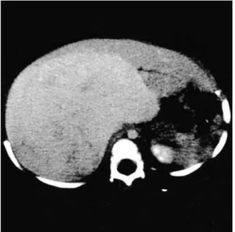
An eleven-month-old boy was admitted to Seoul National University Children’s Hospital, complaining of an abdominal mass, which had been detected at his birth by his family. The patient had been delivered normally at full term with a birth- weight of 3.2 kg. No significant information was available about his past medical history. Physical examination was nor- mal, except for the abdominal mass. The mass was palpable on the whole abdomen, extending to 13 cm below the xyphoid process. It was freely movable, firm, and non-tender. No icterus was found. No significant finding was noted during laboratory examination. Alpha-fetoprotein was 13 ng/dL, and serum HBsAb was positive. On abdominal CT, a 16×12 cm homo- geneously enhanced mass replaced the right lobe of the liver completely and compressed the remainder of the left lobe (Fig. 1). Radiological and clinical impressions were hepato- blastoma. The liver needle biopsy was performed, and the diag- nosis was ‘‘consistent with hepatoblastoma, fetal type’’. The boy was treated through several cycles of chemotherapeutic regimens. After chemotherapy, however, there was no change in size and shape of the mass on radiological studies. Then, the mass excision was performed. On laparotomy, there was much bleeding from the cut surface of the mass. The mass
Han-Seong Kim�, Young A Kim, Chong Jai Kim, Yeon-Lim Suh*, Ja-June Jang, Je G. Chi
Department of Pathology, Seoul National University College of Medicine, Seoul; *Department of Diagnostic Pathology, Sungkyunkwan University College of Medicine, �Department of Pathology, Ilsan Paik Hospital, Inje University College of Medicine, Goyang, Korea
Address for correspondence Je G. Chi, M.D.
Department of Pathology, Seoul National University College of Medicine, 28 Yongon-dong, Chongno-gu, Seoul 110-799, Korea
Tel : +82.2-760-3546, Fax : +82.2-741-6195 E-mail : [email protected]
746 J Korean Med Sci 2003; 18: 746-50
ISSN 1011-8934
Copyright � The Korean Academy of Medical Sciences
Telangiectatic Focal Nodular Hyperplasia of the Liver : A Case Detected at Birth
A case of telangiectatic focal nodular hyperplasia (FNH) was detected at birth and was surgically removed. Grossly, the lesion was a solitary nodule and showed vague nodularity, appearing as an adenoma-like mass with fine fibrous septa, but having no macroscopic scar. On microscopic scale, the mass typically had neither fibrous central scar nor hyperplastic nodules different from the usual FNHs. The hepatic plates were separated by sinusoidal dilatation, sometimes alternating with areas of marked ectasia. Instead of large fibrous scar, thin fibrous septa were often found, and contained abnormal tortuous large arteries. These high-pressure vessels were connected directly into the adjacent sinusoids and made marked dilation of sinusoids. Bile ductular proliferation was also noted in the thin fibrous septa. To our knowledge, this is considered to be the first reported case of telangiectatic FNH detected at birth.
Key Words : Telangiectasia; Focal Nodular Hyperplasia; Infant, Newborn; Liver
Received : 14 August 2002 Accepted : 16 October 2002
could not completely be removed, because a part of the tumor encircled the inferior vena cava. The postoperative course was unremarkable. The patient had been followed up with no sign of recurrence for two years.
Materlals and Methods
The surgically resected tumor was fixed in 10% neutral formalin, and was embedded in paraffin blocks. Hematoxylin and eosin, PAS, D-PAS, reticulin, and Masson trichrome stains were performed. Immunohistochemical study was performed.
Serial sections (3 m-thick) were cut from paraffin blocks, and were deparaffinized in xylene. For antigen retrieval, sample slides were heated for 5 min in a microwave oven, and then were incubated in 0.3% hydrogen peroxide for 30 min to block endogenous peroxidase activity. The primary antibod- ies used were anti-CK19, CD34 and factor VIII-related anti- gen (DAKO, U.S.A.). Slides were incubated with the primary antibodies overnight at 4℃and washed in PBS, and then incubated with biotinylated goat anti-rabbit, anti-mouse immunoglobulin at room temperature for 20 min and washed again, prior to treatment with avidin-complex peroxidase.
Slides were developed with 3,3′diaminobenzidine tetrahy- drochloride for 4 min, and finally, sections were counterstained with Harris hematoxylin. Buffer was substituted for the prima- ry negative controls.
Pathologic Findings
On macroscopic examination, the lesion was a solitary mass.
The specimens measured 16×10×5 cm and weighed 380 g.
The external surface shows smooth, glistening, and red-brown appearance. On the cut surface, the mass had ill-defined bor- ders, and was not encapsulated. Although vague nodularity and thin fibrous bands were noted in parenchyma, no macro- scopically visible central scar was found (Fig. 2). Consistency of the tumors was slightly softer than that of non-neoplastic hepatic tissue. In the parenchyma of lesions, large blood vessels
Fig. 1.CT finding. Enhanced CT shows the huge mass that replaces right lobe completely and compressed the left lobe.
Fig. 2.Gross appearance. Note the solid cut surface with thin fibrous bands without macroscopic scar.
Fig. 3.Light microscopic findings. An abnormal artery (arrow) is found in short fibrous septa with direct irrigation of surrounding dilat- ed sinusoids, making characteristic telangiectatic appearance (H&E, ×100).
748 H.-S. Kim, Y.A. Kim, C.J. Kim, et al.
were easily found.
On low power microscopic examination, there was neither a central scar nor hyperplastic nodular appearance. Instead, thin and short fibrous septa were found throughout the tumor.
The septa contained abnormally enlarged tortuous arteries with hypertrophied muscular media, but without intimal prolif- eration. These abnormal arteries drained directly into the adja- cent sinusoids, and made sinusoidal dilatation (Fig. 3). The sinusoids were congested with red blood cells, which made characteristic ‘telangiectatic’ appearance (Fig. 4A). Also, thin branching fibrous stroma contains proliferating small bile duc- tules (Fig. 4A inlet).
Hepatic plates showed generally one cell thickness, while hypertrophic hepatic cords revealed two to three cells’ width occasionally. Sometimes microvesicular fatty changes are noted in hepatocytes. However, neither necrosis nor mitosis was seen.
The hepatic plates were separated by dilated sinusoids, which sometimes alternated with peliotic areas of marked ectasia.
The sinusoids were always lined by flat endothelial cells and were filled with pools of red blood cells. Also, Kupffer cells and extramedullary hematopoiesis were noted in the sinsusoids.
Moderate amount of lymphocytic infiltration was noted in the parenchyma. The endothelial cells lining dilated sinusoids were strongly positive for CD34 staining, and weakly reactive for factor VIII-related antigen. Bile ductular proliferation was easi- ly noted with CK19 immunohistochemistry (Fig. 4B).
DISCUSSION
Focal nodular hyperplasia of the liver in the pediatric patient
is a rare benign lesion that usually presents as an asymptomatic abdominal mass, and must be diagnosed differentially from other common benign or malignant lesions. One large series have reported 72 cases from birth to 20 yr and only three cases from birth to 2 yr during 30 yr (8). Sixteen cases of FNH including our case have been reported in Republic of Korea (9-19). They ranged in age from 1 to 75 yr, of which seven were children (11 months and 15 yr). The male to female ratio was 1:1. Our case was discovered at birth by the parents.
Unusual feature of lesions in the radiological studies and biop- sy findings may mislead us to erroneous initial diagnosis. No radiological change was noted in the contour or the size of the mass even after several cycles of chemotherapy. Surgical explo- ration proved the lesion is a benign lesion associated with vas- cular abnormality.
Telangiectatic variant of FNH was described first by Wanless et al. (7). In their series, telangiectatic FNHs were usually asso- ciated with multiple FNH syndromes. In a recent study, a total of 47 FNHs of the telangiectatic type were identified in 16 patients (15.4% of their series) (4). This form presented as a solitary tumor in 10 patients. In the other six patients, it was the only histologic type identified (20 of 20 lesions of the same patient) or one of the morphologic variants among other types of FNH (1 of 2, 1 of 4, 1 of 15, 13 of 15, and 1 of 30 lesions from five patients). Therefore, the telangiectatic type seems to be more prevalent in multiple FNH cases than in solitary cases, although some previous reports and our case were solitary lesions (5, 6).
On the macroscopic scale, the aspects of telangiectatic FNH are dissimilar to a classical form with its less prominent lobu- lation and no macroscopic central scar. The remaining lesions
Fig. 4. (A) The sinusoids are dilated and congested with red blood cells (H&E, ×100). Inlet: proliferating cholangioles are noted in the thin fibrous septa (arrows, H&E, ×400), (B) CK19 immunohistochemical finding. Proliferating small bile ductules are noted in the thin fibrous septa (×200).
A B
showed a homogeneous parenchyma resembling adenoma. In terms of size, telangiectatic FNH seems variable from less than 2 cm to 19 cm (4). In a manner similar to these studies, our case was ill defined and non-encapsulated lesion, and had a vaguely lobular architecture on cut surfaces. It measured 16 cm in diameter.
Microscopically, telangiectatic FNH differs from the typical form by the absence of nodular architecture and by its mild architectural distortion. The lobulated appearance found in gross examination could result from the alternating of solid and ectatic areas. The hepatic cords generally consist of single or two cells in width and almost regular. However, they are separated by characteristically dilated sinusoids, which are irrigated directly by high-pressure arterial blood from adjacent abnormal arteries. These vessels are generally located in short and slender fibrous stroma. The arteries have a moderately thickened muscular wall. However, the intimal proliferation is not easily seen, while the latter features a usual characteristic of classical FNH. The very helpful feature in the diagnosis of FNH is the presence of areas of bile ductular proliferation because liver cell adenoma usually lacks bile ducts (20). Mild to marked degree of cholangiolar proliferation was found in 100% of telangiectatic FNHs, including our case (4).
Telangiectatic FNH must be differentiated from other hep- atic nodules showing vague nodularity and prominent vascular pattern. Hepatic adenoma is a rarely occurring benign tumor of the liver reported primarily in teenage girls taking oral con- traceptives. It is usually a solitary nodular lesion, often with large blood vessels over its surface (21). Microscopically the tumor is composed of sheets of neoplastic cells in two-cell thickness, separated by compressed sinusoidal spaces lined by endothelial cells and some Kupffer cells. Areas of necrosis and hemorrhage may be present, and foci of dilated sinusoids may impart a ‘‘pelioid’’ appearance. However, hepatic adenoma has neither bile ductular element in the tumor parenchyma, as well as diffuse sinusoidal dilatation, compared to telang- iectatic FNH. Infantile hemangioendothelioma (IHE) is the most common liver tumor in the first year of life. IHE may be single or multicentric, and is composed of thin vascular channels. Microscopically, IHE lesion is composed of vascular channels lined by a single continuous layer of plump endothe- lial cells in a supporting fibrous stroma (type 1 IHE). In about 20% of cases, large pleomorphic and hyperchromatic cells are present along poorly formed vascular spaces, often displaying tufting or branching (type 2 IHE) (22). In contrast to telang- iectatic FNH, IHE consists of pure vascular channels having fibrous stroma devoid of hepatocytes and bile ducts. Hepatic cords and cholangioles are found only in the periphery of IHE.
Also, endothelial cells of IHE are plump and pleomorphic, especially in type 2 lesion, in comparison with flat and bland looking endothelial cells of telangiectatic FNH. Hepatobla- stoma is the most common malignant liver tumor under 2 yr of age. A variety of histologic patterns are noted, which consist- ed of epithelial and mixed mesenchymal components (22). The
fetal pattern is composed of small, round, uniform cells with abundant cytoplasm and distinct cytoplasmic membranes. The cells are arranged into thin trabeculae, usually two- or three-cell thickness, with alternating light and dark areas. The embryon- al pattern shows sheets of irregular, angulated cells with a high nucleocytoplasmic ratio, increased nuclear chromatin, and indistinct cytoplasmic membranes. Pseudorosette, ribbon and acinar formation are common features. The mixed epithelial and mesenchymal type of tumor contains varying amounts of fetal and embryonal type cells admixed with primitive mes- enchyme, consisted of spindle cells, and osteoid and cartilagi- nous tissue. The small-cell undifferentiated or anaplastic pat- tern is composed of small round cells that have a scanty cyto- plasm and hyperchromatic nuclei. Telangiectatic FNH does not show characteristic neoplastic proliferation of hepatoblas- toma, such like ‘light and dark’ pattern, tubular or rosette-like arrangement, and mesenchymal components.
However, in the biopsy specimen, it does not seem easy to discriminate the telangiectatic FNH from other hepatic nod- ules, due to unusual radiological and histological findings and the rarity of the lesion. Telangiectatic FNH should be consid- ered as a differential diagnosis in the biopsy diagnosis of solid hepatic nodules, which show bland-looking hepatic cord with marked sinusoidal dilatation and mild bile ductular prolif- eration.
In conclusion, telangiectatic FNH is a benign hyperplastic parenchymal lesion of the liver associated with malformed arter- ies. Although precise pathogenesis should be elucidated by further studies, the direct connection between this high-pres- sure large vessels and sinusoids is considered to be a underly- ing pathology that produces the unique telangiectatic appear- ance of this lesion. To our knowledge, our case is considered to be the first reported telangiectatic FNH of congenital origin.
REFERENCES
1. International Working Party. Terminology of nodular hepatocellular lesions. Hepatology 1995; 22: 983-93.
2. Ndimbie OK, Goodman ZD, Chase RL, Ma CK, Lee MW. Heman- gioma with localized nodular proliferation of the liver, a suggestion on the pathogenesis of focal nodular hyperplasia. Am J Surg Pathol 1990; 14: 142-50.
3. Wanless IR, Mawdsley C, Adams R. On the pathogenesis of focal nodular hyperplasia of the liver. Hepatology 1985; 5: 1194-200.
4. Nguyen BN, Flejou JF, Terris B, Belghiti J, Degott, C. Focal Nodu- lar Hyperplasia of the Liver: A comprehensive pathologic study of 305 lesions and recognition of new histologic forms. Am J Surg Pathol 1999; 23: 1441-54.
5. Oligny LL, Lough J. Hepatic sinusoidal ectasia. Hum Pathol 1992;
23: 953-6.
6. Peterfy CG, Rosenthall L. Large telangiectatic focal nodular hyper- plasia presenting with normal radionuclide studies: case report. J Nucl Med 1990; 31: 2037-9.
′
750 H.-S. Kim, Y.A. Kim, C.J. Kim, et al.
7. Wanless IR, Albrecht S, Bilbao J, Frei JV, Heathcote EJ, Roberts EA, Chiasson D. Multiple focal nodular hyperplasia of the liver associated with vascular malformations of various organs and neoplasia of the brain: a new syndrome. Mod Pathol 1989; 2: 456-62.
8. Ishak KG, Goodman Z, Stocker JT: Tumors of the liver and intrahep- atic bile ducts. In Atlas of Tumor Pathology, 3rd series, Fascicle 31.
Washington, DC, Armed Forces Instititue of Pathology, 2001.
9. Chung SK, Son HS, Lee MH, Lee SY, Bahk YW. Focal nodular hy- perplasia: pitfalls in hepatic scintigram. Korean J Nucl Med 1985;
19: 91-2.
10. Song JH, Dong SH, Kim HJ, Kim BH, Chang YW, Lee JI, Chang R, Hong SW, Lee JH. A case of focal nodular hyperplasia of the liver. Korean J Gastroenterol 1994; 25: 1034-40.
11. Ryu JS, Moon DH, Shin SH. Focal nodular hyperplasia diagnosed by 99m-Tc pyrophosphate liver SPECT. Korean J Nucl Med 1994;
28: 157-8.
12. Cha KS, Lee YH, Ryu KH, Choi BJ, Hwang IS, Kim SY, Kim YJ, Cho MK, Kim YI. A case of focal nodular hyperplasia of the liver.
Korean J Int Med 1996; 50: 416-21.
13. Lim CS, Lee ST, Kim DG, Ahn DS, Yu LC, Cho BH. A case of focal nodular hyperplasia of the liver. Korean J Hepatol 1997; 7: 337-43.
14. Cheon JE, Kim WS, Kim IO, Jang JJ, Seo JK, Yeon KM. Radiolog- ical features of focal nodular hyperplasia of the liver in children. Pedi- atr Radiol 1998; 28: 878-83.
15. Bong JD, Oh JY, Cho ST, Chung IK, Kim HS, Park SH, Lee MH, Kim SJ, Yang SH. Focal nodular hyperplasia of the liver mimicking hepatocellular carcinoma. Korean J Gastroenterol 1998; 31: 541-6.
16. Ko YY, Lee KJ, Ko JH, Kwon OY, Jung ST, Kim SD, Kim YS, Kim JH, Hahm KB, Cho SW, Joo HJ. A case of focal nodular hyperplasia of the liver. Korean J Gastroenterol 1998; 32: 554-8.
17. Lee SW, Cho HS, Choi CS, Nam ES, Kim JS, Park CY. A case of childhood multiple focal nodular hyperplasia of the liver. Korean J Pediatr Hematol Oncol 2000; 7: 153-9.
18. Park UH, Cho CM, Lee YD, Lee SY, Tak WY, Kweon YO, Kim SK, Choi YH, Chung JM, Bae HI, Ryeom HG. A case of focal nodular hyperplasia of the liver by surgical resection and histologic exami- nation. Korean J Hepatol 2000; 6: 524-9.
19. Kim TH, Choi EK, Park BH, Kim JA, Choi SC, Kim HC, Nah YH, Yun KJ. A case of focal nodular hyperplasia of the liver treated with hepatic artery embolization. Korean J Gastroenterol 2000; 36:
267-71.
20. Friedman LS, Gang DL, Hedberg SE, Isselbacher KJ. Simultaneous occurrence of hepatic adenoma and focal nodular hyperplasia: report of a case and review of the literature. Hepatology 1984; 4: 536-40.
21. Ishak KG. Benign tumors and pseudotumors of the liver. Appl Pathol 1988; 6: 82-104.
22. Stocker JT. Hepatic tumors in children, Liver disease in children. St.
Louis: The C.V. Mosby company, 1994.