MPTP 유발 파킨슨병 동물 모델에 대한 봉독약침의 신경보호 효과 및 항염증 효과
박원
*
․김재규***
․김종인**
․최도영**
․고형균**
*
키즈앤맘 한의원**
경희대학교 한의과대학 침구학교실***
부산대학교 한의학전문대학원 침구학교실목적 : 이 연구는 MPTP 유발 파킨슨병 동물 모델에서 봉독약침의 신경보호 효과 및 항염증 효과를 확 인하기 위해 시행되었다.
방법 : C57BL/6 mice에 신경독소인 1-methyl-4-phenyl-1,2,3,6-tetrahydropyridine(MPTP)를 하루에 2시 간 간격으로 MPTP-HCl(20mg/kg per dose)을 4번 복강 내 주입하여 중뇌 흑질의 도파민 신경세포를 파괴 한 파킨슨병 동물 모델을 유발하였다. 실험군은 MPTP군, MPTP 현종 BVA군, MPTP 곡지 BVA군, MPTP 신수 BVA군의 4군으로 하였다. 마지막 MPTP 투여 2시간 후에 1차로 봉독약침을 시술하고, 그 후 48시간 간격으로 총 5차 연속 시술하였다. 봉독약침액의 농도는 0.2mg/Kg으로 하였고, 경혈은 양측 현종(GB
39
), 곡 지(LI11
), 신수(BL23
)를 사용했고, 주입량은 각 경혈당 양측으로 각 20㎕씩 주입하였다.항염증작용을 알아보기 위해 TH, MAC-1, iNOS HSP70을, 세포사멸에 대한 신경세포의 보호효과를 알아 보기 위해 caspase-3을 면역조직화학법을 사용하여 실시하였다.
결과 : 실험 결과 MPTP 유발 파킨슨병 동물 모델에서 현종․곡지․신수혈에 대한 봉독약침은 TH- Immunoreactivity neuron의 감소와 microglial activation을 억제하였다. 봉독약침군 모두 효과를 보였으나 그 중 현종과 신수혈에서 특히 억제작용이 컸다. MAC-1에서는 현종혈이 억제작용이 컸다. HSP70-IR neuron
1)
Neuroprotective and Anti-inflammatory Effects of Bee Venom Acupuncture on MPTP-induced Mouse
Park Won * , Kim Jae-kyu *** , Kim Jong-in ** , Choi Do-young ** and Koh Hyung-kyun **
* Kiz&Mom Oriental Medicine Clinic
** Dept. of Acupuncture & Moxibustion, College of Oriental Medicine, Kyung Hee University
*** Dep. of Acupuncture and Moxibustion, Pusan National University School of Korean Medicine
․Acceptance : 2010. 5. 19. ․Adjustment : 2010. 5. 27. ․Adoption : 2010. 5. 28.
․Corresponding author : Koh Hyung-kyun, Department of Acupuncture and Moxibustion, College of Oriental Medicine, Kyung Hee University, 1 Hoegi-dong, Dongdaemun-gu, Seoul, Republic of Korea
Tel. 82-2-958-9133 E-mail : [email protected]
국문초록
Original Article
은 곡지에서 유의한 억제작용을 보였으나, iNOS neuron은 모든 군에서 유의한 차이를 보이지 않았다.
또한 세포사멸억제여부 실험에서 봉독약침은 모두 억제작용을 보였으나 특히 곡지자침군에서 caspase-3 발현을 유의하게 억제하였다.
결론 : 이러한 결과는 봉독약침이 MPTP 투여로 인한 중뇌 흑질의 염증에 의한 도파민 신경세포 손상을, 염증을 억제함으로써 항염 효과를 나타냄을 알 수 있으며, 신경세포를 보호하는 활성이 있음을 보여줌과 동 시에 세포사멸을 억제하는 활성이 있다고 사료된다.
핵심 단어 : Bee venom Acupuncture, Parkinson disease, MPTP(1-methyl-4- phenyl-1,2,3,6-tetrahy- dropyridine), macrophageantigencomplex-1, inducible nitricoxidesynthase, apoptosis, Heatshock- protein
Ⅰ. Introduction
Bee venom acupuncture(BVA) has been tradi- tionally used in Oriental Medicine to relieve pain
1)
and to treat chronic inflammatory diseases such as arthritis, rheumatic diseases, immune disorders2)
, and neurodegenerative diseases such as multiple sclerosis3-6)
.BVA exhibits analgesic, anti-arthritic, and anti- inflammatory effects that are attributable to not only bioactive bee venom(BV) compounds including peptides(melittin, apamin, and adolapin), enzymes (phospholipase A2)
7-9)
, biologically active amines (histamine, epinephrine), several other nonpeptide components including lipids, carbohydrates and free amino acids10)
, but also the mechanical effect of acupuncture stimulation.Recent studies have suggested that BV has anti- inflammatory properties that inhibit the production of inflammatory cytokines and NO in neurodeg- enerative diseases
11,12)
.Several clinical trials and anecdotal reports have recently suggested that BVA may be an effective treatment for demyelinating diseases of the central nervous system(CNS) such as Parkinson’s disease (PD) and multiple sclerosis
3)
, although the under- lying mechanism of this action remains unknown.PD is one of the most common movement dis- orders. The primary neuropathological basis of PD is a severe deficiency of dopamine in the striatum,
resulting from specific loss of nigrostriatal dopa- minergic(DA) neurons, the cell bodies of which reside in the substantia nigra pars compacta(SNpc) and the nerve terminals of which project to the striatum
13)
. While different mechanisms, includeing environmental toxins and genetic factors, initiate DA neuronal damage in the SNpc and striatum in PD, there is unequivocal evidence that activation of neuroinflammatory cells14-17)
leads to apoptosis cell death18)
, which aggravates the neuro-degenerative process.Brain inflammation is the common final pathway in PD
19)
. Central to this inflammation is the acti vation of microglia, which act as intrinsic immune effectors when the brain is injured20,21)
. Microglia are known to be activated in the PD-affected brain, and MPTP(1-methyl-4-phenyl-1,2,3,6- tetrahydropy- ridine) - induced microglial activation is associated with oxidative stress22)
.Several animal models of PD have been de- veloped, including a mouse model in which a parkinsonian pathology develops in response to the administration of the neurotoxin MPTP. These mice display a strong microglial response
23,24)
, accumulation of cytokines, and elevation of reactive oxygen species(ROS)25)
, which peak prior to the death of DA neurons, suggesting a pivotal role for the microglial response in the cascade of deleterious events that ultimately leads to DA neuronal death in the SNpc of the MPTP mouse model with PD26)
.The heat shock proteins(HSPs) represent an
2 4 6 8 10h 1day
Fig. 1. MPTP injection and BVA schedule important cellular protective mechanism against a variety of stresses and insults
27-29)
. Adaptive pro- tection was assessed by the expression of heat shock protein(HSP) 70-IR neurons.We examined whether (a) BVA inhibits the loss of tyrosine hydroxylase(TH)-positive neurons as a result of its inhibition of microglial activation or induction of HSP70 synthesis and (b) the effect of BVA is acupoint dependent and (c) the apoptosis is associated with the mechanism of BVA. Microglial activation and neuroinflammation were measured by the expression of macrophage antigen complex (MAC)-1 and inducible nitric oxide synthase(iNOS)- IR neurons. Apoptosis was measured by the ex- pression of caspase 3-IR neurons in relation to MPTP-induced DA neuronal loss in the SNpc.
Ⅱ. Materials and Methods
1. Animals and MPTP administration
Six-week-old male C57BL/6 mice(Samtaco Co, Korea), weighing 20~25g, wereusedinallexperiments.
Before experiments, the mice were acclimated for 2 weeks in cages at 21°C and were provided with water and food adlibitum. Animal experiments were carried out in accordance with the National Institute of Health’s Guide for Care and Use of Laboratory
Animals, and experimental procedures were approved by the Institutional Animal Care and Use Com- mittee, Kyung Hee University. At the beginning of the experiment, the animals were randomly divided into four groups: MPTP group, MPTP GB
39
BVA group, MPTP LI11
BVA group and MPTP BL23
BVA group. All mice(six per group) received an intraperitoneal(i.p.) injection of × 4(Sigma, St. Louis, MO, USA) in saline every 2hr, over an 8hr period, in 1day
30)
. MPTP was dissolved in 5㎕ saline and an i.p. injection was performed as previously described30)
using a 30-㎕ Hamilton syringe with a 30-gauge needle. The animals were sacrificed at one day after the last BVA(Fig. 1).2. Treatment with diluted bee venom
BV was diluted to doses of 0.2mg/kg in 40㎕ of normal saline; 20㎕ of each dose were subcut- aneously administered bilaterally into each GB
39
, LI11
and BL23
.GB
39
, LI11
and BL23
began 2 hr after the last MPTP i.p. injection and then resumed at 48 hr intervals for a total of 5 times until the mice were sacrificed, 10 days after the last MPTP injection.For this procedure, the mice in the MPTP+BVA groups were immobilized, and BV was administered into GB
39
, LI11
and BL23
.3. Tissue preparation and immuno- histochemistry
The mice in all groups were sacrificed by anesthesia with pentobarbital sodium(60mg/kg, i.p.) 10 days after the last MPTP injection and perfused transcardially with paraformaldehyde(4% in 0.1 M phosphate buffer, pH 7.4). The brains were isolated, post-fixed in the same fixative overnight, sub- sequently cryoprotected with 30% sucrose in 0.05M phosphate-buffered saline(PBS, pH 7.4) for 48h, and sectioned coronally into 30μm slices for histological analysis.
For the immunohistochemistry, brain sections were incubated with one of the following anti- bodies: (1) rabbit anti-TH antibody(Chemicon; 1 : 4,000), (2) rabbit anti-MAC-1 antibody(Serotec; 1 : 500), (3) rabbit anti-heat shock protein(HSP70) antibody(Chemicon; 1 : 2,000), (4) mouse anti-iNOS (Upstate; 1 : 2,000) and (5) rabbit anti-caspase 3 antibody(Cell Signaling Technology; 1 : 1,000). Brain sections were treated with primary antibody at room temperature for 16hr, followed by bio- tinylated secondary antibody(1 : 200; Vector) for 1h.
Then the sections were incubated with ABC solution(1 : 100; Vector) and finally developed in DAB or Ni-DAB solution. Sections were dehydrated with alcohol and xylene, and then mounted with Permount solution.
4. Quantitative analysis
SNpc neuronal counts were manually performed by technicians who were blinded to the treatment schedule. TH-IR cells in the SNpc were bilaterally counted using a confocal microscope(Multiscan, Ful- lerton, CA, USA) for at least three TH-immuno- stained mesencephalic sections at the widest dimen- sion of the SNpc at AP-3.16(Franklin and Paxinos, 1996), lateral to the roots of the third cranial nerve separating the medial and lateral SNpc.
Evaluation of the neuronal staining intensity was performed by measuring the optical density of MAC-1, iNOS, HSP70, and caspase 3-IR neurons
in 10 sections from the SNpc.
The optical density of the stained neurons was quantitatively assessed by microdensitometry using an image analyzer(Multiscan, Fullerton, CA, USA).
Before the densitometry measurement, the voltage- related change in optical density was evaluated.
The optimal voltage was then obtained from the linear portion of the S-shaped voltage-related optical density curve
31)
. During full measurement of the optical density, the optical voltage was maintained at a constant level.5. Statistical analysis
Means and S.D. were calculated for the estimated numbers of TH-IR neurons and the optical density of MAC-1-IR, iNOS-IR, HSP70-IR, and caspase- 3-IR positive neurons. To rule out a possible change in SNpc volume as an influencing factor, the numbers of TH-IR neuron is expressed as a ratio of controls per area of SNpc. Statistical analy- sises were performed using analysis of variance (ANOVA). A Bonferroni multiple comparison test was used to compare individual means. Differences between the means of experimental groups were considered significant at
p
<0.05.Ⅲ. Results
1. BVA inhibits the MPTP-induced neuronal loss of TH-IR neurons in the CPu
Immunohistochemical staining with TH antibody was performed on the brain samples collected from each group 10 days after the last MPTP injection.
TH-IR neurons were counted bilaterally at least three TH-immunostained mesencephalicsections. TH- IR neurons were plentiful in the MPTP+BVA groups (Fig. 2A).
The survival TH-IR neurons in the CPu of the MPTP group were 77~83% compared with the
Fig. 2. Effects of BVA on MPTP-induced neuronal loss of tyrosine hydroxylase(TH)-immunoreactive(IR) neurons in the caudeus-putamen(CPu)
(A) TH-IR neurons on day 10 after the 4thMPTPinjection.
(B) Levels of the number of TH-IR neurons on day 10.
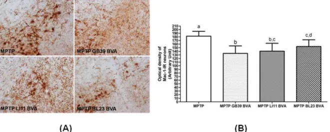
Fig. 3. Effects of BVA on MPTP-induced microglial activation of MAC-1-immunoreactive(IR) neurons in the substantia nigra pars compacta(SNpc)
(A) Mac-1-IR neurons on day 10 after the 4thMPTPi.p.injection.
(B) The optical density levels of MAC-1-IR neurons on day 10.
other groups. In the MPTP GB
39
and BL23
BVA group, the survival TH-IR neurons was 30%greater than in the MPTP group, which was statistically significant(Fig. 2B).
The levels are expressed as the average number of TH-IR caudeus-putamen neurons per section in the MPTP group. The average number was signifi- cantly greater in all MPTP+BVA groups than in the MPTP group. Values are means±S.D.(Bonferroni’s multiple range test, α = 0.05).
2. BVA inhibits the MPTP-induced activation of MAC-1-IR neurons in the SNpc
The SNpc is relatively rich in microglia com- pared with other brain regions
32,33)
. Previous studies have suggested that activation of microglia may trigger or participate in the neurodegenerative pro- cesses in PD19)
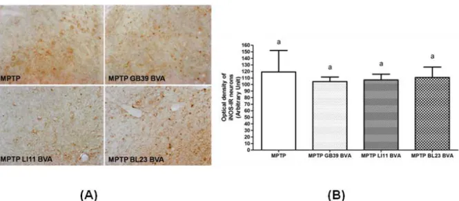
. To determine whether the beneficial effect of BVA was associated with inhibition of theFig. 4. Effects of BVA on MPTP-induced microglial activation of iNOS-immunoreactive(IR) neurons in the substantia nigra pars compacta(SNpc)
(A) INOS-IR neurons of on day 10 after the 4thMPTPinjection.
(B) The optical density levels of iNOS-IR neurons on day 10.
MPTP-induced glial response, we examined the expression of MAC-1, a marker of microglial activation, 10days after the last MPTP injection. In the MPTP group, marked expression of MAC-1 and an increase in dendritic processes surrounding the MAC-1-IR neurons were observed. In contrast, the optical density and expression of activated microglia were decreased in the MPTP+BVA groups, compared with the MPTP group(Fig. 3A).
The activation of MAC-1-IR neurons was de- creased in the MPTP BVA group compared with the MPTP group. In particular, the activation of MAC-1-IR neurons was significantly decreased in the MPTP GB
39
BVA group compared with the MPTP group(Fig. 3B)Optical density was measured in six sections throughout the entire rostrocaudal extent of the SNpc. The levels are expressed as the average optical density of MAC-1-IR neurons per section.
The optical density was statistically significantly lower in all MPTP+BVA groups than in the MPTP group. Data are expressed as mean±S.D. of the average optical density for each section. The means with same letter over the bars is not significantly different(Bonferroni’s multiple range test, α = 0.05).
3. Effects of BVA on MPTP-induced microglial activation of iNOS-IR neurons in the substantia nigra pars compacta(SNpc)
Because iNOS-derived nitric oxide plays a major role in inflammation-mediated neurodegeneration, we measured the expression of iNOS in the SNpc.
To date, one of the best characterized cytotoxic mechanisms induced by proinflammatory cytokines in PD is the activation of iNOS, which mediates the synthesis of high levels of nitric oxide shown to be toxic to neurons
4,7,20)
. MPTP i.p. injection to mice produced a robust gliosis in the SNpc associated with significant up-regulation of iNOS, and these changes paralleled MPTP-induced DA neuroinflam- mation10,31)
.Optical density was measured in six sections throughout the entire rostrocaudal extent of the SNpc. The levels are expressed as the average optical density of iNOS-IR neurons per section(Fig.
4A). The expression and the optical density did not show any significant differences among all groups.
Data are expressed as mean±S.D. of the average optical density for each section. The means with
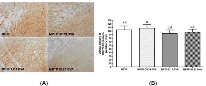
Fig. 5. Effects of BVA on MPTP-induced microglial activation of HSP70-immunoreactive(IR) neurons in the substantia nigra pars compacta(SNpc)
(A) HSP70-IR neurons on day 10 after the 4thMPTPi.p.injection.
(B) The optical density levels of HSP70-IR neurons on day 10
same letter over the bars is not significantly differ- ent(Bonferroni’s multiple range test, α = 0.05).
4. Effects of BVA on MPTP-induced microglial activation of HSP70- immunoreactive(IR) neurons in the substantia nigra pars compacta(SNpc)
Activation of heat shock protein(HSP) synthesis in neurons is an important mechanism for adaptive protection of cells in MPTP-induced neurotoxicity
29,34)
. To define the temporal relationship between the effect of BVA and HSP synthesis, HSP70 was assessed in MPTP-induced DA cell loss on day 10 day after the last MPTP injection. The optical density was decreased in the MPTP LI11
BVA group than in the MPTP group(Fig. 5).Optical density was measured in six sections throughout the entire rostrocaudal extent of the SNpc. The levels are expressed as the average optical density of HSP70-IR neurons per section.
The optical density was lower in the MPTP LI
11
BVA group than in the MPTP group. Data are expressed as mean±S.D. of the average optical density for each section. The means with same letter over the bars is not significantly different (Bonferroni’s multiple range test, α = 0.05).
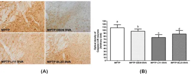
5. Effects of BVA on the expression of caspase 3–IR neurons in MPTP- induced SNpc neurodegeneration
Apoptosis represents a morphologically and bio- chemically distinct form of programmed cell death that was originally recognized to play a consi- derable role in developmental cell death
18)
. Apoptotic cell death was found to be initiated within 72h of the first injection of the neurotoxin, and to peak 24 h after the last MPTP injection35)
. Caspase 3-IR neurons are effectors of SNpc neuron apoptosis36)
, and caspase 3-IR neuron expression is more sensitive to the pathological process in the SNpc, which is induced by MPTP injection37)
.The optical density of caspase 3-IR neurons was significantly decreased in the MPTP LI
11
BVA group compared to the MPTP and other MPTP BVA groups(Fig. 6).Optical density was measured in six sections throughout the entire rostrocaudal extent of the SNpc. The levels are expressed as the average optical density of caspase 3-IR neurons per section.
The optical density was lower in all MPTP+BVA groups than in the MPTP group where the density of MPTP LI
11
BVA was the lowest. Data are expressed as mean±S.D. of the average opticalFig 6. Effects of BVA on MPTP-induced microglial activation of caspase 3-immunoreactive(IR) neurons in the substantia nigra pars compacta(SNpc)
(A) caspase 3-IR neurons on day 10 after the 4thMPTPi.p.injection.
(B) The optical density levels of caspase 3-IR neurons on day 10.
density for each section. The means with different letter over the bars is significantly different in every pair(Bonferroni’s multiple range test, α = 0.05) In the Bonferroni’s multiple range test, the difference between MPTP group and MPTP LI
11
BVA group was the largest(mean difference = 31.920±2.689,
p
=0.000) among every pairwise com- parision.Ⅳ. Discussion
PD is the result of a quite specific and pro- gressive neurodegeneration of pigmented nigrostriatal dopaminergic neurons. The symptoms of PD are only apparent after the loss of at least 50% of the dopaminergic neurons in the substantia nigra pars compacta(SNpc), which leads to a reduction of over 80% in the dopamine(DA) levels in the striatum
38,39)
. The cause of PD remains unclear, but several theories have been proposed regarding the possible factors behind the neuronal degeneration. These include environmental toxins, genetic factors and mitochondrial dysfunction as well as free radical- mediated cell death and oxidative stress40-43)
. Recently, however, there has been increasing recognition of the possible major role of neuroinflammation in thepathogenesis of PD
44)
, induced by exposure to either infectious agents or toxicants with proinflammatory characteristics.Microglia are resident immunocompetent and phagocytic cells in the central nervous system (CNS), and are thought to mediate the innate defense system and thus serve a critical role in normal CNS function
45)
. They are activated in the event of infection, inflammation, trauma, ischemia and neurodegeneration in the CNS46)
. It has been shown that glial cells are activated in the PD brain and MPTP-induced glial activation in association with oxidative stress22)
. MPTP is a neurotoxin that induces Parkinsonian features in humans, rodents and non-human primates and has been demon- strated to cause rapid and selective DA neuro- toxicity47)
. Recent studies suggested that BV has anti-inflammatory properties that inhibit production of inflammatory cytokines and NO in the activated microglia11,12)
. Acupuncture also inhibits microglial activation and inflammatory events in the MPTP- induced mouse model48)
.The heat shock proteins(HSPs) are so named due to the fact that their synthesis was initially found to be enhanced in response to an increase in temperature
49)
. HSPs are a group of highly con- served stress proteins that play an important role in maintaining body self-stability. The main func-tions of HSPs are to promote cellular tolerance against stress factors, to maintain normal cellular physiological function, and to increase cellular defense and adaptation to deadly stimulation
50)
. It is known that a plethora of insults other than heat stress have been found to increase the expression of HSPs including neurotoxicants, drugs of abuse, environmental stress, metals, oxidative stress and many other pathophysiological states and non- stressful conditions. Previous data from in vivo and in vitro studies strongly suggest that HSPs play a neuroprotective role in MPTP-induced neurotoxicity34)
. Moxibustion and electroacupuncture can induce the synthesis of heat shock protein 7051,52)
. However, to date no studies have examined the relationship between BVA and HSPs.In the present study, BVA resulted in pre- servation of TH-IR neurons on day 10 after MPTP i.p. injection. A strong relationship was observed between the damage incurred to DA neurons and the intensity of MAC-1 expression that was present in the activated microglia of the SNpc.
BVA also decreased the expression of caspase 3-IR neurons, which are responsible for developmental cell death
11)
.Therefore, along with the cited findings for BVA, our results supported the hypothesis that BVA may attenuate inflammatory activities in the SNpc induced by MPTP administration. Considering the role of microglia in mediating neurodegeneration, these results suggest that BVA might be an attractive alternative treatment to suppress the development or progression of chronic inflammatory diseases of the central nervous system, such as PD, multiple sclerosis, and amyotrophic lateral sclerosis.
This study showed that MPTP-induced mouse model to examine whether BVA inhibits the loss of tyrosine hydroxylase(TH)-positive neurons as a result of its inhibition of microglial activation or induction of HSP70 and the effect of BVA is acupoint dependent. Microglial activation was measured by the expression of MAC-1. Due to the strain-dependent sensitivity to MPTP
53)
, we used C57BL/6 mice.TH is the rate-limiting enzyme in the synthesis of the catecholamine neurotransmitters, such as dopamine, epinephrine, and norepinephrine. It converts L-tyrosine to L-dihydroxyphenylalanine(L-DOPA), the rate-limiting step in the synthesis of dopa- mine
54)
. Since TH is a rate-limiting enzyme for the biosynthesis of dopamine, TH activity is progres- sively decreased following the loss of dopamine neurons in the substantia nigra in patients with PD55)
. TH immunohistochemistry has widely been used as an important method of detecting the injury or death of dopaminergic fibers and cell bodies56,57)
.Previous studies using rats showed that acu- puncture at the GB
34
, LR3
and ST36
acupoints reduced the degeneration of dopaminergic neurons induced by 6-hydroxydopamine(6-OHDA)58)
. GB34
and LR
3
also protected against MPTP induced dopaminergic neuronal cell damage48)
.In our study, BVA inhibited MPTP-induced loss of TH-IR neurons and activation of MAC-1-IR neurons in the SNpc. These results demonstrate that BVA possesses a potent suppressive effect on microglial activation and suggest that BVA may offer substantial therapeutic potential for the treat- ment of neurodegenerative diseases that are accom- panied by microglial activation. HSP70-IR cells were lower in the MPTP LI
11
BVA group than in the MPTP group, but there are no significant differences between the groups.V. Conclusions
We found that BVA inhibited MPTP-induced neuronal loss of tyrosine hydroxylase(TH)-positive neurons as a result of its inhibition of microglial activation or induction of HSP70 in the SNpc of a mouse PD model. And the effect of BVA is acupoint dependent.
In addition, BVA suppressed microglial activ- ation, which was associated and colocalized with an increase in MAC-1, iNOS and HSP-70 expression.
Furthermore, BVA prevented MPTP-induced apo-
ptosis of DA neurons via caspase-3 inhibition.
Ⅵ. References
1. Lee JD, Park HJ, Chae Y, Lim S. An Overview of Bee Venom Acupuncture in the Treatment of Arthritis. Evid Based Complement Alternat Med.
2005 ; 2 : 79-84.
2. Goldberg A, Confino-Cohen R. Effectiveness of maintenance bee venom immunotherapy administered at 6-month intervals. Ann Allergy Asthma Immunol. 2007 ; 99 : 352-7.
3. Castro HJ, Mendez-Lnocencio JI, Omidvar B, Om- idvar J, Santilli J, Nielsen HS Jr Pavot AP, Richert JR, Bellanti JA. A phase I study of the safety of honeybee venom extract as a possible treatment for patients with progressive forms of multiple sclerosis. Allergy Asthma Proc. 2005 ; 26 : 470-6.
4. Mirshafiey A. Venom therapy in multiple sclerosis.
Neuropharmacology. 2007 ; 53(3) : 353-61.
5. Shinto L, Calabrese C, Morris C, Sinsheimer S, Bourdette D. Complementary and alternative medicine in multiple sclerosis: survey of licensed naturopaths. J Altern Complement Med. 2004 ; 10 : 891-7.
6. Wesselius T, Heersema DJ, Mostert JP, Heerings M, Admiraal-Behloul F, Talebian A, Van Buchem MA, De Keyser J. A randomized crossover study of bee sting therapy for multiple sclerosis.
Neurology. 2005 ; 65 : 1764-8.
7. Eiseman JL, von Bredow J, Alvares AP. Effect of honeybee (Apis mellifera) venom on the course of adjuvant-induced arthritis and depression of drug metabolism in the rat. Biochem Pharmacol. 1982 ; 31(6) : 1139-46.
8. Kwon YB, Lee HJ, Han HJ, Mar WC, Kang SK, Yoon OB, Beitz AJ, Lee JH. The water-oluble fraction of bee venom produces anti-nociceptive and anti-inflammatory effects on rheumatoid arthritis in rats. Life Sci. 2002 ; 71(2) : 191-204.
9. Schmidt JO. Biochemistry of insect venoms. Annu
Rev Entomol. 1982 ; 27 : 339-68.
10. Lariviere WR, Melzack R. The bee venom test: a new tonic-pain test. Pain. 1996 ; 66(2-3) : 271-7.
11. Moon DO, Park SY, Lee KJ, Heo MS, Kim KC, Kim MO, Lee JD, Choi YH, Kim GY. Bee venom and melittin reduce proinflammatory mediators in lipopolysaccharide-stimulated BV2 microglia. Int Immunopharmacol. 2007 ; 7(8) : 1092-101.
12. Han S, Lee K, Yeo J, Kweon H, Woo S, Lee M, Baek H, Kim S, Park K. Effect of honey bee venom on microglial cells nitric oxide and tumor necrosis factor-alpha production stimulated by LPS. J Ethnopharmacol. 2007 ; 111(1) : 176-81.
13. Dauer W, Przedborski S. Parkinson’s disease : mech- anisms and models. Neuron. 2003 ; 39 : 889-909.
14. Dehmer T, Lindenau J, Haid S, Dichgans J, Schulz JB, Deficiency of inducible nitric oxide synthase protects against MPTP toxicity in vivo. J Neurochem. 2000 ; 74 : 2213-6.
15. Hunot S, Hirsch EC, Neuroinflammatory processes in Parkinson’ disease Ann Neurol. 2003 ; 53 : Suppl 3 S49-58 ; discussion S58-60.
16. Wilms H, Zecca L, Rosenstiel P, Sievers J, Deuschl G, Lucius R. Inflammation in Parkinson’s diseases and other neurodegenerative diseases : cause and therapeutic implications. Curr Pharm Des. 2007 ; 13 : 1925-8.
17. Wu DC, Teismann P, Tieu K, Vila M, Jackson- Lewis V, Ischiropoulos H, Przedborski S. NADPH oxidase mediates oxidative stress in the 1- methyl- 4-phenyl-1,2,3,6-tetrahydropyridine model of Parkinson’s disease. Proc Natl Acad Sci USA.
2003 ; 100 : 6145-50.
18. Eberhardt O, Schulz JB, Apoptotic mechanisms and antiapoptotic therapy in the MPTP model of Parkinson’s disease. Toxicol Lett. 2003 ; 139 : 135-51.
19. Teismann P, Schulz JB. Cellular pathology of Parkinson’s disease : astrocytes, microglia and inflammation. Cell Tissue Res. 2004 ; 318(1) : 149-61.
20. Aloisi F. Immune function of microglia. Glia. 2001 ; 36(2) : 165-79.
21. Nakajima K, Kohsaka S. Microglia: activation and
their significance in the central nervous system. J Biochem. 2001 ; 130(2) : 169-75.
22. Kohutnicka M, Lewandowska E, Kurkowska- Jastrzebska I, Czlonkowski A, Czlonkowska A.
Microglial and astrocytic involvement in a murine model of Parkinson’s disease induced by 1-methyl -4-phenyl-1,2,3,6-tetrahydropyridine(MPTP).
Immunopharmacology. 1998 ; 39(3) : 167-80.
23. McGeer PL, McGeer EG. Glial reactions in Par- kinson’s disease. Mov Disord. 2008 ; 23 : 474-83.
24. Przedborski S, Vila M. The 1-methyl-4-phenyl- 1,2,3,6-tetrahydropyridine mouse model : a tool to explore the pathogenesis of Parkinson’s disease.
Ann N Y Acad Sci. 2003 ; 991 : 189-98.
25. Cassarino DS, Fall CP, Swerdlow RH, Smith TS, Halvorsen EM, Miller SW, Parks JP, Parker WD Jr, Bennett JP Jr. Elevated reactive oxygen species and antioxidant enzyme activities in animal and cellular models of Parkinson’s disease. Biochim Biophys Acta. 1997 ; 1362 : 77-86.
26. Teismann P, Vila M, Choi DK, Tieu K, Wu DC, Jackson-Lewis V, Przedborski S, COX-2 and neurodegeneration in Parkinson’s disease. Ann NY Acad Sci. 2003 ; 991 : 272-7.
27. Wu C. Heat shock transcription factors: structure and regulation. Annu Rev Cell Dev Biol. 1995 ; 11 : 441-69.
28. Wegele H, Muller L, Buchner J. Hsp70 and Hsp90―a relay team for protein folding. Rev Physiol Biochem Pharmacol. 2004 ; 151 : 1-44.
29. Morimoto RI, Santoro MG. Stress-inducible re- sponses and heat shock proteins: new pharma- cologic targets for cytoprotection. Nat Biotechno.l 1998 ; 16(9) : 833-8.
30. Jackson-Lewis V, Przedborski S. Protocol for the MPTP mouse model of Parkinson’s disease. Nat Protoc. 2007 ; 2(1) : 141-51.
31. Burke RE, Cadet JL, Kent JD, Karanas AL, Jackson-Lewis V. An assessment of the validity of densitometric measures of striatal tyrosine hydroxylase-positive fibers : relationship to apomorphine-induced rotations in 6-hydroxy- dopamine lesioned rats. J Neurosci Methods. 1990 ; 35(1) : 63-73.
32. Lawson LJ, Perry VH, Dri P, Gordon S.
Heterogeneity in the distribution and morphology of microglia in the normal adult mouse brain.
Neuroscience. 1990 ; 39(1) : 151-70.
33. Kim WG, Mohney RP, Wilson B, Jeohn GH, Liu B, Hong JS. Regional difference in susceptibility to lipopolysaccharide-induced neurotoxicity in the rat brain: role of microglia. J Neurosci. 2000 ; 20(16) : 6309-16.
34. Freyaldenhoven TE, Ali SF. Role of heat shock proteins in MPTP-induced neurotoxicity. Ann N Y Acad Sci. 1997 ; 825 : 167-78.
35. Tatton NA, Kish SJ, In situ detection of apoptotic nuclei in the substantia nigra compacta of 1-methyl-4-phenyl-1,2,3,6-tetrahydropyridinetreate d mice using terminal deoxynucleotidyl transferase labelling and acridine orange staining.
Neuroscience. 1997 ; 77 : 1037-48.
36. Eberhardt O, Coelln RV, Kugler S, Lindenau J, Rathke-Hartlieb S, Gerhardt E, Haid S, Isenmann S, Gravel C, Srinivasan A, Bahr M, Weller M, Dichgans J, Schulz JB. Protection by synergistic effects of adenovirus-mediated X- chromosome- linked inhibitor of apoptosis and glial cell line- derived neurotrophic factor gene transfer in the 1-methyl-4-phenyl-1,2,3,6-tetrahydropyridine model of Parkinson’s disease. J Neurosci. 2000 ; 20 : 9126-34.
37. Hartmann A, Hunot S, Michel PP, Muriel MP, Vyas S, Faucheux BA, Mouatt-Prigent A, Turmel H, Srinivasan A, Ruberg M, Evan GI, Agid Y, Hirsch EC. Caspase-3 : A vulnerability factor and final effector in apoptotic death of dopaminergic neurons in Parkinson’s disease. Proc Natl Acad Sci USA. 2000 ; 97 : 2875-80.
38. Lang AE, Lozano AM. Parkinson’s disease. First of two parts. N Engl J Med. 1998 ; 339(15) : 1044-53.
39. Deumens R, Blokland A, Prickaerts J. Modeling Parkinson’s disease in rats : an evaluation of 6-OHDA lesions of the nigrostriatal pathway. Exp Neurol. 2002 ; 175(2) : 303-17.
40. Schapira AH. Evidence for mitochondrial dysfunc- tion in Parkinson’s disease―a critical appraisal.
Mov Disord. 1994 ; 9(2) : 125-38.
41. Ben-Shachar D, Zuk R, Glinka Y. Dopamine neuro- toxicity : inhibition of mitochondrial respiration. J Neurochem. 1995 ; 64(2) : 718-23.
42. Hoehn MM, Yahr MD. Parkinsonism : onset, progression, and mortality. Neurology. 1998 ; 50(2) : 318-34.
43. Rosenberg RN. Mitochondrial therapy for Parkinson disease. Arch Neurol. 2002 ; 59(10) : 1523.
44. Mc Geer PL, Yasojima K, Mc Geer EG. Inflam- mation in Parkinson’s disease. Adv Neurol. 2001 ; 86 : 83-9.
45. Kim SU, de Vellis J. Microglia in health and disease. J Neurosci Res. 2005 ; 81(3) : 302-13.
46. Beyer M, Gimsa U, Eyupoglu IY, Hailer NP, Nitsch R. Phagocytosis of neuronal or glial debris by microglial cells: upregulation of MHC class II expression and multinuclear giant cell formation in vitro. Glia. 2000 ; 31(3) : 262-6.
47. Langston JW, Forno LS, Tetrud J, Reeves AG, Kaplan JA, Karluk D. Evidence of active nerve cell degeneration in the substantia nigra of humans years after 1-methyl-4-phenyl-1,2,3,6- tetrahydropyridine exposure. Ann Neurol. 1999 : 46(4) : 598-605.
48. Kang JM, Park HJ, Choi YG, Choe IH, Park JH, Kim YS, Lim S. Acupuncture inhibits microglial activation and inflammatory events in the MPTP- induced mouse model. Brain Res. 2007 ; 1131(1) : 211-9.
49. Tissieres A, Mitchell HK, Tracy UM. Proteinsyn the sis in salivary glands of Drosophila melanogaster:
relation to chromosome puffs. J Mol Biol. 1974 ; 84(3) : 389-98.
50. Takumida M, Anniko M. Heat shock protein 70 delays gentamicin-induced vestibular hair cell death. Acta Otolaryngol. 2005 ; 125(1) : 23-8.
51. Kobayashi K. Induction of heat-shock protein(hsp) by moxibustion. Am J Chin Med. 1995 ; 23(3-4) :
327-30.
52. Sun N, Shi J, Chen L, Liu X, Guan X. Influence of electroacupuncture on the mRNA of heat shock protein 70 and 90 in brain after cerebral ischemia/
reperfusion of rats. J Huazhong Univ Sci Technolog Med Sci. 2003 : 23(2) : 112-5.
53. Boyd JD, Jang H, Shepherd KR, Faherty C, Slack S, Jiao Y, Smeyne RJ. Response to 1-methyl-4- phenyl-1,2,3,6-tetrahydropyridine (MPTP) differs in mouse strains and reveals a divergence in JNK signaling and COX-2 induction prior to loss of neurons in the substantia nigra pars compacta.
Brain Res. 2007 ; 1175 : 107-116.
54. Asanuma M, MiyazakiI,OgawaN.Dopamine-orL- DOPA-induced neurotoxicity : the role of dopamine quinone formationand tyrosinase in a model of Parkinson’s disease. Neurotox Res 2003 ; 5(3) : 165-76.
55. Haavik J, Toska K. Tyrosine hydroxylase and Parkinson’s disease. Mol Neurobiol. 1998 : 16(3) : 285-309.
56. Hurley FM, Costello DJ, Sullivan AM. Neuro- protective effects of delayed administration of growth/ differentiation factor-5 in the partial lesion model of Parkinson’s disease. Exp Neurol.
2004 ; 185(2) : 281-9.
57. Oiwa Y, Yoshimura R, Nakai K, Itakura T.
Dopaminergic neuroprotection and regeneration by neurturin assessed by using behavioral, bio- chemical and histochemical measurements in a model of progressive Parkinson’s disease. Brain Res. 2002 ; 947(2) : 271-83.
58. Park HJ, Lim S, Joo WS, Yin CS, Lee HS, Lee HJ, Seo JC, Leem K, Son YS, Kim YJ, Kim CJ, Kim YS, Chung JH. Acupuncture prevents 6- hydroxydopamine-induced neuronal death in the nigrostriatal dopaminergic system in the rat Parkinson’s disease model. Exp Neurol. 2003 ; 180(1) : 93-8.