INTRODUCTION
Parkinson’s disease (PD) is a neurodegenerative disease characterized by impaired movement, muscular rigidity, rest-ing tremor, bradykinesia, and postural instability (Przedborski, 2017). Along with nigrostriatal dopaminergic degeneration and cytoplasmic aggregate inclusion, persistent neuroinflamma-tion is a neuropathological feature of PD and contributes to the progression of PD (Liu et al., 2019; Pajares et al., 2020). Therefore, neuroinflammation is considered a potential thera-peutic target in the treatment of PD.
Microglia are usually primed by pathological conditions, resulting in an amplified immune response to secondary in-flammatory challenges (Perry and Teeling, 2013; Neher and Cunningham, 2019). The pathological significance of primed microglia both in aging and in neurodegenerative diseases has previously been studied (Perry and Holmes, 2014; Norden
et al., 2015). Microglial priming has been particularly observed in PD patients and rodent models (Langston et al., 1999; Perry and Holmes, 2014). Elevated levels of pro-inflammatory cy-tokines such as tumor necrosis factor-α (TNF-α), interleukin-1β (IL-interleukin-1β), and IL-18 have been observed in the striatum and cerebrospinal fluid of PD patients and in the substantia nigra (SN) of PD animal models (Mogi et al., 1996; Hébert et al., 2003). Primed microglia aggravated nigrostriatal degeneration during subsequent immune challenge and further increased the release of pro-inflammatory cytokines such as IL-1β com-pared to non-primed cells (Pott Godoy et al., 2008; Dwyer et al., 2020). Several studies have demonstrated that IL-1β is a key component of the inflammatory response that leads to PD onset or exacerbation (Koprich et al., 2008; Pott Godoy et al., 2008).
The activity of IL-1β is particularly controlled by a cytosolic multimolecular complex termed ‘inflammasome,’ which con-Microglial priming is the process of microglial proliferation and activation in response to neurodegeneration and abnormal protein accumulation. Priming makes microglia susceptible to secondary inflammatory stimuli and causes exaggerated inflammatory responses. In the present study, we established a microglial priming model in mice by administering a single injection of 1-methyl-4-phenyl-1,2,3,6-tetrahydropyridine (MPTP, 20 mg/kg). MPTP induced microglial activation without dopaminergic degeneration; however, subsequent treatment with a sub-toxic dose of lipopolysaccharides (LPS) induced an amplified inflammatory response and caused nigrostriatal dopaminergic degeneration. These pathological and inflammatory changes, including microglial activa-tion and dopaminergic cell loss in the substantia nigra (SN) area were reversed by papaverine (PAP) administraactiva-tion. In addiactiva-tion, MPTP/LPS enhanced interleukin-1β (IL-1β) expression and processing via nod-like receptor protein 3 (NLRP3) inflammasome ac-tivation in the SN region of mice. However, PAP treatment suppressed inflammasome acac-tivation and subsequent IL-1β maturation. Moreover, PAP inhibited nuclear factor-κB (NF-κB) and enhanced cAMP-response element binding protein (CREB) activity in the SN of MPTP/LPS mice. These results suggest that PAP inhibits the activation of NLRP3 inflammasome by modulating NF-κB and CREB signaling pathways, which results in reduced microglial activation and neuronal cell death. Thus, PAP may be a potential candidate for the treatment of Parkinsons’s disease, which is aggravated by systemic inflammation.
Key Words: Parkinson’s disease, Microglial priming, Systemic inflammation, Papaverine, NLRP3 inflammasome, Neuronal cell death
Abstract
*
Corresponding Author E-mail: [email protected]Tel: +82-2-6986-6270, Fax: +82-2-6986-7014
Received Feb 26, 2021 Revised Mar 23, 2021 Accepted Mar 26, 2021 Published Online May 1, 2021
Copyright © 2021 The Korean Society of Applied Pharmacology https://doi.org/10.4062/biomolther.2021.039 Open Access
This is an Open Access article distributed under the terms of the Creative Com-mons Attribution Non-Commercial License (http://creativecomCom-mons.org/licens- (http://creativecommons.org/licens-es/by-nc/4.0/) which permits unrestricted non-commercial use, distribution, and reproduction in any medium, provided the original work is properly cited.
www.biomolther.org
Papaverine Exerts Neuroprotective Effect by Inhibiting NLRP3
Inflammasome Activation in an MPTP-Induced Microglial Priming
Mouse Model Challenged with LPS
Yea-Hyun Leem, Jin-Sun Park, Jung-Eun Park, Do-Yeon Kim and Hee-Sun Kim
*
Department of Molecular Medicine and the Ewha Medical Research Institute, School of Medicine, Ewha Womans University, Seoul 07804, Republic of Korea
tains nod-like receptor protein 3 (NLRP3), adaptor protein ASC, and pro-caspase-1 (Martinon et al., 2002; Seok et al., 2021). The inflammasome proceeds in two steps: the prim-ing step first upregulates the expression of inflammasome components, including NLRP3 and pro-IL-1β/IL-18, and the activation step then induces caspase-1-dependent proteolytic maturation of IL-1β and IL-18 (Swanson et al., 2019). NLRP3, caspase-1, and IL-1β levels are found to be elevated in PD pa-tients and several studies have reported the role of inflamma-somes in PD pathogenesis (Mogi et al., 2000; von Herrmann et al., 2018). Martinez et al. (2017) reported that NLRP3 causes inflammatory changes and nigral cell loss in rotenone-exposed mice. The dopaminergic degeneration in 1-methyl-4-phenyl-1,2,3,6-tetrahydropyridine (MPTP)-treated mice was rescued by suppressing inflammasomes, wherein the chemi-cal inhibition or genetic deletion of NLRP3 mitigated neuroin-flammation and inhibited α-synuclein aggregation (Gordon et al., 2018; Lee et al., 2019a). Wang et al. (2016) reported that caspase-1 induces truncation and aggregation of α-synuclein, which is a PD-associated protein. Thus, it is essential to devel-op therapeutic agents that can control NLRP3 inflammasome and the subsequent IL-1β processing to prevent neuroinflam-mation and dopaminergic neuronal cell death.
In the present study, we examined the effect of papaver-ine (PAP; 6,7-dimethoxy-1-veratryl-isoquinolpapaver-ine) in an MPTP-induced microglial priming mouse model challenged with lipo-polysaccharide (LPS). PAP is a non-narcotic opium alkaloid isolated from Papaver somniferum, and is clinically used to treat conditions associated with spasm and dysmotility of the gastrointestinal tract (Han et al., 2010; Zhu et al., 2014). PAP is a selective inhibitor of phosphodiesterase 10A (PDE10A) that is highly expressed in the striatum, SN, and olfactory tubercle (Wilson and Brandon, 2015). In this study, we demonstrated that PAP inhibits microglial activation and dopaminergic neu-ronal cell death by modulating NLRP3 inflammasome activa-tion in MPTP/LPS mice; this indicates the therapeutic potential of PAP in PD, which is exacerbated by systemic inflammation.
MATERIALS AND METHODS
Animals
Adult male C57BL/6 mice (9 weeks of age) were purchased from Orient Bio Inc. (Seongnam, Korea), a branch of Charles River Laboratories. Mice were maintained at 21°C under a 12 h light:12 h dark cycle and had ad libitum access to water and rodent chow. Every effort was made to minimize animal suffering. All experiments were performed in accordance with the National Institutes of Health (NIH, Bethesda, MD, USA) and Ewha Womans University (Seoul, Korea) guidelines for laboratory animal care and use, and the study was approved by the Institutional Animal Care and Use Committee of the Medical School of Ewha Womans University (#EUM 20-022).
MPTP-induced microglia priming and drug administration
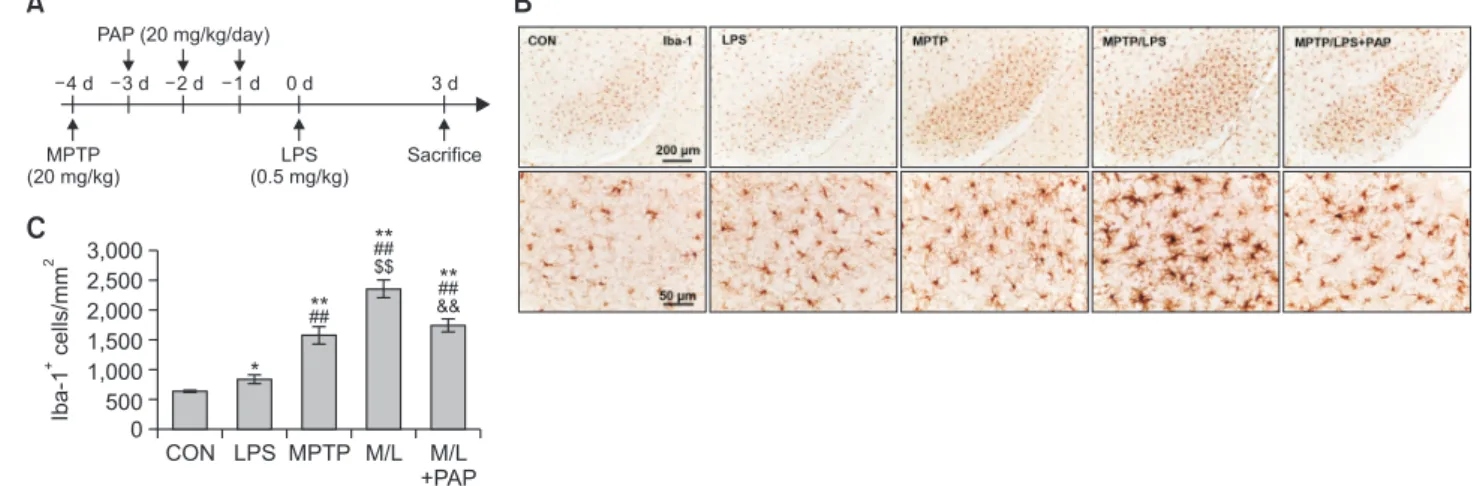
Mice were randomly divided into the following five groups (N=10-16): 1. CON (control); 2. MPTP; 3. LPS; 4. MPTP/LPS, MPTP+LPS; 5. MPTP/LPS+PAP, MPTP+LPS+papaverine. Mice were administered MPTP (20 mg/kg, i.p.), followed by 3 con-secutive days of PAP administration (20 mg/kg, i.p.). Mice were administered LPS (0.5 mg/kg, i.p.) 1 d after the last treatment with PAP and were sacrificed 3 days after LPS administration. LPS and PAP were obtained from Sigma-Aldrich (St. Louis, MO, USA). MPTP was purchased from Tokyo Chemical Industry Co (Tokyo, Japan). A schematic of this procedure is shown in Fig. 1A.
Histological analysis
Mice were anesthetized with sodium pentobarbital (80 mg/ kg; Hanlim Pharm Co. Ltd., Seoul, Korea) to induce rapid and prolonged anesthesia. The mice were then perfused transcar-dially, their brains were removed, and 40 µm-thick sections were prepared using a cryotome. For immunohistochemistry (IHC) analysis, the sections were subjected to endogenous peroxidation inactivation with 3% hydrogen peroxide (H2O2),
and non-specific binding was blocked with 4% bovine serum albumin (BSA). The sections were first incubated overnight
CON 3,000 2,500 2,000 1,500 1,000 500 Iba-1 cells/mm + 2 0 LPS MPTP M/L M/L +PAP
A
B
C
MPTP (20 mg/kg) LPS (0.5 mg/kg) Sacrifice 4 d 3 d 2 d 1 d 0 d PAP (20 mg/kg/day) 3 d * ## ** $$ ##** && ## **Fig. 1. PAP attenuated hyperreactive microglia in the brains of MPTP/LPS-treated mice. (A) The experimental procedure: Mice were
sub-jected to a single injection of MPTP (20 mg/kg), followed by administration of PAP (20 mg/kg/day) for three days. The mice were thentreat-ed with a low dose of LPS (i.p., 0.5 mg/kg), and sacrifictreat-ed three days later. (B) Photomicrographs of the immunoreactivity of SN Iba-1+ cells
(N=5-8 in each group). (C) Quantification of the immunoreactivity of SN Iba-1+ cells. Statisctical analyses were performed for all data using
ANOVA with post-hoc test of the LSD. Data are presented as mean ± SEM. *p<0.05, vs. CON; **p<0.01, vs. CON; ##p<0.01, vs. LPS;
with primary antibodies and were then incubated with biotinyl-ated secondary antibodies for 1 h at 25°C, on the following day. The sections were subsequently incubated with avidin-biotin-horse radish peroxidase (HRP) complex reagent solu-tion for 1.5 h (Vector Laboratories, Burlingame, CA, USA) and a peroxidase reaction was then performed using diaminoben-zidine tetrahydrochloride (Vector Laboratories). For double immunofluorescence (IF) analysis, non-specific binding was blocked, and the sections were incubated with primary bodies followed by fluorochrome-conjugated secondary anti-bodies. The tissue was then mounted with an antifade reagent (Vector Laboratories). Digital images of IHC and IF staining were captured using a Leica DM750 microscope (Leica Micro-systems, Nussloch, Germany). Quantification was performed using ImageJ (NIH Image Engineering, Bethesda, MD, USA) and AxioVision (Carl Zeiss Microscopy GmbH, Jena, Ger-many). The following primary antibodies were used in this study: tyrosine hydroxylase (TH), IL-1β, anti-NLRP3, anti-ASC antibodies from Cell Signaling Technology, Inc. (Danvers, MA, USA), anti-ionized calcium-binding adap-tor molecule 1 (anti-Iba1) from Wako (Osaka, Japan), and biotinylated and fluorophore-conjugated secondary antibodies from Vector Laboratories. Anti-CD11b (OX42) was obtained from Bio-Rad (Hercules, CA, USA).
Preparation of protein extracts and western blot analysis
Tissues collected from SN were homogenized in ice-cold radioimmuno precipitation assay (RIPA) lysis buffer (10 mM Tris-Cl [pH 7.4], 300 mM NaCl, 1% Triton X-100, 0.1% sodium dodecyl sulfate [SDS], 0.1% sodium deoxycholate, 1 mM eth-ylenediaminetetraacetic acid) containing a protease inhibitor cocktail (Complete Mini, Roche, Mannheim, Germany). Sub-sequently, the samples were vortexed vigorously and incu-bated for 30 min at 4°C. The samples were then centrifuged at 20,000×g for 30 min, and the supernatant was collected. Protein samples (30-50 µg) were separated by SDS-PAGE, transferred to a nitrocellulose membrane, and incubated with primary antibodies according to the manufacturer’ s in-structions for dilution. The membranes were then thoroughly washed with TBST (tris-buffered saline and tween 20) and incubated with HRP-conjugated secondary antibodies (Bio-Rad; 1:3,000 dilution in TBST). Subsequently, the blots were developed using an enhanced chemiluminescence detection kit (Thermo Fisher Scientific, Waltham, MA, USA). For quan-tification, the density of specific target bands was normalized against β-actin using ImageJ software, version 1.37 (NIH). The following primary antibodies were used in this study: IL-1β, caspase-1, NLRP3, IkB, p-IkB, anti-CREB, and anti-p-CREB from Cell Signaling Technology, Inc. Antibody against β-actin was purchased from Sigma-Aldrich.Statistical analysis
Statistical analyses were performed using SPSS for Win-dows (version 18.0; SPSS Inc., Chicago, IL, USA). The dif-ferences among the groups were analyzed using a one-way analysis of variance (ANOVA). Post-hoc comparisons were conducted using the least significant difference (LSD) test. All values are presented as the mean ± standard error of the mean (SEM). A value of p<0.05 was considered statistically significant.
RESULTS
PAP attenuated hyperreactive microglia in the SN of mice
cotreated with MPTP and LPS
We first established an in vivo microglia priming model, by administering a single injection of MPTP (20 mg/kg) followed by a sub-toxic dose of LPS (0.5 mg/kg) (Fig. 1A). The number of Iba-1+ cells in the MPTP (primed/pre-activated microglia)
or LPS (the second inflammatory stimulator) group were sig-nificantly higher than the CON group, and Iba-1+ cells were
significantly enhanced in the MPTP group compared to the LPS group. The number of Iba-1+ cells in the SN of the MPTP/
LPS group (hyperreactive microglia) increased substantially compared to the LPS and MPTP group, while PAP signifi-cantly reduced Iba-1+ cells (Fig. 1B, 1C; F4, 26=33.23, p<0.01).
Cytoarchitecture revealed increased hypertrophy of microglial processes and soma in the MPTP/LPS group than in the LPS or MPTP groups, which in turn was reversed by PAP (Fig. 1B). The microglial morphology of the LPS group was similar to that of the CON group. This result indicated that the phenotype of MPTP-triggered primed microglia shifted from pre-activated (mildly reactive) to hyperreactive following LPS treatment, while PAP treatment alleviated the exaggerated inflammatory response. Therefore, our experimental paradigm was relevant for exploring the effects of PAP in a primed microglia model.
PAP alleviated nigrostriatal dopaminergic degeneration
in mice cotreated with MPTP and LPS
When we examined the effect of MPTP or LPS treatment on the survival of dopaminergic neurons, the number of TH+
dopaminergic cells did not change significantly compared with the control group (Fig. 2). However, the number of TH+ cells in
the SN of the MPTP/LPS group was profoundly reduced com-pared to the other groups, including the CON, LPS, and MPTP groups; cell death was then recovered by PAP treatment (Fig. 2A, 2B; F4, 27=8.362, p<0.01). In addition, striatal TH+
immu-noreactivity corresponded well with the results from SN (Fig. 2A, 2C; F4, 27=9.09, p<0.01). The results indicated that primed/
pre-activated microglia induced only by MPTP or the second inflammatory stimulus (LPS) did not produce nigrostriatal do-paminergic degeneration, whereas hyperreactive microglia caused nigrostriatal dopaminergic degeneration in MPTP/LPS mice. In addition, PAP probably has an ameliorating effect on dopaminergic degeneration.
PAP reduced microglial IL-1β expression in the SN of mice
cotreated with MPTP and LPS
Next, we examined the effect of MPTP and LPS on the ex-pression of IL-1β, which plays a major role in microglial priming and the exacerbation of neuronal loss (Koprich et al., 2008; Pott Godoy et al., 2008). We found that the IL-1β+
immunoreactivi-ties of the LPS and MPTP groups were significantly enhanced compared to the CON group, and the IL-1β+ fluorescence
in-tensities of the MPTP/LPS group were higher than the LPS or MPTP group (Fig. 3A, 3B; F4, 15=20.97, p<0.01). Moreover, the
area of IL-1β that overlapped with OX42-positive microglial cells significantly increased in the MPTP/LPS group compared to the LPS or MPTP group (Fig. 3A, 3C; F4, 15=17.54, p<0.01). This
result suggests that hyperreactive microglia produce a multi-plied inflammatory response to the subsequent LPS treatment, thereby amplifying IL-1β protein expression in our experimental paradigm. In addition, PAP treatment significantly reduced
IL-1β expression in the MPTP/LPS group, which corroborates the anti-inflammatory effect of PAP.
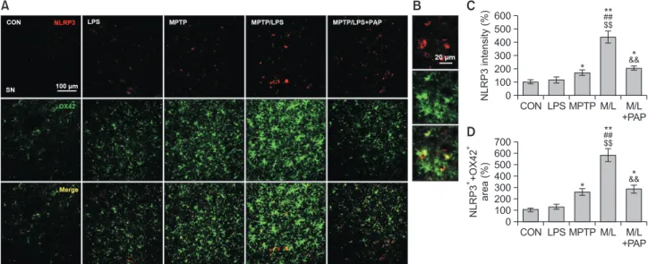
PAP inhibited microglial NLRP3 expression in the SN of
mice cotreated with MPTP and LPS
The changes in IL-1β expression prompted us to measure the expression of microglial NLRP3 to identify the involvement of inflammasomes. We found that NLRP3+ fluorescence
inten-sities of the MPTP/LPS group were considerably augmented compared to the LPS or MPTP group, which was then re-versed by PAP treatment (Fig. 4A-4C; F4, 15=26.75, p<0.01).
In addition, the area of NLRP3+ that was merged with OX42+
fluorescent signal, corresponded to the NLRP3 expression data (Fig. 4A, 4D; F4, 15=46.11, p<0.01). This result indicated
that hyperreactive microglia strongly induced NLRP3 protein expression, whereas PAP dampened microglial NLRP3 ex-pression.
PAP reduced the ASC specks in SN of mice cotreated with
MPTP and LPS
The number of ASC specks, a hallmark of inflammasome activation, in the LPS and MPTP group increased significantly compared to the CON group, while ASC specks of the MPTP/ LPS group were markedly enhanced relative to the LPS or MPTP group (Fig. 5A-5C; F4, 23=13.21, p<0.01). Moreover,
ASC specks in the MPTP/LPS group were reduced by PAP treatment. This result indicates that hyperreactive microglia accelerate inflammasome activation during the subsequent
in-A
CON 120 100 80 60 40 20 SNpc TH cells/mm + 2 0 LPS MPTP M/L M/L +PAPB
$$ ##** && CON 120 100 80 60 40 20 Striatum TH optical density (%) + 0 LPS MPTP M/L M/L +PAPC
$$ ##** &&Fig. 2. PAP alleviated nigrostriatal dopaminergic degeneration in MPTP/LPS-treated mice. (A) Photomicrographs of the immunoreactivity of
SN and striatal TH+ cells (N=6-8 in each group). (B, C) Quantification of the immunoreactivity of SN and striatal TH+ cells. Data are
present-ed as mean ± SEM. **p<0.01, vs. CON; ##p<0.01, vs. LPS; $$p<0.01, vs. MPTP; &&p<0.01, vs. M/L. TH, tyroxine hydroxylase; SNpc, Sub
-stantia nigra par compacta.
A
CON 700 600 500 400 300 200 100 IL-1 intensity (%) 0 LPS MPTP M/L M/L +PAPB
$$ ##** & CON 1,600 1,400 1,200 1,000 800 600 400 200 IL-1 OX42 area (%) + + + 0 LPS MPTP M/L M/L +PAPC
$$ ##** & ** ## ** * ** ## ** **Fig. 3. PAP reduced IL-1β expression in microglia of MPTP/LPS-treated mice. (A) Photomicrographs of the double fluorescent staining of
SN OX42+ and IL-1β+ Particles (N=4 in each group). (B) Quantification of the fluorescent intensity of SN IL-1β+ particles. (C) Quantification
of the area of SN OX42+ and IL-1β+ particles. Data are presented as mean ± SEM. *p<0.05, vs. CON; **p<0.01, vs. CON; ##p<0.01, vs. LPS;
flammatory stimuli, while PAP has an inflammasome-inhibiting effect.
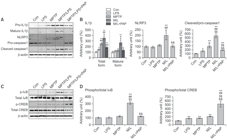
PAP suppressed the expression and activation of NLRP3
inflammasome by modulating NF-κB/CREB signaling in
the SN of mice cotreated with MPTP and LPS
To further investigate the mechanism underlying inflamma-some inhibition by PAP, we performed western blot analysis to determine the protein expression levels of NLRP3 inflamma-some components and its related signaling molecules such as NF-κB and CREB. The results showed that total IL-1β protein levels (pro- and mature IL-1β) in the LPS or MPTP group in-creased significantly compared to the CON group. The cytokine levels in the MPTP/LPS group were higher than in the LPS or MPTP group, which was reversed by PAP treatment (Fig. 6A, 6B; F4, 15=25.21, p<0.01). In accordance with this, mature
IL-1β levels in the MPTP/LPS group were significantly augmented relative to the other groups, which was however significantly attenuated by PAP (Fig. 6A, 6B; F4, 5=8.12, p<0.01). Mature
IL-1β levels in the LPS and MPTP groups were comparable to those in the CON group. PAP significantly inhibited the NLRP3 protein expression induced by MPTP/LPS, while the levels of NLRP3 in the LPS and MPTP groups were similar to those in the CON group (Fig. 6A, 6B; F4, 15=10.87, p<0.01). Caspase-1
activity (cleaved/pro-caspase-1) was profoundly enhanced in the MPTP/LPS group compared to the LPS or MPTP group, which was reversed by PAP (Fig. 6A, 6B; F4, 15=38.52, p<0.01).
With regard to the signaling pathway, MPTP/LPS signifi-cantly increased NF-κB activity (p-IkB/total IkB), which was inhibited by PAP. However, there was no significant differ-ence among the CON, LPS, and MPTP groups (Fig. 6C, 6D; F4, 15=22.58, p<0.01). Conversely, single or combined
treat-ment with MPTP and LPS significantly increased CREB ac-tivity (p-CREB/total CREB), which was augmented by PAP (Fig. 6C, 6D; F4, 15=22.58, p<0.01). These data imply that PAP
suppressed IL-1β expression and inflammasome-dependent IL-1β maturation by inhibiting NF-κB and upregulating CREB signaling in MPTP/LPS-treated mice.
C
CON 600 500 400 300 200 100 NLRP3 intensity (%) 0 LPS MPTP M/L M/L +PAP $$ ##** &&* *D
CON 700 600 500 400 300 200 100 NLRP3 +OX42 area (%) + + 0 LPS MPTP M/L M/L +PAP $$ ##** &&* *A
B
Fig. 4. PAP inhibited NLRP3 expression in microglia of MPTP/LPS-treated mice. (A) Photomicrographs of the double fluorescent staining of
SN OX42+ and NLRP3+ particles (N=4 in each group). (B) The high-magnitude of image. (C) Quantification of the fluorescent intensity of SN
NLRP3+ particles. (D) Quantification of the area of SN OX42+ and NLRP3+ particles. Data are presented as mean ± SEM. *p<0.05, vs. CON;
**p<0.01, vs. CON; ##p<0.01, vs. LPS; $$p<0.01, vs. MPTP; &&p<0.01, vs. M/L. CO N 700 600 500 400 300 200 100 ASC specks (%) 0 LPS MPTP M/ L M/L+P AP
C
$$ ##** & ** ** *A
B
Fig. 5. PAP reduced ASC specks in MPTP/LPS-treated mice. (A) Photomicrographs of the double fluorescent staining of SN ASC
+ andDAPI+ particles (N=5-7 in each group). (B) The high-magnitude of image. In non-assembly, ASC is localized to the cytoplasm and nucleus of
the cell (white arrow head). During inflammasome activation, one large ASC speck (around 1 µm in diameter) assembles in the perinuclear area of the cell (yellow arrow). (C) Quantification of the number of SN ASC specks. Data are presented as mean ± SEM. *p<0.05, vs. CON;
DISCUSSION
In the current study, we demonstrated that MPTP-induced microglial priming exacerbated nigrostriatal dopaminergic de-generation and amplified the inflammatory response to sub-sequent systemic inflammation induced by a sub-toxic dose of LPS. The priming effects of MPTP were alleviated by the administration of PAP. Thus, PAP inhibited microglial activa-tion and dopaminergic neuronal cell death in MPTP/LPS-treat-ed mice. PAP administration particularly suppressMPTP/LPS-treat-ed NLRP3 inflammasome-dependent IL-1β processing, which potentially contributes to the anti-inflammatory and neuroprotective ef-fects of PAP.
PAP, which is a selective PDE10 inhibitor, inhibits phospho-diesterase activity and increases the intracellular cAMP level (Zagorska et al., 2018). Previous studies have reported that cAMP negatively regulates NLRP3 inflammasome activation (Lee et al., 2012; Yan et al., 2015). Yan et al. (2015) demon-strated that cAMP binds to NLRP3 and subsequently induces NLRP3 ubiquitination/degradation in bone marrow macro-phages and in mouse models of MPTP-induced neuroinflam-mation, LPS-induced systemic inflamneuroinflam-mation, and MSU-in-duced peritoneal inflammation. Consistent with these studies, we showed that PAP inhibited NLRP3 protein levels in MPTP/ LPS-induced neuroinflammation in mice. This effect is likely to be associated with PAP-induced increase in cAMP levels.
Corroborating this, our group recently reported that PAP in-creased the intracellular level of cAMP in LPS-stimulated mi-croglia (Lee et al., 2019b). Moreover, we demonstrated that PAP inhibited microglial activation and dopaminergic neuronal cell death via PKA/CREB signaling.
Another mechanism underlying the inhibition of NLRP3 in-flammasome by PAP could possibly be NF-κB inhibition. In response to activation of receptors such as toll-like recep-tors (TLR2/4) and TNF receprecep-tors by priming signals, NF-κB is activated and upregulates the expression of inflammasome components such as NLRP3 and pro-IL1β/IL-18 (Seok et al., 2021). In the present study, we found that PAP inhibited NF-κB activity, resulting in reduced expression of NLRP3 and IL-1β. The inhibition of NF-κB may also be associated with increased CREB signaling, because p-CREB is known to inhibit NF-κB-mediated transcription by competing for limited amounts of coactivator CREB binding protein (CBP) (Wen et al., 2010). Taken together, the present study suggests two possible mechanisms of PAP in MPTP/LPS-treated mouse brains. First, PAP may suppress microglial inflammasome activation by promoting cAMP-mediated NLRP3 degradation. Second, PAP-mediated inhibition of NF-κB activity may block inflam-masome activation by downregulating the synthesis of its components. The inhibition of NF-κB may also be associated with CREB phosphorylation induced by PAP. Further studies are necessary to clarify the mechanism underlying
inflamma-Con 700 600 500 400 300 200 100 Arbitrary unit (%) 0 LPS MPTP M/ L M/L+P AP $$ ##** * Con 250 200 150 100 50 Arbitrary unit (%) 0 LPS MPTP M/ L M/L+ PAP $ ##** && Total form 500 400 300 200 100 Arbitrary unit (%) 0 Mature form
B
IL1 NLRP3 Cleaved/pro-caspase1 Con LPS MPTP M/L M/L+PAP Pro-IL1 Mature IL1 NLRP3 Pro-caspase1 Cleaved caspase1 -actin Con LPS MPTP MPTP/LPSMPTP/LPS+P APA
D
Con 400 300 200 100 Arbitrary unit (%) 0 LPS MPTP M/ L M/L+ PAP $$ ## ** && Phospho/total I B p-I B Total I B p-CREB Total CREB -actin Con LPS MPTP MPTP/LPSMPTP/LPS+P APC
Con 700 600 500 400 300 200 100 Arbitrary unit (%) 0 LPS MPTP M/ L M/L+P AP ** ** Phospho/total CREB # ** && $$ ** * ## ## ** ** $ ## ** && &&**** $ # ** & & ** && $$ ** ##Fig. 6. Effects of PAP on the expression of NLRP3 inflammasome components and the activities of NF-κB and CREB. (A, B)
Photomicro-graphs (A) and quantification (B) of western blot analysis of the protein expression of IL-1β, NLRP3, and caspase-1 in the SN (N=4 in each group). (C, D) Photomicrographs (C) and quantification (D) of western blot analysis of protein expression of p-IkB, IkB, p-CREB, and CREBin the SN (N=4 in each group). Data are presented as mean ± SEM. *p<0.05, vs. CON; **p<0.01, vs. CON; #p<0.05, vs. LPS; ##p<0.01, vs.
some inhibition by PAP.
Previous studies have reported the therapeutic effects of PAP in various neuropathological conditions. PAP improved cognitive impairment in a Huntington’s disease mouse model by increasing GluA1 and CREB phosphorylation (Giralt et al., 2013). PAP is a potent PDE10 inhibitor that activates cAMP/ PKA signaling in striatopallidal and striatonigral medium spiny neurons, which indicates the therapeutic potential of PAP in neuropsychotic disorders such as schizophrenia (Zagorska et al., 2018). Recently, our group reported the therapeutic ef-fects of PAP in PD mouse models (Lee et al., 2019b; Leem et al., 2020). PAP exerts neuroprotective and anti-inflamma-tory effects in an MPTP-induced PD mouse model (Lee et al., 2019b). In addition, PAP inhibits α-synuclein aggregation by modulating neuroinflammation and matrix metalloprotein-ase-3 expression in the subacute MPTP/probenecid mouse model of PD (Leem et al., 2020).
Our study reports for the first time the therapeutic effect of PAP in an MPTP-induced microglial priming model challenged with LPS, and the involvement of NLRP3 inflammasome in this mechanism. Considering that PAP is a non-addictive opi-um alkaloid with few side effects, it is a potential therapeutic candidate for PD and other neurodegenerative diseases that are associated with microglial priming and activation.
CONFLICT OF INTEREST
The authors have no conflict of interest to declare.
ACKNOWLEDGMENTS
This research was supported by the National Research Foundation of Korea (NRF) grant funded by the Korea govern-ment (MSIT) (2016R1A6A3A11930120, 2018R1A2B6003074 and 2020R1I1A1A01057922).
REFERENCES
Dwyer, Z., Rudyk, C., Thompson, A., Farmer, K., Fenner, B., Fortin, T., Derksen, A., Sun, H. and Hayley, S.; CLINT (Canadian LRRK2 in inflammation team) (2020) Leucine-rich repeat kinase-2 (LRRK2) modulates microglial phenotype and dopaminergic
neurodegen-eration. Neurobiol. Aging 91, 45-55.
Giralt, A., Saavedra, A., Carretón, O., Arumí, H., Tyebji, S., Alberch, J. and Pérez-Navarro, E. (2013) PDE10 inhibition increases GluA1 and CREB phosphorylation and improves spatial and recognition memories in a Huntington’s disease mouse model. Hippocampus 23, 684-695.
Gordon, R., Albornoz, E. A., Christie, D. C., Langley, M. R., Kumar, V., Mantovani, S., Robertson, A. A. B., Butler, M. S., Rowe, D. B., O’Neill, L. A., Kanthasamy, A. G., Schroder, K., Cooper, M. A. and Woodruff, T. M. (2018) Inflammasome inhibition prevents alpha-synuclein pathology and dopaminergic neurodegeneration in
mice. Sci. Transl. Med. 10, eaah4066.
Han, X., Lamshöft, M., Grobe, N., Ren, X., Fist, A. J., Kutchan, T. M., Spiteller, M. and Zenk, M. H. (2010) The biosynthesis of
papav-erine proceeds via (S)-reticuline. Phytochemistry 71, 1305-1312.
Hébert, G., Arsaut, J., Dantzer, R. and Demotes-Mainard, J. (2003) Time-course of the expression of inflammatory cytokines and ma-trix metalloproteinases in the striatum and mesencephalon of mice injected with 1-methyl-4-phenyl-1,2,3,6-tetrahydropyridine, a
dopa-minergic neurotoxin. Neurosci. Lett. 349, 191-195.
von Herrmann, K. M., Salas, L. A., Martinez, E. M., Young, A. L., How-ard, J. M., Feldman, M. S., Christensen, B. C., Wilkins, O. M., Lee, S. L., Hickey, W. F. and Havrda, M. C. (2018) NLRP3 expression in mesencephalic neurons and characterization of a rare NLRP3 polymorphism associated with decreased risk of Parkinson’s
dis-ease. NPJ Parkinsons Dis. 4, 24
Koprich, J. B., Reske-Nielsen, C., Mithal, P. and Isacson, O. (2008) Neuroinflammation mediated by IL-1beta increases susceptibility of dopamine neurons to degeneration in an animal model of
Par-kinson’s disease. J. Neuroinflammation 5, 8.
Langston, J. W., Forno, L. S., Tetrud, J., Reeves, A. G., Kaplan, J. A. and Karluk, D. (1999) Evidence of active nerve cell degeneration in the substantia nigra of humans years after
1-methyl-4-phenyl-1,2,3,6-tetrahydropyridine exposure. Ann. Neurol. 46, 598-605.
Lee, E., Hwang, I., Park, S., Hong, S., Hwang, B., Cho, Y., Son, J. and Yu, J. W. (2019a) MPTP-driven NLRP3 inflammasome activation in microglia plays a central role in dopaminergic
neurodegenera-tion. Cell Death Differ. 26, 213-228.
Lee, G. S., Subramanian, N., Kim, A. I., Aksentijevich, I., Goldbach-Mansky, R., Sacks, D. B., Germain, R. N., Kastner, D. L. and Chae, J. J. (2012) The calcium-sensing receptor regulates the NLRP3
in-flammasome through Ca2+ and cAMP. Nature 492, 123-127.
Lee, Y. Y., Park, J. S., Leem, Y. H., Park, J. E., Kim, D. Y., Choi, Y. H., Park, E. M., Kang, J. L. and Kim, H. S. (2019b) The phosphodies-terase 10 inhibitor papaverine exerts anti-inflammatory and neuro-protective effects via the PKA signaling pathway in neuroinflamma-tion and Parkinson’s disease mouse models. J. Neuroinflammaneuroinflamma-tion 16, 246.
Leem, Y. H., Park, J. S., Park, J. E., Kim, D. Y., Kang, J. L. and Kim, H. S. (2020) Papaverine inhibits α-synuclein aggregation by modu-lating neuroinflammation and matrix metalloproteinase-3 expres-sion in the subacute MPTP/P mouse model of Parkinson’s
dis-ease. Biomed. Pharmacother. 130, 110576.
Liu, C. Y., Wang, X., Liu, C. and Zhang, H. L. (2019) Pharmacological targeting of microglial activation: new therapeutic approach. Front. Cell. Neurosci. 13, 514.
Martinez, E. M., Young, A. L., Patankar, Y. R., Berwin, B. L., Wang, L., von Herrmann, K. M., Weier, J. M. and Havrda, M. C. (2017) Edi-tor’s highlight: Nlrp3 is required for inflammatory changes and ni-gral cell loss resulting from chronic intragastric rotenone exposure
in mice. Toxicol. Sci. 159, 64-75.
Martinon, F., Burns, K. and Tschopp, J. (2002) The inflammasome: a molecular platform triggering activation of inflammatory caspases
and processing of proIL-beta. Mol. Cell 10, 417-426.
Mogi, M., Harada, M., Narabayashi, H., Inagaki, H., Minami, M. and Nagatsu, T. (1996) Interleukin (IL)-1 beta, IL-2, IL-4, IL-6 and transforming growth factor-alpha levels are elevated in ventricular cerebrospinal fluid in juvenile parkinsonism and Parkinson’s
dis-ease. Neurosci. Lett. 211, 13-16.
Mogi, M., Togari, A., Kondo, T., Mizuno, Y., Komure, O., Kuno, S., Ichinose, H. and Nagatsu, T. (2000) Caspase activities and tumor necrosis factor receptor R1 (p55) level are elevated in the
substan-tia nigra from parkinsonian brain. J. Neural Transm. (Vienna) 107,
335-341.
Neher, J. J. and Cunningham, C. (2019) Priming microglia for innate
immune memory in the brain. Trends Immunol. 40, 358-374.
Norden, D. M., Muccigrosso, M. M. and Godbout, J. P. (2015) Microg-lial priming and enhanced reactivity to secondary insult in aging, and traumatic CNS injury, and neurodegenerative disease. Neuro-pharmacology 96, 29-41.
Pajares, M., Rojo, A. I., Manda, G., Bosca, L. and Cuadrado, A. (2020) Inflammation in Parkinson’s disease: mechanisms and therapeutic
implications. Cells 9, 1687.
Perry, V. H. and Holmes, C. (2014) Microglial priming in
neurodegen-erative disease. Nat. Rev. Neurol. 10, 217-224.
Perry, V. H. and Teeling, J. (2013) Microglia and macrophages of the central nervous system: the contribution of microglia priming and systemic inflammation to chronic neurodegeneration. Semin. Im-munopathol. 35, 601-612.
Pott Godoy, M. C., Tarelli, R., Ferrari, C. C., Sarchi, M. I. and Pitossi, F. J. (2008) Central and systemic IL-1 exacerbates neurodegenera-tion and motor symptoms in a model of Parkinson’s disease. Brain
131, 1880-1894.
Przedborski, S. (2017) The two-century journey of Parkinson disease
research. Nat. Rev. Neurosci. 18, 251-259.
Seok, J. K., Kang, H. C., Cho, Y. Y., Lee, J. S. and Lee, J. Y. (2021) Therapeutic regulation of the NLRP3 inflammasome in chronic
in-flammatory diseases. Arch. Pharm. Res. 44, 16-35.
Swanson, K. V., Deng, M. and Ting, J. P. (2019) The NLRP3 inflam-masome: molecular activation and regulation to therapeutics. Nat. Rev. Immunol. 19, 477-489.
Wang, W., Nguyen, L. T., Burlak, C., Chegini, F., Guo, F., Chataway, T., Ju, S., Fisher, O. S., Miller, D. W., Datta, D, Wu, F., Wu, C. X., Landeru, A., Wells, J. A., Cookson, M. R., Boxer, M. B., Thomas, C. J., Gai, W. P., Ringe, D., Petsko, G. A. and Hoang, Q. Q. (2016) Caspase-1 causes truncation and aggregation of the Parkinson’s disease-associated protein α-synuclein. Proc. Natl. Acad. Sci. U.S.A. 113, 9587-9592.
Wen, A. Y., Sakamoto, K. M. and Miller, L. S. (2010) The role of the
transcription factor CREB in immune function. J. Immunol. 185,
6413-6419.
Wilson, L. S. and Brandon, N. J. (2015) Emerging biology of
PDE10A. Curr. Pharm. Des. 21, 378-388.
Yan, Y., Jiang, W., Liu, L., Wang, X., Ding, C., Tian, Z. and Zhou, R. (2015) Dopamine controls systemic inflammation through inhibition
of NLRP3 inflammasome. Cell 160, 62-73.
Zagorska, A., Partyka, A., Bucki, A., Gawalskax, A., Czopek, A. and Pawlowski, M. (2018) Phosphodiesterase 10 inhibitors - novel perspectives for psychiatric and neurodegenerative drug
discov-ery. Curr. Med. Chem. 25, 3455-3481.
Zhu, W., Liu, S., Zhao, J., Liu, S., Jiang, S., Li, B., Yang, H., Fan, C. and Cui, W. (2014) Highly flexible and rapidly degradable papaverine-loaded electrospun fibrous membranes for preventing vasospasm