NTRODUCTION
The healing of cutaneous wounds is a complex process that requires the integration of biological and molecular events such as cell migration and proliferation, extracellular matrix (ECM) deposition, angiogenesis, and remodeling (1). How- ever, this orderly progression is impaired in many chronic diseases, such as, diabetes (2). During the past two decades, substantial progress has been made with respect to our under- standing of the pathophysiology of wound healing; and new therapeutic methods have been developed. However, enhance- ment of wound healing remains a common challenge in the fields of plastic and reconstructive surgery.
Adipose-derived stromal cells (ADSCs) are abundant in fatty tissue, and are also referred to stromal vascular fraction (SVF), processed lipoaspirate cells (PLA), and adipose tissue- derived stem cells (ATSCs). In addition, ADSCs have multi- lineage mesodermal potential like bone marrow mesenchy- mal stem cells (BMSCs) (3, 4). ADSCs and BMSCs have been studied in various fields with respect to the effect on wound healing and their clinical applications (3-5). Wu et al. (6) reported that BMSCs enhance wound healing, whereas Kim et al. (7) reported that ADSCs also promote wound healing.
In this study, we examined effects of ADSCs applied in wound healing splint model (8), and determinated whether factors present in cell extracts of ADSCs enhanced wound healing.
MATERIALS AND METHODS Animal care
All protocols were approved by the Institutional Animal Care and Use Committee in Songeui Campus, The Catholic University of Korea (CUMC-2008-0155-01). All animals were treated in compliance with the National Research Coun- cil criteria as outlined in the ‘‘Guide for the Care of Laborato- ry Animals’’ prepared by the Institute of Laboratory Animal Resources and published by the National Institute of Health.
Isolation of adipose-derived stem cells
Epididymal fat pads of six 8-weeks male BALB/c mice were obtained and washed with phosphate buffer saline (PBS;
Sigma Chemical Co, St. Louis, MO, USA) with 100 units/mL
746
Jin Soo Lim and Gyeol Yoo
Department of Plastic Surgery, College of Medicine, The Catholic University of Korea, Seoul, Korea
Address for Correspondence Gyeol Yoo, M.D.
Department of Plastic Surgery, College of Medicine, The Catholic University of Korea, Yeouido St. Mary’s Hospital, 49 Yuksam-gil, Yeongdeungpo-gu, Seoul 150-713, Korea
Tel : +82.2-3779-1198, Fax : +82.2-780-9167 E-mail : [email protected]
The authors wish to acknowledge the financial support of the Catholic Institute of Cell therapy Basic Science Programs Foundation made in the program year of 2007.
Effects of Adipose-derived Stromal Cells and of their Extract on Wound Healing in a Mouse Model
In this study, the authors investigated the effects of adipose-derived stromal cells (ADSCs) and of their extract on wound healing. After creating wound healing splint model on the backs of mice, ADSCs and their extract were applied. Wound healing rates were calculated at 3, 5, 7, 10, and 14 days after the wounding, and tissues were harvested at 7 and 14 days for histological analysis. Wound healing rates were significantly higher at 7, 10, and 14 days in the cell group than in the control, but in the cell extract group wound healing rates were significantly decreased (P<0.05).
Histological scores and capillary densities in the cell group were significantly higher at 2 weeks (P<0.05). In the cell group, thick inflammatory cell infiltration and many capillaries were observed at 1 week, and thick epithelium and numerous large cap- illaries were observed at 2 weeks. The present study suggests that ADSCs accel- erate wound healing as known, and the effects of ADSCs on wound healing may be due to replacing insufficient cells by differentiation of ADSCs in the wound and secreting growth factors by differentiated cells, and not due to the effect of factors within ADSCs.
Key Words : Adipose-derived Stromal Cell; Cell Extract; Wound Healing
Received : 7 May 2009 Accepted : 24 September 2009
ⓒ 2010 The Korean Academy of Medical Sciences.
This is an Open Access article distributed under the terms of the Creative Commons Attribution Non-Commercial License (http://creativecommons.org/licenses/by-nc/3.0) which permits unrestricted non-commercial use, distribution, and reproduction in any medium, provided the original work is properly cited.
penicillin and 100 units/mL streptomycin (Gibco BRL, Grand Island, NY, USA). After mincing the fat pads, the minced fat tissue was digested at 37℃for 1 hr with intermittent shak- ing in PBS with 0.03% collagenase (Sigma) and neutralized with same amount of basic media (Dulbecco’s Modified Eagle’s Media high glucose [DMEM-hg]; Gibco) containing 10%
fetal bovine serum (FBS; Gibco), 100 units/mL penicillin and 100 units/mL streptomycin (Gibco). After filtration through a 25-mm filter and centrifugation (250 g, 10 min), the float- ing fat and adipocytes were removed. The cells in precipitated layer were cultured in basic media in humidified air with 5%
(v/v) CO2at 37℃. The non-adherent cells were washed with PBS after 24 hr and the media was changed every 3 days.
Before the cells were confluent, the cells were subcultured after isolation with 0.25% trypsin-EDTA (Gibco). The cells thus obtained were regarded as ADSCs, which were used for the experiments at passages 1-5.
Wound healing model and ADSCs transplantation
The experimental mice were randomly divided into three groups and the excisional splinting model was generated as described previously (8). In brief, after hair removal from the dorsal surface and anesthesia, two 6-mm full thickness exci- sional skin wounds were created on each side of the midline.
For control group (n=8), 60 mL of PBS was injected intrader- mally around the wound at four injection sites and 40 mL of PBS was applied onto the wound bed. For cell group (n=8), 60 mL of PBS containing 0.6 mL×106cells was injected intra- dermally around the wound at four injection sites. PBS (40
mL) containing 0.4×10⁶cells was also applied onto the wound bed. For cell extract group (n=8), after 100 mL of PBS contain- ing 1×10⁶cells was frozen and thawed rapidly three times, 60 mL was injected intradermally around the wound at four injection sites and 40 mL was applied onto the wound bed.
A donut shaped silicone splint was placed so that the wound was centered within the splint. An immediate-bonding adhe-
sive (Krazy Glue�; Elmer’s Inc., Columbus, OH, USA) was used to fix the splint to the skin, followed by interrupted sutures to stabilize its position and Tegaderm (3M Health Care, 3M, St. Paul, MN, USA) was placed over the wounds (Fig. 1). The animals were housed individually. We tested the adhesive on the skin in mice prior to this experiment and did not find any skin irritation or allergic reaction.
Wound analysis
Digital photographs of wounds were taken at days 3, 5, 7, 10, and 14. Time to wound closure was defined as the time at which the wound bed was completely re-epithelialized and filled with new tissue. Wound area was measured by tracing the wound margin and calculated using an image analysis pro- gram (Image J; National Institute of Health, Bethesda, MD, USA). The investigators who measured the wound were blind- ed. The wound healing rate was calculated as follows: (Area of original wound-Area of remaining wound)/Area of origi- nal wound ×100.
Histological examination
Mice were sacrificed at 7 and 14 days, when skin samples including the wound and 5 mm surrounding skin, were har- vested. The specimens were fixed in 10% neutral buffered formalin for at least 25 hr at room temperature. After fixa- tion, perpendicular sections to the anterior-posterior axis of the wound were embedded in paraffin. Five mm-thick sections were stained with hematoxylin and eosin. As part of the his- tological evaluation, all slides were examined in blind fashion under ×40 to ×100 magnifications. Each slide was given a histological score ranging from 1 to 12: 1-3, none to min- imal cell accumulation, no granulation tissue or epithelial travel; 4-6, thin, immature granulation, dominated by inflam- matory cells with a few fibroblasts, capillaries, or collagen deposition, and minimal epithelial migration; 7-9, moder- ately thick granulation tissue, dominant inflammatory cells, more fibroblasts and collagen deposition, extensive neovas- cularization, and minimal to moderate migrating epithelium;
10-12, thick, vascular granulation tissue dominated by fibrob- lasts and extensive collagen deposition, and epithelium par- tially to completely covering the wound (9).
Immunohistochemical examination
Five mm-thick sections were placed on Superfrost/Plus slides (Fisher Scientific, Pittsburgh, PA, USA). The sections were defaraffinized, rehydrated through graded alcohols, pre- treated with 3% hydrogen peroxide in PBS for 15 min to block endogenous peroxidase activity, and then blocked for non-specific protein binding with 3% rabbit serum (Sigma) in PBS. Primary rat anti-mouse CD31 antibodies (platelet endothelial cell adhesion molecule-1 [PECAM-1], BD Phar-
Fig. 1. Two 6-mm circular full-thickness skin defects on the back of mouse with silicone rings.
mingenTM, San Diego, CA, USA) are diluted 1:50 in PBS and applied on the sections. The slides were incubated with the antibody overnight at 4℃in a humid chamber. After three washes with PBS for 3 min each, sections were flooded and incubated for 40 min at 36℃with anti-rat IgG-peroxi- dase (Sigma) diluted 1 in 500 in PBS. The slides were washed in PBS. Then, the detected antigens were visualized by incu- bation with 3,3′-diaminobenzidine tetra-hydrochloride solu- tion (DAB peroxidase substrate, Sigma). Finally, sections were counterstained with Mayers hematoxylin and mounted.
Capillary density was assessed morphometrically by exam- ining three fields per section of the wound between the edges in six successive sections after immunofluorescence staining for endothelial cells with anti-CD31 antibody. Capillaries, identified by positive staining for CD31 and appropriate mor- phology, were counted by a single observer in blind fashion under ×400 magnification. Capillary density was calculated as mean number of capillaries per high power field (HPF) (10).
Statistical analysis
All results were expressed as mean±SEM. Statistical anal- ysis was performed with ANOVA followed by Duncan’s new Multiple Range Test using SPSS for Windows (Version 15.0).
P values <0.05 were considered to denote statistical signifi- cance.
RESULTS Wound healing rates
Wound healing rates were measured at 3, 5, 7, 10, and 14 days after the wounding. The rates were 29.7±9.5%, 32.2±9.3%, 45.6±16.4%, 61.0±19.1%, and 89.6± 6.6%, respectively, in the control group; 13.8±10.3%, 28.6
±15.3%, 57.7±18.6%, 80.1±5.8%, and 98.3±2.5%
in the cell group; and 10.0±4.0%, 16.2±10.2%, 22.4± 20.6%, 53.8±28.5%, and 92.0±4.2% in the cell extract group, respectively. In all groups, wound healing rates in- creased with time. Wound healing rates in the two experi- mental groups were significantly lower than in the control group at 3 days after wounding, but were significantly high- er at 7, 10, and 14 days after wounding (P<0.05).
In the cell extract group, wound healing rates were signifi- cantly lower at 3, 5, and 7 days after the wounding and were not significantly different from those of the control at 10 days.
Furthermore, wound healing rates were lower in the cell extract group than in the cell group from 5 days after wounding (P<
0.05) (Fig. 2).
Histology and immunohistochemistry
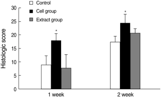
The histological scores of wounds were 3.3±0.6 (control group), 4.5±0.7 (cell group), and 3.1±0.6 (cell extract group) at 1 week after the wounding and 5.2±0.8 (control group), 8.0±1.9 (cell group), and 6.2±0.9 (cell extract group) at 2 weeks. The cell group had significantly higher scores than the other two groups at 1 and 2 weeks after wound- ing (P<0.05), and no significant difference was observed bet- ween the control and cell extract groups (Fig. 3).
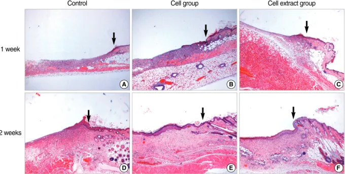
Histological evaluation of week 1 and 2 wounds disclosed that the cell group had enhanced cellularity (Fig. 4) and in- creased vasculature (also shown in Fig. 5). Granulation tissue in the cell group appeared to be thicker and larger than other groups (Fig. 4). In addition, the cell group wounds appeared to have increased reepithelialization. However, in the cell extract group, celluarity and granulation tissue were similar to those of the control at 1 and 2 weeks after wounding (Fig.
4), and decreased vascularity (also shown in Fig. 5).
Mean capillary densities were 9.0±3.6/HPF (control group), 9.8±2.4/HPF (cell group), and 9.3±6.2/HPF (cell extract group) at 1 week, and there was no significant difference bet- ween the groups. At 2 weeks, mean capillary densities were
*
Fig. 2. Changes of wound healing rates of the control, the cell, and the cell extract groups at 3, 5, 7, 10, and 14 days after wounding.
*P<0.05 compared to control; �P<0.05 compared to the cell group;
�P<0.05 compared to the cell extract group.
Wound helaing rate (%)
100 80 60 40 20
0
3 5 7 10 14
Days
Control Cell group Extract group
*,�
*,�
*,�
*
*
*,�
Fig. 3. Histological scores of wound in the control, the cell group, and the cell extract groups at 1 and 2 weeks after wounding.
*P<0.05 compared to control and the cell extract group.
Histologic score
30 25 20 15 10 5 0
1 week 2 week
Control Cell group Extract group
*
*
17.3±3.0/HPF (control group), 24.5±6.7/HPF (cell group), and 19.1±4.9/HPF (cell extract group) and all groups show-
ed higher capillary densities than those at 1 week after the wounding. In particular, capillary density was significantly
Fig. 4. Micrographs of wound bed and wound margin in each group at 1 and 2 weeks after wounding (H&E stain, 40). Wound edges are indicated by arrows. At 1 week after wounding, the control group shows thin granulation tissue containing many neutrophils and lympho- cytes (A), the cell group shows thick inflammatory cell infiltrations with many capillaries (B), and the cell extract group has more granula- tion tissues than in the control group (C). At 2 weeks after wounding, the control group shows thick granulation tissues with little re-epithe- lialization (D), the cell group shows slightly thicker epithelium and greater granulation tissue (E), and the cell extract group shows less re- epithelization and granulation tissue than in the cell group at 2 weeks (F).
1 week
A B C
2 weeks
D E F
Control Cell group Cell extract group
Fig. 5. Micrographs of wound bed and wound margin in each group at 1 and 2 weeks after wounding (Immunohistochemical staining for CD31, ×100). Capillaries with endothelial cells are stained brown (thin arrows). Wound edges are indicated by thick arrows. At 1 week, some small numbers of capillaries are observed in granulation tissues in the control group (A) and the cell extract group (C), but large numbers of capillaries are observed in the cell group (B). At 2 weeks after wounding, increased capillary development is noted in the control group (D), the cell group (E), and the cell extract groups (F). Particularly, in the cell group numerous larger capillaries are observed in compari- son with other groups.
1 week
A B C
2 weeks
D E F
Control Cell group Cell extract group
higher in the cell group than those in the other groups at 2 weeks (P<0.05) (Fig. 6).
DISCUSSION
Cutaneous wound healing process requires interactions bet- ween cells in the dermis and epidermis and the release of chem- ical mediators from inflammatory cells, fibroblasts, and ker- atinocytes. The proliferation of mesenchymal cells and cap- illaries, as well as the influx of macrophages into granulation tissue, serves to replace the dermal defect and to provide sub- strates and inducers for re-epithelialization. Because various cell types participate in wound healing process, various cell- based therapies offer promising therapeutic strategies to im- prove wound healing in physiological and pathological con- ditions. In recent studies, the transplantation of BMSCs has been reported to activate the healing process due to their capac- ity to differentiate in the skin epidermis and appendages, thus to mediate dermal regeneration (6). And also several studies have recently demonstrated accelerated rates of wound closure after transplantations of BMSCs (11), mesenchymal stem cells (12), or ADSCs (7). In our experiments, the ADSC-treated group showed significantly more rapid wound healing rates and had higher histological scores than those of the control group. Authors used the wound healing splint model, which is invented to prevent skin contraction around the wound, allow the wounds to heal via granulation and re-epithelial- ization (8). Acceleration of wound healing rates by ADSCs treatment in this model is thought to be caused by the sub- stitution of deficient cells after differentiation of ADSCs as stated in previous reports (6).
Neovascularization is a crucial step in wound healing pro- cess (2), which is necessary to sustain newly formed granula- tion tissue and ensure the survival of keratinocytes. Wu et al.
(6) demonstrated that BMSC-treated wounds have higher capillary densities, and suggested that BMSCs promote angio- genesis. In our study, ADSC-treated wounds had significantly
higher capillary densities than that of the control group, which suggestd that ADSCs, like BMSCs, promote neovasculari- zation. The promoting neovascualrization by ADSCs is con- sidered one of factors which accelerate the wound healing by ADSCs.
Many types of cytokines and growth factors are responsible for inflammation, re-epithelialization, the formation of gran- ulation tissue, and neovascularization during the healing pro- cess. Thus the application or induction of these cytokines and factors is known to accelerate wound healing (13). Kim et al.
(7) reported that paracrine factors secreted by ADSCs induce collagen synthesis and promote wound healing through fibrob- last activation, migration, and proliferation. Gaustad et al.
also reported that cell extracts, which contain growth and dif- ferentiation factors, can be used as an alternative to stimulate surface molecules to promote the differentiation of ADSCs to target cells (14). Cell extract was thought to differentiate mesenchymal stem cells, which are located in tissues around wound, into cells required for the wound healing process. In order to confirm the note of cell extract, we performed this comparative study on the effect of ADSCs and ADSC cell extract on wound healing to investigate whether cell extract from ADSCs could have beneficiary effect on wound healing.
Our results showed that the cell extract group showed rather lower wound healing rates than those of the control group throughout experimental periods, and no significant differ- ence versus the control until 2 weeks. Because ADSCs are not differentiated cells, the promotion of the specific differ- entiation of mesenchymal stem cells by cell extract of ADSCs would not occur. This result was in contrast to a previous report (14). Accordingly, we consider that ADSC cell ext- ract has no effect on wound healing, and that many factors in ADSC cell extract are less helpful in wound healing.
In conclusion, our experiments showed that ADSCs treat- ment enhanced wound healing as previously reported. Fur- thermore, our results suggest that this beneficiary effects of ADSCs on wound healing may be not caused by factors in ADSCs, but rather by the substitution of deficient cells after differentiation of ADSCs.
REFERENCES
1. Martin P. Wound healing--aiming for perfect skin regeneration. Sci- ence 1997; 276: 75-81.
2. Falanga V. Wound healing and its impairment in the diabetic foot.
Lancet 2005; 366: 1736-43.
3. Zuk PA, Zhu M, Ashjian P, De Ugarte DA, Huang JI, Mizuno H, Alfonso ZC, Fraser JK, Benhaim P, Hedrick MH. Human adipose tissue is a source of multipotent stem cells. Mol Biol Cell 2002; 13:
4279-95.
4. De Ugarte DA, Morizono K, Elbarbary A, Alfonso Z, Zuk PA, Zhu M, Dragoo JL, Ashjian P, Thomas B, Benhaim P, Chen I, Fraser J, Hedrick MH. Comparison of multi-lineage cells from human adipose Fig. 6. Capillary densities in wounds of the control, cell group, and
cell extract group at 1 and 2 weeks after wounding.
*P<0.05 compared to the control and cell extract groups.
Capillary density (capillaries/HPF)
35 30 25 20 15 10 5 0
1 week 2 week
Control Cell group Extract group
*
tissue and bone marrow. Cells Tissues Organs 2003; 174: 101-9.
5. Izadpanah R, Trygg C, Patel B, Kriedt C, Dufour J, Gimble JM, Bunnell BA. Biologic properties of mesenchymal stem cells derived from bone marrow and adipose tissue. J Cell Biochem 2006; 99:
1285-97.
6. Wu Y, Chen L, Scott PG, Tredget EE. Mesenchymal stem cells en- hance wound healing through differentiation and angiogenesis. Stem Cells 2007; 25: 2648-59.
7. Kim WS, Park BS, Sung JH, Yang JM, Park SB, Kwak SJ, Park JS.
Wound healing effect of adipose-derived stem cells: a critical role of secretory factors on human dermal fibroblasts. J Dermatol Sci 2007; 48: 15-24.
8. Galiano RD, Michaels J, Dobryansky M, Levine JP, Gurtner GC.
Quantitative and reproducible murine model of excisional wound healing. Wound Repair Regen 2004; 12: 485-92.
9. Jacobi J, Jang JJ, Sundram U, Dayoub H, Fajardo LF, Cooke JP.
Nicotine accelerates angiogenesis and wound healing in genetically diabetic mice. Am J Pathol 2002; 161: 97-104.
10. Yoon YS, Murayama T, Gravereaux E, Tkebuchava T, Silver M, Curry C, Wecker A, Kirchmair R, Hu CS, Kearney M, Ashare A, Jackson DG, Kubo H, Isner JM, Losordo DW. Vegf-c gene therapy augments postnatal lymphangiogenesis and ameliorates secondary lymphedema. J Clin Invest 2003; 111: 717-25.
11. Badiavas EV, Abedi M, Butmarc J, Falanga V, Quesenberry P. Par- ticipation of bone marrow derived cells in cutaneous wound healing.
J Cell Physiol 2003; 196: 245-50.
12. Nakagawa H, Akita S, Fukui M, Fujii T, Akino K. Human mesenchy- mal stem cells successfully improve skin-substitute wound healing.
Br J Dermatol 2005; 153: 29-36.
13. Gharaee-Kermani M, Phan SH. Role of cytokines and cytokine ther- apy in wound healing and fibrotic diseases. Curr Pharm Des 2001;
7: 1083-103.
14. Gaustad KG, Boquest AC, Anderson BE, Gerdes AM, Collas P. Dif- ferentiation of human adipose tissue stem cells using extracts of rat cardiomyocytes. Biochem Biophys Res Commun 2004; 314: 420-7.