MPTP로 유발된 파킨슨병 Mouse 모델에 대한 봉약침의 농도의존적 효과
전형준ㆍ김용석
경희대학교 한의과대학 침구학교실
목적 : 최근 한의학에서 널리 사용되며, 신경계 질환에도 응용되고 있는 봉약침의 농도의존적 효과를 알 아보기 위하여, 대표적인 신경 퇴행성 질환인 파킨슨병의 동물모델을 통해 세포보호효과와 세포사멸 및 신 경염증 기전을 관찰하였다.
방법 : C57BL/6 mice에 신경독소인 1-methyl-4-phenyl-1, 2, 3, 6-tetrahydropyridine(MPTP)를 4번 복강 내 주입하여 중뇌의 흑질 도파민 신경세포를 파괴하여 Parkinson 질병동물 모델을 만든 후, 2개의 군에는 마지막 MPTP 투여 2시간 후에 1차, 그 후로 48시간이 지날 때마다 양측 신수에 각각 0.06mg/kg 농도와 0.6 mg/kg 농도의 봉약침을 시행하여 총 4회 시술한 후, 도파민 세포를 측정하는 TH 면역조직 화학법을 통해 세포의 보존 정도를 관찰하고, 세포사멸과 관련된 양상을 확인하기 위하여 Caspase 3, 신경염증과 관련된 양 상을 확인하기 위하여 iNOS의 발현여부를 면역 조직화학법을 이용하여 관찰하였다.
결과 : 관찰결과 MPTP 투여 후 MPTP 투여군의 흑질의 도파민 세포 수는 감소하였으나 0.6mg/kg 봉약침 을 투여한 경우에는 유의성 있게 세포 수가 유지되었다. Caspase-3와 iNOS 발현억제 실험에서 0.6mg/kg 봉 약침군은 MPTP 투여군과 0.06mg/kg의 봉약침군과 비교하여 Caspase-3, iNOS 발현을 유의하게 억제하였다.
1)
Dose-dependent Effects of Bee Venom
Acupuncture on MPTP-induced Mouse Model of Parkinson’s Disease
Jun Hyung-joon and Kim Yong-suk
Dept. of Acupuncture & Moxibustion, College of Oriental Medicine, Kyung Hee University
․This work was funded by the Kyung Hee University Young Researcher in Medical Science Program in 2007(20071495)
․Acceptance : 2010. 8. 31. ․Adjustment : 2010. 9. 20. ․Adoption : 2010. 9. 20.
․Corresponding author : Kim Yong-suk, Kangnam Korean Hospital Kyung Univ. 994-5 Daechi 2-dong Gangnam-gu, Seoul 135-501, Republic of Korea
Tel. 82-2-3457-9013 E-mail : [email protected]
국문초록
Original Article
결론 : 봉약침은 MPTP 투여로 인한 신경세포 손상에 대하여 농도에 따라 세포사멸 기전과 신경염증 기 전을 억제함으로 신경세포를 보호하는 것으로 추정되며, 추후 적절한 경혈점 및 최적의 봉약침 농도를 찾는 데 지속적인 연구가 필요할 것이다.
핵심 단어 : 파킨슨 병, 봉약침, 농도의존적 효과
Ⅰ. Introduction
Bee venom(BV) is used in oriental medicine to treat various conditions. In recent years it has been reported that bee venom particularly one of its ma- jor constituent, melittin, possesses radioprotec- tive1), antimutagenic2), proinflammatory3), anti-inflam- matory4,5), antinociceptive6) and anticancer effect7,8).
In Korea, many Oriental medical doctors use bee venom acupuncture(BVA) by different dose, and some studies proved that BVA could be effective against various conditions in a dose-dependent manner9,10).
Parkinson’s disease(PD) is characterised by a loss of dopaminergic cells within the substantia nigra pars compacta(SNpc) of the midbrain11). This cell loss results in a reduction of dopamine levels in the striatum, which in turn, triggers a cascade of abnormal neural circuits that menifest in the dis- tinct signs of Parkinson’s disease, namely tremor, rigidity and bradykinesia(slowness of movement)11,12). In PD, several pathogenic factors play important roles in promoting degenerative processes in the nigrostriatal system including oxidative stress, mito- chondrial dysfunction, excitotoxicity, inflammatory process, and apoptosis13).
Several experimental models have been developed for PD; the most frequently used is that produced by 1 methyl 4 phenyl 1, 2, 3, 6 tetrahydropyridine (MPTP) administration in mice14) and MPTP causes a partial lesion of the substantia nigra and a significant reduction in striatal dopamine levels15). In vivo and vitro models of PD using MPTP, some have suggested that the mechanism of cell death operates via an apoptotic mechanism16-18). Exces-
sive activation of microglia, a major component of neuroinflammation, could be a driving force of the PD progression19). Several reports have suggested MPTP induced microglial activation and enhanced, progressive dopaminergic degeneration19-21).
Since there is no research on dose-dependent effect of bee venom acupuncture on Parkinson’s disease, in the present study, we selected BL23 and examined the effect of BVA on MPTP-induced Parkinson’s disease in C57BL/6 mice, and inves- tigated whether BVA could be effective dose- dependently.
Ⅱ. Materials and methods
1. Animals and MPTP administration
6-week old male C57BL/6 mice(Samtako co., Korea), weighing 20~25g, were used in all the experiments.
The mice were acclimated for 2 weeks in cages at 21℃ and were provided with water and food ad libitum. At the beginning of the experiment, the animals were randomly divided into four groups, and animal experiments were carried out in accor- dance with National Institute of Health’s Guide for Care and Use of Laboratory Animals. Experimental procedures were approved by the Institutional Ani- mal Care and Use Committee, Kyunghee University.
Except control group, mice(6per group) of the other groups received an intraperitoneal(i.p.) injection of MPTP(20mg/kg per dose; Sigma, St Louis, MO, USA) in saline at 2h intervals over an 8h period in 1 day. For i.p. injection, MPTP was dissolved in 5㎕
MPTP or Saline, i.p. injection, 4 tines BVA s.c. injection
End(7th day) Start
Fig. 1. MPTP administration and BVA procedure schedule
Control : Saline 5㎕, I.P. at 2h intervals over an 8h period in 1 day.
MPTP : MPTP-HC1, 20mg/kg per dose* 4 in saline at 2h intervals over an 8h period in 1 day.
BVA 1(0.06mg/kg BVA) : 2h after the 4th MPTPinjection, beevenom(20㎕/point administered at BL23 1st, 3rd, 5th and 7th day(total 4times)).
BVA 2(0.06mg/kg BVA) : 2h after the 4th MPTP injection, beevenom(20㎕/point administered at BL23 1st, 3rd, 5th and 7th day(total 4times)).
MPTP : 1-methyl-4-phenyl-1, 2, 3, 6-tetrahydropyridine.
BVA : bee venom acupuncture.
Shinsu(BL23) : located on the lower back, below the spinous process of the 2nd lumbar vertebra, 0.5cm lateral to the posterior midline.
of saline and i.p. injection was performed as pre- viously described22) using a 30㎕-Hamilton syringe with a 30-gauge needle. The control group(6mice) was injected with saline only as same as MPTP procedure. The animals were sacrificed at 7 days after the 4th MPTP or saline injection(Fig. 1).
2. BVA procedure
BV purchased from Sigma(St. Louis, MO, USA) was diluted to doses of 0.06mg/kg BVA(BVA 1) in 40㎕ of saline and of 0.6mg/kg BVA(BVA 2) in 40
㎕ of saline, and 20㎕ of each dose were subcu- taneously administered bilaterally into BL23, located on the lower back, below the spinous process of the 2nd lumbar vertebra, 0.5cm lateral to the post- erior midline, thus totaling 40㎕ of injection for each subject as was done(using 1cc syringe with a 30-gauge needle, Serin co, Korea). This dose of diluted BV was selected because this dose is most frequently used by Oriental medical doctors in Korea.
BVA at BL23 was performed 2h after the 4th MPTP i.p. injection and resumed at 48h intervals until the mice were sacrificed at the 7th days. For this pro- cedure, the mice in BVA 1 and 2 groups were im- mobilized, and BVA was done into BL23 (Fig. 1).
3. Tissue preparation and immuno- histochemistry
On the 7th day after 4th MPTP administration (per 6 mice in each group), mice were anesthetized with pentobarbital sodium(60mg/kg, i.p.) and perfused transcardially with 4% paraformaldehyde in 0.1M phosphate buffer at pH 7.4. Brains were isolated, post-fixed in same fixative overnight, subsequently cryoprotected with 30% sucrose in 0.05M phosphate- buffered saline(PBS, pH 7.4) for 48h, and sectioned coronally into 30㎛ for histological analysis.
For the immunohistochemistry, brain sections were incubated with one of the following antibodies : (1) rabbit anti-tyrosine hydroxylase(TH) antibody(Che- micon; 1 : 4,000); (2) rabbit anti-caspase 3 anti- body(Cell Signaling Technology; 1 : 1,000); (3) mouse anti-inducible nitric oxide synthase(iNOS, Upstate; 1 : 2,000). Brain sections were treated with primary antibody at room temperature for 16 h and then with biotinylated secondary antibody(1 : 200; Vector) for 1h. Then the sections were incubated with ABC solution(1 : 100; Vector) and finally developed in DAB or Ni-DAB solution. Sections were dehy- drated with alcohol and xylene, and then mounted with Permount solution.
4. Quantitative analysis
SNpc neuronal counts were performed manually by researchers blinded of the treatment schedule.
TH-positive SNpc cells were bilaterally counted on at least three TH-immunostained mesencephalic se- ctions at the widest dimension of the SNpc at AP- 3.16(Franklin and Paxinos, 1996) lateral to the roots of the third cranial nerve separating medial and lateral SNpc using a confocal microscope(Multiscan, Fullerton, CA, USA). Evaluation of the staining intensity of stained neurons was performed by measuring the optical density of caspase 3, iNOS- IR neurons in 10 sections from the substantia nigra.
The optical density of the stained neurons was quantitatively assessed by microdensitometry using an image analyzer(Multiscan, Fullerton, CA, USA).
Before the measurement of densitometry, we evalu- ated the voltage-related change in optical density.
We obtained the optimal voltage from the linear portion of S-shaped voltage-related optical den- sity curve. During the full measurement of optical density, the optical voltage was maintained at a constant level.
5. Statistics
Means and S.D. were calculated for the estimated numbers of TH-IR neurons and the optical density of caspase-3-IR, iNOS-IR positive neurons. To rule out a possible change in SNpc volume as an influencing factor, the results of the number of TH-IR neurons were expressed as a ratio of normal controls per area of SNpc. Statistical ana- lyses were performed using analysis of variance (ANOVA). A Bonferroni Multiple Comparison Test was used to compare individual means. Differences between the means of experimental groups were considered significant at p<0.05.
Ⅲ. Results
1. Effects of BVA on MPTP-induced neuronal loss of TH-IR neurons in SNpc
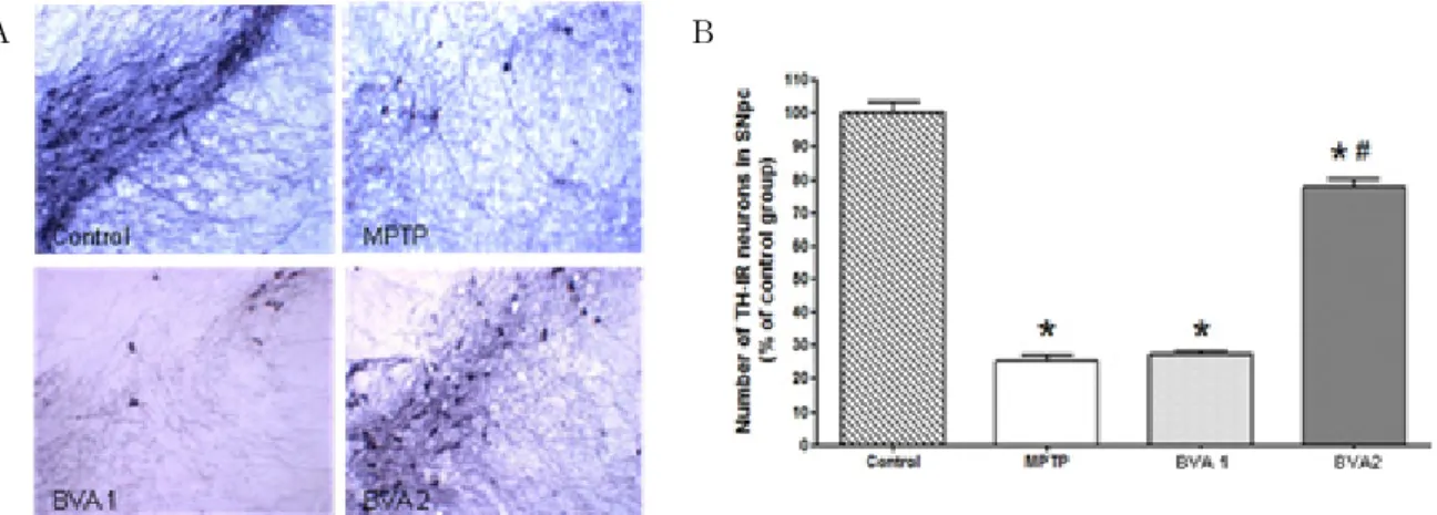
We analyzed the effects of BVA on MPTP- induced neuronal loss of TH-IR neurons in the SNpc by observing photomicrographs of TH-IR neurons of the SNpc and counting the number of TH-IR neurons in the SNpc at 7th day after 4th MPTP i.p. injection.
TH-IR neurons of the SNpc were significantly decreased in the MPTP and BVA 1 groups. But, TH-IR neurons were well preserved in the BVA 2 group than the MPTP and BVA 1 groups(Fig. 2).
2. Effects of BVA on the expression of caspase 3-IR neurons in MPTP- induced SNpc apoptosis
Because caspase-3 appears to be a key player in neuronal apoptosis23), the expression of caspase 3- IR neurons in MPTP-induced SNpc apoptosis was observed by photomicrographs of caspase 3-IR neu- rons of the SNpc and measured by the optical density of caspase-3-IR neurons in the the SNpc at the 7th day after the 4th MPTP i.p. injection.
Caspase 3-IR neurons were not observed in the control group. But, caspase 3-IR cells were observed in the SNpc of MPTP, BVA 1, and BVA 2 groups.
The number of caspase 3-IR cells was significantly decreased in the BVA 2 group than the MPTP and BVA 1 groups(Fig. 3).
3. Effects of BVA on the expression of iNOS-IR neurons in MPTP- induced SNpc neuroinflammation
iNOS expression in the glial cells of the SNpc has been suggested to play a role in the patho- genesis of PD24,25). Reducing NO production by de- creasing iNOS expression might be associated with
A B
Fig. 2. Effects of BVA on MPTP-induced neuronal loss of TH-IR neurons in SNpc
A : is photomicrographs of THIR neurons of the SNpc of MPTP-induced C57BL/6 mice on the 7th day after the 4th MPTP i.p. injection. Control, normal mice injected i.p. with 5㎕ normal saline; MPTP, mice which only received i.p. injection of MPTP; BVA 1, mice treated with bilateral BVA at BL23(0.06mg/kg) after the 4th MPTP i.p. injection; BVA 2, mice treated with bilateral BVA at BL23(0.6mg/kg) after the 4th MPTP i.p. injection. The preservation of TH-IR neurons and the dendritic processes surrounding the TH-IR neurons were observed in the BVA 2 group.
B : shows levels of the number of THIR neurons in the SNpc on the 7th day. TH-IR neurons were counted bilaterally in at least three TH-immunostained mesencephalic sections. The levels where expressed as the ratio of average number of TH-IR nigral neurons per section in the control group. The ratio was significantly greater in the BVA 2(0.6mg/kg) group than in the MPTP and BVA 1(0.06mg/kg) groups. All values represent means S.D. of the average number of TH-IR neurons per section in the control group(n=6).
* : p<0.001, one-way ANOVA, post hoc Bonferroni, compared to the control group.
# : p<0.001, one-way ANOVA, post hoc Bonferroni, compared to the MPTP group.
A B
Fig. 3. Effects of BVA on the expression of caspase 3-IR neurons in MPTP-induced SNpc apoptosis A : is photomicrographs of caspase 3-IR neurons of the SNpc of MPTP-induced C57BL/6 mice on the 7th day after the 4th
MPTP i.p. injection. Caspase 3-IR neurons were not observed in the control group. The expression of caspase 3-IR neurons was increased in the MPTP and the BVA 1(0.06mg/kg) groups compared to the BVA 2(0.6mg/kg) group.
B : shows the optical density of caspase 3IR neurons in the SNpc on the 7th day. The optical density was measured in ten sections throughout the entire rostrocaudal extent of the SNpc. The levels are expressed as the average optical density of caspase 3-IR neurons per section. The optical density was lower in the BVA 2 group than in the MPTP and BVA 1 groups. Data are expressed as mean S.D. of the average optical density for each section.
# : p<0.001, one-way ANOVA, post hoc Bonferroni, compared to the MPTP group.
the neuroprotective effects on MPTP-induced glial activation and apoptosis14).
Therefore, we observed photomicrographs of iNOS- IR neurons of the SNpc and counted the number of NOS-IR neurons in the the SNpc at the 7th day
after 4th MPTP i.p. injection.
iNOS-IR neurons were not observed in the con- trol group. However, iNOS-IR cells were observed in the SNpc of MPTP, BVA 1, and BVA 2 groups.
The number of iNOS-IR cells was significantly
A B
Fig. 4. Effects of BVA on the expression of iNOS-IR neurons MPTP-induced SNpc neuroinflammation A : is photomicrographs of iNOS IR neurons of the SNpc of MPTP-induced C57BL/6 mice on the 7th day after the 4th
MPTP i.p. injection. iNOS-IR neurons were not observed in the control group on the 7th day. The expression of iNOS-IR neurons was increased in the MPTP and BVA 1 groups(0.06mg/kg). However, in the BVA 2 group (0.6mg/kg), the expression of iNOS-IR neurons was significantly decreased.
B : shows optical density levels of iNOSIR neurons in the SNpc on the 7th day. Optical density was measured in ten sections throughout the entire rostrocaudal extent of the SNpc. The levels are expressed as the average optical density of iNOS-IR neurons per section. The optical density was lower in the BVA 2 group than in the MPTP and BVA 1 groups. Data are expressed as mean S.D. of the average optical density for each section.
# : p<0.01, one-way ANOVA, post hoc Bonferroni, compared to the MPTP group.
decreased in the BVA 2 group than the MPTP and BVA 1 groups(Fig. 4).
Ⅳ. Discussion
In the present study, MPTP-induced loss of TH- IR neurons and appearance of caspase 3-IR and iNOS-IR cells. After 7 days of experiment, TH-IR neurons were well preserved in BVA 2 group, and the number of caspase 3-IR and iNOS-IR cells was significantly decreased in BVA 2 group than in MPTP and BVA 1 groups.
The MPTP model in mice is widely used to study neuroprotective effect of drugs because it recapitulates the primary pathological and biochemical features of Parkinson’s disease, such as oxidative stress, mitochondrial dysfunction, and apoptosis26). He et al27) reported a single injection of MPTP (50mg/kg) significantly induced apoptosis in the subventricular zone(SVZ) and rostral migratory stream (RMS) in the brain of adult C57BL/6 mice, and substantial evidence indicates that MPTP-induced
neuronal death includes apoptosis4). Kokovay et al28) suggested that, in the MPTP mouse model of Parkinson’s disease, activated microglia may be directly involved in nigral degeneration.
The exact cause of dopaminergic neuronal loss is still unknown, but recent histological studies per- formed on brains from Parkinsonian patients sug- gest that nigral dopaminergic neurons die by apo- ptosis29-31). In addition to these studies, to treat PD, various kinds of mechanisms, such as micro- glial activation and inflammatory events20), and oxidative stress11) have been investigated. Some suggested that, in MPTP models of Parkinson’s disease, dopaminergic neurons have shown to die via apoptosis14,26,32), but Barcia et al33) suggested that it is still unknown whether inflammatory changes are responsible for active cell death or whether they play a protective role in neurodegeneration or inflammatory changes are related to neuronal loss in Parkinson’s disease.
Apoptosis represents a morphologically and bio- chemically distinct form of programmed cell death first recognized to play a considerable role in developmental cell death. Later on, apoptosis was
also found to underlie non-physiological cell death, such as in trauma, stroke or neurodegenerative disorders32).
Excessive activation of microglia, a major com- ponent of neuroinflammation, could be a driving force of the PD progression19). Peripheral microglia may amplify iNOS response to MPTP and expres- sion of iNOS and production of NO by microglia has been shown to mediate dopaminergic neuro- degeneration in the MPTP model21).
In this study, we observed the changes of H- positive neurons and the expression of capspase-3, iNOS for examining the impact of MPTP and effects of BVA against PD mouse model.
Caspases, a family of cysteine proteases, are integral parts of the apoptotic pathway: caspase-3 in particular, when activated, has many cellular targets that produce the morphologic features of apoptosis34), and appears to be a key player in neuronal apoptosis23), and is also an important mediator of apoptotic cell death35).
iNOS is not or is minimally expressed in the brain. However, in pathological conditions, iNOS expression can increase in brain glial cells and invading macrophages in response to a variety of injuries21). Recently, it has been shown that in mice deficient in iNOS, the loss of H-positive neurons is almost completely prevented21,24). iNOS expression in the glial cells of the SNpc has also been sug- sted to play a role in the pathogenesis of PD24,25), and that glial activation in the SNpc, which is accompanied by the up-regulation of iNOS, may have a pivotal role in PD36). Previous studies in- cate that inhibition of iNOS displays neurootective effects in the MPTP model of PD37).
BV consists of several biologically active peptides, including melittin, apamin, adolapin and mast cell degranulating peptide38) and is used in traditional medicine to treat various conditions, but the effect of BVA on MPTP-induced apoptosis or neuroin- ammation in Parkinson’s disease mouse model has not been studied yet.
In the present study, BVA prevented MPTP- nduced cell death of dopaminergic neurons, and
could be effective in PD by inhibition of apoptosis, e.g. by inhibition of caspase. And expression of iNOS was reduced in the BVA 2 group, so we suggest the protective role of BVA on MPTP- nduced glial activation and neuroinflammation may be associated with reducing NO production by decreasing iNOS expression.
These results support the hypothesis that ap- tosis could contribute to cell death in PD, we proved that BVA could be effective against MPTP-induced apoptosis and neuroinflammation in nigrostriatal dopaminergic neuron in C57BL/6 mice in a dose-dependent manner.
In addition to the dose-dependent effects of BVA further studies should also be performed on different acupoints.
It is known that BV induces apoptosis in mammary carcinoma7), human rheumatoid arthritis synovial fibroblast39), human leukemic cells40) and human melanoma8). But in this study, we observed that BVA prevented apoptosis inducing dopamin- gic neuronal loss by contraries. In consideration of opposite result in this study, it is an important area of further study to examine the correlation between BVA and apoptosis. Therefore we should verificate these effects of BVA using more various dose, and find out the most effective dose.
Ⅴ. Conclusions
In conclusion, we found that BVA protects neuronal loss and inhibits neuroinflammation and apoptosis in the SNpc of a mouse PD model in a dose-dependent manner. BVA could be a useful treatment strategy in neurodegenerative diseases such as Parkinson’s disease.
Ⅵ. References
1. Varanda EA, Tavares DC. Radioprotection:
Mechanism and radioprotective agents including honey bee venom. Venom Anim Toxins. 1998 ; 4(1) : 5-21.
2. Varanda EA, Monti R, Tavares DC. Inhibitory effect of propolis and bee venom on the mutagencity of some direct - and indirect- acting mu tagens. Teratog Carcinog Mutagen.
1999 ; 19(6) : 403-13.
3. Sumikura H, Andersen OK, Drewes, AM, Arendt-Nielsen LA. Comparison of hyperalgesia and neurogenic inflammation induced by melittin and capsaicin in humans. Neurosci Lett. 2003 ; 337(3) : 147-50.
4. Nam KW, Je KH, Lee JH, Han HJ, Lee HJ, Kamg SK, Mar W. Inhibition of COX-2 activity and proinflammatory cytokines(TNF- alpha and IL-1beta) production by water-soluble subfrac- tionated parts from bee(Apis Mellifera) venom.
Arch Pharm Res. 2003 ; 26(5) : 383-8.
5. Yoon SY, Kim HW, Roh DH, Kwon YB, Jeong TO, Han HJ, Lee HJ, Choi SM, Ryu YH, Beitz AJ, Lee JH. The anti-inflammatory effect of peripheral bee venom stimulation is mediated by central muscarinic type 2 receptors and acti- vation of sympathetic preganglionic neurons.
Brain Res. 2005 Jul 12 ; 1049(2) : 210-6.
6. Kim HW, Kwon YB, Ham TW, Roh DH, Yoon SY, Lee HJ, Han HJ, Yang IS, Beits AJ, Lee JH. Acupoint stimulation using bee venom at- tenuates formalin-induced pain behavior and spinal cord fos expression in rats. J Vet Med Sci.
2003 ; 65(3) : 349-55.
7. Orsolic N, Sver L, Verstovsek S, Terzic S, Basic I. Inhibition of mammary carcinoma cell proliferation in vitro and tumor growth in vivo by bee venom. Toxicon. 2003 ; 41(7) : 861-70.
8. Tu WC, Wu CC, Hsieh HL, Chen CY, Hsu SL.
Honeybee venom induces calcium-dependent but caspase-independent apoptotic cell death in human melanoma A2058 cells. Toxicon. 2008 Aug 1 ; 52(2) : 318-29.
9. Moon DO, Park SY, Lee KJ, Heo MS, Kim KC, Kim MO, Lee JD, Choi YH, Kim GY. Bee venom and melittin reduce proinflammatory
mediators in lipopolysaccharide-stimulated BV2 microglia. Int Immunopharmacol. 2007 Aug ; 7(8) : 1092-101.
10. Kim KW, Shin YS, Kim KS, Chang YC, Park KK, Park JB, Choe JY, Lee KG, Kang MS, Park YG, Kim CH. Suppressive effects of bee venom on the immune responses in collagen- induced arthritis in rats. J phymed. 2008 Apr ; 16 : 1-9.
11. Ma J, Shaw VE, Mitrofanis J. Does melatonin help save dopaminergic cells in MPTP-treated mice? Parkinsonism Relat Disord. 2008 Sep ; 13 : 1-8.
12. Blandini F, Nappi G, Tassorelli C, Martignoni E.
Functional changes of the basal ganglia cir- cuitry in Parkinson’s disease. Prog Neurobiol.
2000 ; 62 ; 70.
13. Ferger B, Leng A, Mura A, Hengerer B, Feldon J. Genetic ablation of tumor necrosis factor- alpha(TNF-alpha) and pharmacological inhibition of TNF-synthesis attenuates MPTP toxicity in mouse striatum. J Neurochem. 2004 ; 89 : 822.
14. Xu BB, Liu CQ, Gao X, Zhang WQ, Wang SW, Cao YL. Possible mechanisms of the protection of ginsenoside Re against MPTP-induced apo- ptosis in substantia nigra neurons of Parkin- son’s disease mouse model. J Asian Nat Prod Res. 2005 Jun ; 7(3) : 215-6, 218.
15. Heikkila RE, Hess A, Duvoisin RC. Dopaminergic neurotoxicity of 1-methyl-4-phenyl-1,2,5,6-tetrahy- dropyridine in mice. Science. 1884 ; 224 : 1451-3.
16. Mochizuki H, Nakamura N, Nishi K, Mizuno Y.
Apoptosis induced by i-methyl-4-phenylpyridiumion MPP+) in ventral mesencephalic striatal co-culture in rat. Neurosci Lett. 1994 ; 170 : 191-4.
17. Spooren WPJM, Gentsch C, Wiessner C. TUNEL- positive cells in the substantia nigra of C57BL/6 mice after a single bolus of 1-methyl-4-phenyl- 1,2,3,6-tetrahydropyridine. Neuroscience. 1998 ; 85 : 649-51.
18. Tatton NA, Kish S. In situ detection of apo- ptotic nuclei in the substantia nigra compacta of 1-methyl-4-phenyl-1,2,3,6-tetrahydropyridine-tr eated mice using terminal deoxylnucleotidyl trans-
ferase labeling and acridine orange staining.
Neuroscience 1997 ; 77 : 1037-48.
19. Gao HM, Hong JS. Why neurodegenerative diseases are progressive: uncontrolled inflam- mation drives disease progression. Trends Im- munol. 2008 Aug ; 29(8) : 357-65.
20. Kang JM, Park HJ, Choi YG, Choe IH, Park JH, Kim YS, Lim S. Acupuncture inhibits microglial activation and inflammatory events in the MPTP- induced mouse model. Brain Res. 2007 Feb 2 ; 1131(1) : 211-9.
21. Liberatore GT, Jackson-Lewis V, Vukosavic S, Mandir AS, Vila M, McAuliffe WG, Dawson VL, Dawson TM, Przedborski S. Inducible nitric oxide synthase stimulates dopaminergic.
22. Jackson-Lewis V, Przedborski S. Protocol for the MPTP mouse model of Parkinson’s disease.
Nat Protoc. 2007 ; 2(1) : 141-51.
23. Salvesen GS, Dixit, VM. Caspases : intracellular signaling by proteolysis. Cell. 1997 ; 91 : 443-6.
24. Dehmer T, Lindenau J, Haid S, Dichgans J, Schulz JB. Deficiency of inducible nitric oxide synthase protects against MPTP toxicity in vivo. J Neurochem. 2000 May ; 74(5) : 2213-6.
25. Hunot S, Boissière F, Faucheux B, Brugg B, Mouatt-Prigent A, Agid Y, Hirsch EC. Nitric oxide synthase and neuronal vulnerability in Parkinson’s disease. Neuroscience. 1996 May ; 72(2) : 355-63.
26. Geng X, Tian X, Tu P, Pu X. Neuroprotective effects of echinacoside in the mouse MPTP model of Parkinson’s disease. Eur J Pharmacol.
2007 Jun 14 ; 564(1-3) : 66-74.
27. He XJ, Nakayama H, Dong M, Yamauchi H, Ueno M, Uetsuka K et al. Evidence of apo- ptosis in the subventricular zone and rostal migratory stream in the MPTP mouse model of Parkinson disease. J Neuropathol Exp Neurol.
2006 ; 65 : 873-82.
28. Kokovay E, Cunningham LA. Bone marrow- derived microglia contribute to the neuroin- flammatory response and express iNOS in the MPTP mouse model of Parkinson’s disease.
Neurobiol Dis. 2005 Aug ; 19(3) : 471-8.
29. Anglade P, Vyas S, Javoy-Agid F, Herrero MT, Michel PP, Marquez J, Mouatt-Prigent A, Ru- berg M, Hirsch EC, Agid Y. Apoptosis and autophagy in nigral neurons of patients with Parkinson’s disease. Histol Histopathol. 1997 Jan ; 12(1) : 25-31.
30. Kingsbury AE, Mardsen CD, Foster OJ. DNA fragmentation in human substantia nigra: apo- ptosis or perimortem effect? Mov Disord. 1998 Nov ; 13(6) : 877-84. Links.
31. Tatton NA, Maclean-Fraser A, Tatton WG, Perl DP, Olanow CW. A fluorescent double-labeling method to detect and confirm apoptotic nuclei in Parkinson’s disease. Ann Neurol. 1998 Sep ; 44(3 Suppl 1) : S142-8.
32. Eberhardt O, Schulz JB. Apoptotic mechanisms and antiapoptotic therapy in the MPTP model of Parkinson’s disease. Toxicol Lett. 2003 Apr 4 ; 139(2-3) : 135-51.
33. Barcia C, Fernández Barreiro A, Poza M, Herrero MT. Parkinson’s disease and inflam- matory changes. Neurotox Res. 2003 ; 5(6) : 411-8.
34. Cohen GM, Caspases: the executioners of apo- ptosis. Biochem J. 1997 ; 326 : 1-16.
35. Thomas M, Le WD. Minocycline: neuroprotective mechanisms in Parkinson’s disease. Curr Pharm Des. 2004 ; 10(6) : 680.
36. Hunot S, Brugg B, Ricard D, Michel PP, Muriel MP, Ruberg M, Faucheux BA, Agid Y, Hirsch EC. Nuclear translocation of NF-kappaB is increased in dopaminergic neurons of patients with parkinson disease. Proc Natl Acad Sci USA. 1997 Jul 8 ; 94(14) : 7531-6.
37. Molina JA, Jiménez-Jiménez FJ, Ortí-Pareja M, Navarro JA. The role of nitric oxide in neuro degeneration. Potential for pharmacological inter- vention. Drugs Aging. 1998 Apr ; 12(4) : 251-9.
38. Lariviere WR, Melzack R. The bee venom test:
a new tonic-pain test. Pain. 1996 Aug ; 66(2-3) : 271-7.
39. Hong SJ, Rim GS, Yang HI, Yin CS, Koh HG, Jang MH, Kim CJ, Choe BK, Chung JH. Bee venom induces apoptosis through caspase-3 acti-
vation in synovial fibroblasts of patients with rheumatoid arthritis. Toxicon. 2005 Jul ; 46(1) : 39-45.
40. Moon DO, Park SY, Choi YH, Kim ND, Lee C,
Kim GY. Melittin induces Bcl-2 and caspase- 3-ependent apoptosis through downregulation of Akt phosphorylation in human leukemic U937 cells. Toxicon. 2008 Jan ; 51(1) : 112-20.