MPTP 유발 파킨슨 병 동물 모델에서의 신수혈(BL
23
) 봉독약침의 항염증 효과김찬영ᆞ이재동ᆞ이상훈ᆞ고형균 경희대학교 한의과대학 침구학교실
목적 : 파킨슨 병은 기저핵 흑질의 치밀부에서 도파민성 신경세포의 퇴행으로 인하여 발생하는 질병으로 신경 염증이 주요 병인으로 밝혀져 있다. 이 연구는 MPTP 유발 파킨슨 병 동물 모델에서 신수혈(BL
23)에 대한 봉독 약침의 항염증 효과 및 그 기전을 확인하기 위해 시행되었다.
방법 : C57
BL/6쥐를 무처치군, MPTP+saline군, MPTP+BVA(0.06mg/kg)군, MPTP+BVA(0.6mg/kg)군의 4 군으로 나눈 뒤 무처치군을 제외한 모든 그룹에 총 8시간 동안 2시간 간격으로 MPTP-HCl(20mg/kg per dose×4)을 복강내로 주입하였다. MPTP+BVA 군에서 봉독약침은 마지막 MPTP 주입 2시간 후부터 48시간 간격으로 신수혈(BL
23)에 양측으로 각 20㎕씩 주입하였고 MPTP+saline군에서는 봉독약침 대신 Saline을 주 입하였다. 마지막 MPTP 주입 후 7일째에 쥐의 뇌를 적출한 후 면역조직화학법을 시행하였다.
결과 : MPTP 유발 파킨슨 병 동물 모델에서 신수혈에 대한 봉독약침은 농도 의존적으로 TH- Immunoreactivity neuron의 감소와 microglial activation을 억제하였다. HSP70-IR neuron은 모든 군에서 나 타나지 않았다.
결론 : 봉독약침이 용량의존적으로 microglial activation을 억제하는 효과를 통해 도파민성 신경세포의 파 괴를 억제함으로써 항염 효과를 나타냄을 알 수 있었다. 이 결과는 봉독약침이 microglial activation 억제를 통해 임상적으로 파킨슨 병과 같은 신경 퇴행성 질병에 있어 유용한 치료수단이 될 수 있음을 시사한다.
1)
Anti-inflammatory Effect of Bee Venom Acupuncture at Sinsu (BL 23 ) in a MPTP Mouse
Model of Parkinson Disease
Kim Chan-young, Lee Jae-dong, Lee Sang-hoon and Koh Hyung-kyun
Dept. of Acupuncture & Moxibustion, College of Oriental Medicine, Kyung Hee University
․Acceptance : 2009. 7. 7. ․Adjustment : 2009. 7. 27. ․Adoption : 2009. 7. 28.
․Corresponding author : Koh Hyung-kyun, Department of Acupuncture and Moxibustion, College of Oriental Medicine, Kyung Hee University, 1 Hoegi-dong, Dongdaemun-gu, Seoul, Republic of Korea
Tel. 82-2-958-9200 E-mail : [email protected]
국문초록
Original Article
핵심단어 : Bee venom, Acupuncture, Parkinson disease, MPTP(1-methyl-4-phenyl-1,2,3,6-tetrahydropyridine), Sinsu(BL
23: Shen Shu), Heat shock protein(HSP)
Ⅰ. Introduction
Parkinson disease(PD) is one of the most com- mon movement disorders. The cardinal symptoms of the disease include a paucity of spontaneous movement, akinesia, bradykinesia, rigidity, and a characteristic tremor at rest. In the early 1960s, PD was shown to result largely from the degeneration of dopaminergic neurons in the substantia nigra pars compacta(SNpc)
1). Brain inflammation is the common final pathway in PD
2). Central to this inflammation is the activation of microglia, which act as intrinsic immune effectors when the brain is injured
3,4). Microglia are known to be activated in the PD-affected brain, and MPTP-induced microglial activation is associated with oxidative stress
5).
Bee venom contains a variety of peptides and proteins with a range of molecular weights, including melittin, apamin, adolapin, mast cell degranulating peptide and phospholipase A2
6-8). In addition, it also contains biologically active amines(histamine, epi- nephrine) and several other non-peptide components including lipids, carbohydrates and free amino acids
9). Bee venom(BV) acupuncture(BVA) has been traditionally used in Oriental medicine to relieve pain and to treat chronic inflammatory diseases such as rheumatoid arthritis
10), and neuro- degenerative diseases such as multiple sclerosis
11,12). Recent studies have suggested that BV has anti- inflammatory properties that inhibit the production of inflammatory cytokines and nitric oxide(NO) in neurodegenerative diseases
13,14).
Several animal models of PD have been developed, including a mouse model in which a parkinsonian pathology develops in response to the adminis- tration of the neurotoxin 1-methyl-4-phenyl-1,2,3,6-
tetrahydropyridine (MPTP). MPTP causes many of the hallmark features of PD in humans and nonhuman primates, and it induces SNpc-specific dopaminergic cell loss in other mammalian species including mice
15).
The heat shock proteins(HSPs) are so named due to the fact that their synthesis was initially found to be enhanced in response to an increase in temperature
16). HSPs represent an important cellular protective mechanism against a variety of stresses and insults
17-19). Recent studies have suggested that HSP may be involved in neuronal cell death in
PD
20,21)and may play a neuroprotective role in
MPTP-induced neurotoxicity
22). Thus, HSPs may represent an important molecular target for neuro- protective strategies in PD treatment, and geld- anamycin is known to induce the expression of HSP70 and protect against MPTP-induced dopa- minergic neurotoxicity in mice
23).
In this study, we used an MPTP-induced neuro- inflammation mouse model to examine whether if (a) BVA inhibits the loss of tyrosine hydroxylase (TH)-positive neurons as a result of its inhibition of microglial activation or induction of HSP70 synthesis and (b) the effect of BVA is dose dependent. Microglial activation was measured by the level of macrophage antigen complex(MAC-1) expression.
Ⅱ. Materials and Methods
1. Animals and MPTP administration
Six-week old male C57BL/6 mice(Samtaco Co.,
Korea), weighing 20-25g, were used in all experiments.
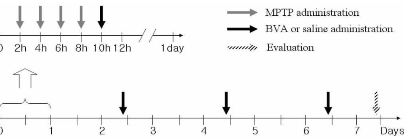
Fig. 1. MPTP & BVA administration schedule
At the beginning of the experiment, the animals were randomly divided into four groups: a naïve group, an MPTP+saline group, an MPTP+BVA(0.06mg/kg) group and an MPTP+BVA(0.6mg/kg) group. All mice except those in the naïve group received an intraperitoneal(i.p.) injection of MPTP-HCl(20mg/kg per dose×4; Sigma, St. Louis, MO, USA) in saline at 2h intervals over an 8h period in 1day.
In the MPTP+BVA groups, BVA administered at BL23(bilateral; 20µl per point) began 2h after the last MPTP injection and then resumed at 48h. In the MPTP+saline group, saline was injected instead of BV. All mice were sacrificed seven days after the last MPTP injection. Mice in the naïve group were sacrificed on the same day.
The mice were acclimated for 2 weeks in cages at 21°C and were provided with water and food ad libitum. At the beginning of the experiment, the animals were randomly divided into four groups: A naïve group, an MPTP+saline group, an MPTP+
BVA(0.06mg/kg) group and an MPTP+BVA(0.6mg/kg) group. Animal experiments were carried out in accordance with the National Institute of Health’s Guide for Care and Use of Laboratory Animals, and experimental procedures were approved by the Institutional Animal Care and Use Committee at the Kyunghee University. Except for the naïve group, the mice(six per group) in the other groups received an intraperitoneal(i.p.) injection of MPTP-HCl(20mg/
kg per dose×4; Sigma, St. Louis, MO, USA) in saline every 2h, over an 8h period, in 1 day24). For i.p. injection, MPTP was dissolved in 5㎕ saline and an i.p. injection was performed as previously described
24)using a 30-㎕ Hamilton syringe with a 30-gauge needle. The animals were sacrificed at seven days after the last MPTP injection(Fig. 1).
2. Treatment with diluted bee venom
Bee venom (Sigma, St. Louis, MO, USA) was diluted to doses of 0.06mg/kg in 40µl of saline and
0.6mg/kg in 40µl of saline; 20µl of each dose were subcutaneously administered bilaterally into each BL23 point, located at the depression lateral to the lower border of spinous process of the second lumbar vertebra
25). Thus, total injection volume of 40µl was administered to each subject, as described previously
26).
BVA at BL
23began 2h after the last MPTP i.p.
injection and then resumed at 48h intervals until the mice were sacrificed, seven days after the last MPTP i.p. injection. For this procedure, the mice in the MPTP+BVA groups were immobilized, and BV was administered into BL
23. For the MPTP+saline group, 40µl of saline was injected in place of BV.
3. Tissue preparation and immunohistochemistry
The mice in all groups were sacrificed by anesthesia with pentobarbital sodium (60mg/kg, i.p.) 7days after the last MPTP i.p. injection and perfused transcardially with paraformaldehyde (4%
in 0.1M phosphate buffer, pH 7.4). The brains were
isolated, post-fixed in the same fixative overnight,
subsequently cryoprotected with 30% sucrose in
0.05M phosphate-buffered saline(PBS, pH 7.4) for
48h, and sectioned coronally into 30μm slices for histological analysis.
For the immunohistochemistry, brain sections were incubated with one of the following antibodies:
(1) rabbit anti-tyrosine hydroxylase(TH) antibody (Chemicon; 1 : 4,000), (2) rabbit anti-MCA-1 antibody (Serotec; 1 : 500) and (3) rabbit anti-heat shock protein (HSP70) antibody (Chemicon; 1 : 2,000). Brain sections were treated with primary antibody at room temperature for 16h, followed by biotinylated secondary antibody (1 : 200; Vector) for 1h. Then the sections were incubated with ABC solution (1 : 100; Vector) and finally developed in DAB or Ni-DAB solution. Sections were dehydrated with alcohol and xylene, and then mounted with Permount solution.
4. Quantitative analysis
SNpc neuronal counts were manually performed by technicians who were blinded to the treatment schedule. TH-IR cells in the SNpc were bilaterally counted using a confocal microscope (Multiscan, Fullerton, CA, USA) for at least 3 TH- immunostained mesencephalic sections at the widest dimension of the SNpc at AP-3.16
27), lateral to the roots of the third cranial nerve separating the medial and lateral SNpc. Evaluation of the neuronal staining intensity was performed by measuring the optical density of MAC-1-IR and HSP70-IR neurons in 10 sections from the substantia nigra. The optical density of the stained neurons was quantitatively assessed by microdensitometry using an image analyzer(Multiscan, Fullerton, CA, USA). Before the densitometry measurement, the voltagerelated change in optical density was evaluated. The optimal voltage was then obtained from the linear portion of the S-shaped voltage-related optical density curve
28). During full measurement of the optical density, the optical voltage was maintained at a constant level.
5. Statistical analysis
Means and standard deviation(SD) were calculated for the estimated numbers of TH-IR neurons and the optical density of MAC-1-IR and HSP70-IR positive neurons. To rule out a possible change in SNpc volume as an influencing factor, the number of TH-IR neurons are expressed as a ratio of normal controls per area of SNpc. Statistical analyses were performed using analysis of variance (ANOVA). A Bonferroni Multiple Comparison Test was used to compare individual means. Differences between the means of experimental groups were considered significant at p<0.05.
Ⅲ. Results
1. BVA inhibits the MPTP-induced neuronal loss of TH-IR neurons in the SNpc in a dose dependent manner
Immunohistochemical staining with TH antibody was performed on the brain samples collected from each group 7days after the last MPTP injection.
TH-IR neurons were counted bilaterally at least three TH-immunostained mesencephalic sections.
TH-IR neurons were plentiful in the SN of the
naïve MPTP+Saline
MPTP+BVA(0.06) MPTP+BVA(0.06)
(A)
(B)
Fig. 2. Effects of BVA at BL
23on MPTP- induced loss of tyrosine hydroxylase (TH)-immu- noreactive (IR) neurons in the substantia nigra pars compacta (SNpc)
(A) shows photomicrographs of TH-IR neurons of the SNpc of MPTP-induced C57BL/6 mice 7days after the last injection of MPTP. Naïve, naïve mice; MPTP+saline, mice treated with bilateral saline acupuncture at BL23
after MPTP administration; MPTP+BVA(0.6), mice treated with bilateral BVA at BL23(0.6mg/kg) after MPTP admin- istration ; MPTP+BVA(0.06), mice treated with bilateral BVA at BL23(0.06mg/kg) after MPTP administration.
(B) shows levels of the number of TH-IR neurons in the SNpc 7days after the last injection of MPTP. The levels are expressed as the ratio of the average number of TH-IR neurons per section in the naïve group. All values represent means±S.D. of the average number of TH-IR neurons per section in the naïve group(n=6).
Means designated by the same letter are not significantly different according to the Bonferroni Multiple Comparison test(α=0.05).
naïve group(Fig. 2A).
The survival ratios of TH-IR neurons in the SNpc of the MPTP+saline and MPTP+BVA(0.06) groups were only 40% and 44%, compared with the naïve group. In contrast, in the MPTP+BVA(0.6) group, the survival ratio of TH-IR neurons was 89% which was significantly different from the survival ratios of the MPTP+saline and MPTP+
BVA(0.06) groups(Fig. 2B).
2. BVA at BL
23inhibits the MPTP- induced activation of MAC-1-IR neurons in the SNpc in a dose dependent manner
The SNpc is relatively rich in microglia compared with other brain regions
29,30). Previous studies have suggested that activation of microglia
(B)
naïve MPTP+Saline
MPTP+BVA(0.06) MPTP+BVA(0.06)
(A)
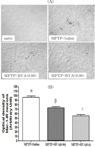
Fig. 3. Effects of BVA at BL23 on MPTP-i nduced microglial activation of MAC-1-IR neurons in the SNpc
(A) shows photomicrographs of MAC-1-IR neurons of the SNpc of MPTP-induced C57BL/6 mice on 7days after the last MPTP injection. Naïve, naïve mice; MPTP+saline, mice treated with bilateral saline acupuncture at BL23after MPTP administration; MPTP+BVA(0.6), mice treated with bilateral BVA at BL23(0.6mg/kg) after MPTP admin- istration; MPTP+BVA(0.06), mice treated with bilateral BVA at BL23(0.06mg/kg) after MPTP administration.
(B) shows the optical density levels of MAC-1-IR neurons in the SNpc. Optical density was measured in six sections throughout the entire rostrocaudal extent of the SNpc. The levels are expressed as the average optical density of MAC-1-IR neurons per section. Data are expressed as mean±S.D. of the average optical density for each section. Means designated by different letters are significantly different according to the Bonferroni Multiple Comparison test(α=0.05).
may trigger or participate in the neurodegenerative
processes in PD
2). To determine whether the
beneficial effect of BVA was associated with
inhibition of the MPTP-induced glial response, we
examined the expression of MAC-1, a marker of
microglial activation, 7days after last the MPTP injection. In the naïve group, activated microglia were not observed. In the MPTP+saline group, marked expression of MAC-1 and an increase in dendritic processes surrounding the MAC-1-IR neurons were observed. In contrast, the optical density and expression of activated microglia were significantly decreased in the MPTP+BVA groups in a dose dependent manner, compared with the MPTP+saline group(Fig. 3A). In particular, the activation of MAC- 1-IR neurons was significantly decreased in the MPTP-BVA(0.6) group compared with the MPTP+
BVA(0.06) group(Fig. 3B).
3. HSP70-IR neurons were not observed in the SNpc of any group
Heat shock proteins(HSPs) play a neuroprotective role in MPTP-induced neurotoxicity
22). To determine whether the beneficial effect of BVA was associated with HSPs, we examined the expression of HSP70,
naïve MPTP+Saline
M PTP+BVA(0.06) M PT P+BVA (0.06)
Fig. 4. Expression of HSP70-IR neurons of the SNpc of MPTP-induced C57BL/6 mice 7days after the last MPTP injection, and the relationship with BVA at BL
23Photomicrographs of HSP70-IR neurons of the SNpc of MPTP-induced C57BL/6 mice, taken 7days after the last MPTP injection. Naïve, naïve mice; MPTP+saline, mice treated with bilateral saline acupuncture at BL23 after MPTP administration; MPTP+BVA(0.6), mice treated with bilateral BVA at BL23(0.6mg/kg) after MPTP admin- istration; MPTP+BVA(0.06), mice treated with bilateral BVA at BL23(0.06mg/kg) after MPTP administration.
HSP70-IR neurons were not observed in the substantia nigra of any group.
7days after last MPTP injection. HSP70-IR cells were not observed in the substantia nigra of any group(Fig. 4).
Ⅳ. Discussion
Parkinson disease(PD), first described by James Parkinson in 1817, as paralysis agitans, or the
‘shaking palsy’, is the result of a quite specific and progressive neurodegeneration of pigmented nigro- striatal dopaminergic neurons. The symptoms of PD are only apparent after the loss of at least 50% of the dopaminergic neurons in the substantia nigra pars compacta(SNpc), which leads to a reduction of over 80% in the dopamine(DA) levels in the stri- um
31,32).
The cause of PD remains unclear, but several theories have been proposed regarding the possible factors behind the neuronal degeneration. These include environmental toxins, genetic factors and mitochondrial dysfunction as well as free radical- mediated cell death and oxidative stress
33-36). Re- cently, however, there has been increasing recognition of the possible major role of neuroinflammation in the pathogenesis of PD
37), induced by exposure to either infectious agents or toxicants with proin- flammatory characteristics.
Microglia are resident immunocompetent and
phagocytic cells in the central nervous system
CNS), and are thought to mediate the innate
defense system and thus serve a critical role in
normal CNS function
38). They are activated in the
event of infection, inflammation, trauma, ischemia
and neurodegeneration in the CNS
39). It has been
shown that glial cells are activated in the PD brain
and MPTP-induced glial activation in association
with oxidative stress
5). MPTP is a neurotoxin that
induces Parkinsonian features in humans, rodents
and non-human primates and has been demon-
strated to cause rapid and selective DA neuro-
toxicity
40). Recent studies suggested that BV has anti-
inflammatory properties that inhibit production of
inflammatory cytokines and NO in the activated microglia
13,14). Acupuncture also inhibits microglial activation and inflammatory events in the MPTP- induced mouse model
41).
The heat shock proteins(HSPs) are so named due to the fact that their synthesis was initially found to be enhanced in response to an increase in temperature
16). HSPs are a group of highly conserved stress proteins that play an important role in main- taining body self-stability. The main functions of HSPs are to promote cellular tolerance against stress factors, to maintain normal cellular physi- ological function, and to increase cellular defense and adaptation to deadly stimulation
42). It is known that a plethora of insults other than heat stress have since been found to increase the expression of HSPs including neurotoxicants, drugs of abuse, environmental stress, metals, oxidative stress and many other pathophysiological states and non-stressful conditions. Previous data from in vivo and in vitro studies strongly suggest that HSPs play a neuro- protective role in MPTP-induced neurotoxicity
22). Moxibustion and electroacupuncture can induce the synthesis of heat shock protein 70
43,44). However, to date no study had examined the relationship between BVA and HSPs.
In the present study, we used an MPTP-induced neuroinflammation mouse model to examine whether (a) BVA inhibits the loss of tyrosine hydroxylase(TH)-positive neurons as a result of its inhibition of microglial activation or induction of HSP70 and (b) the effect of BVA is dose de- pendent. Microglial activation was measured by the expression of MAC-1. Due to the strain-dependent sensitivity to MPTP
45), we used C57BL/6 mice.
TH is the rate-limiting enzyme in the synthesis of the catecholamine neurotransmitters, such as dopa- mine, epinephrine, and norepinephrine. It converts L-tyrosine to L-dihydroxyphenylalanine(L-DOPA), the rate-limiting step in the synthesis of dopa- mine
46). Since TH is a rate-limiting enzyme for the biosynthesis of dopamine, TH activity is progres- sively decreased following the loss of dopamine neurons in the substantia nigra in the patients with
Parkinson disease
47). TH immunohistochemistry has widely been used as an important method of detecting the injury or death of dopaminergic fibers and cell bodies
48,49).
Previous studies using rats showed that acu- puncture at the GB
34, LR
3and ST
36acupoints reduced the degeneration of dopaminergic neurons induced by 6-hydroxydopamine(6-OHDA)
50). GB
34and LR
3also protected against MPTP induced dopaminergic neuronal cell damage
41). For the present study, we chose the Sinsu(BL
23) acupoint because PD is a brain disease, and in Oriental medicine the brain is the sea of marrow nourished by Kidney essence.
BL
23is the Kidney transport point that is the specific point on the back where the qi of the Kidney is infused
51). Acupuncture at BL
23lessens the memory loss and decreased immune responses that accompany aging. BL
23can also increase the release of 5-HT
52). However, no study has examined the effect of BL
23on Parkinson disease to date.
In our study, BVA inhibited MPTP-induced loss of TH-IR neurons and activation of MAC-1-IR neurons in the SNpc, in a dose dependent manner.
These results demonstrate that BVA possess a potent suppressive effect on microglial activation and suggest that BVA may offer substantial thera- peutic potential for the treatment of neurode- generative diseases that are accompanied by micro- glial activation.
HSP70-IR cells were not observed in the SNpc of any of the tested groups. However, since the only observation was made 7days after the last MPTP injection, further research, including obser- vation at different time points, is required.
Ⅴ. Conclusions
In conclusion, we found that BVA at BL
23inhibited MPTP-induced neuronal loss of TH-IR
neurons and microglial activation in the SNpc of a
mouse PD model, and that this suppression occurred
in a dose dependent manner. However, HSP70-IR neurons were not observed in the SNpc 7days after the fourth MPTP injection.
Ⅵ. References