Received January 5, 2016, Revised April 20, 2016, Accepted for publication May 23, 2016
Corresponding author: Fatma Pelin Cengiz, Department of Dermatology, Adnan Menderes Boulevard, Bezmialem Vakif University Hospital, Fatih 34710, Istanbul, Turkey. Tel: 90-5067015406, Fax: 90-2124531700, E-mail:
fpelinozgen@hotmail.com
This is an Open Access article distributed under the terms of the Creative Commons Attribution Non-Commercial License (http://creativecommons.
org/licenses/by-nc/4.0) which permits unrestricted non-commercial use, distribution, and reproduction in any medium, provided the original work is properly cited.
Copyright © The Korean Dermatological Association and The Korean Society for Investigative Dermatology
Ann Dermatol Vol. 29, No. 1, 2017 https://doi.org/10.5021/ad.2017.29.1.26
ORIGINAL ARTICLE
Dermoscopic Features of Small, Medium, and Large-Sized Congenital Melanocytic Nevi
Fatma Pelin Cengiz, Nazan Emiroglu, Dilek Biyik Ozkaya, Ozlem Su, Nahide Onsun
Department of Dermatology, Bezmialem Vakif University, Istanbul, Turkey
Background: Congenital melanocytic nevi (CMN) are pres- ent at birth. It is well known that the presence of large-sized congenital nevus in early life could predict a major risk of de- veloping melanoma. Objective: To investigate the clinical and dermoscopic features of the CMN, to search for and high- light any differences between small-sized, medium-sized, large-sized CMN. Methods: A nonrandomized observat- ional study was performed. A total of 108 melanocytic nevi were analysed by clinical and dermoscopic examination.
Results: Of the subjects, 57.4% were aged less than 16 years, 42.6% were aged 16 and more. Of the nevi, 26 had reticular pattern (24.1%), 35 had globular pattern (32.4%), 13 had re- ticular-globular pattern (12.0%), 16 had homogeneous pat- tern (14.8%), 6 had reticular-homogeneous pattern (5.6%), 2 had globular-homogeneous pattern (1.9%), 7 had cobble- stone pattern (6.5%), 3 had reticular patchy pattern (2.8%).
Atypical dots and globules, focal hypopigmentation and peri- follicular hypopigmentation are the most common dermo- scopic features of CMN. The rarest dermoscopic feature is the blue-whitish veil. Conclusion: Most of the dermoscopic features related with dysplastic nevi up to the present, such as atypical dots and globules, focal hypopigmentation, peri- follicular hypopigmentation were observed in CMN, in our study. Congenital nevus and dysplastic nevi may share the
same dermoscopic features, therefore it is important to know it is found at birth or not. (Ann Dermatol 29(1) 26∼32, 2017) -Keywords-
Dermoscopy, Nevus
INTRODUCTION
Congenital melanocytic nevi (CMN) are defined as nevi present at birth or appearing during subsequent months1. CMN are present at birth on the skin of 1% to 6% of new- borns and are usually classified according to their size as small (<1.5 cm), medium (1.5∼19.9 cm), and large/giant (>20 cm)2-5. Most of the CMN are smaller than 1.5 cm3. CMN may not exist at birth, because of the embryonic ab- sence of pigmentation and may appear clinically after the pigment developments months to years after birth2,6,7. Furthermore, these lesions are described as ‘congenital ne- vus-like nevi’ or ‘tardive congenital nevi’ by some authors 2,6. The estimated prevalence of CMN ranges from 0.5% to 31.7%8. Small CMN have an estimated incidence of 1 in 100 live births, whereas large CMN is 1 in 20,000 live births9-11. They occur in all races and ethnic groups, and males and females have an equal risk. Many of the nevi termed “dysplastic” or “atypical” are actually small CMN12. CMN may develop due to a mutation that affects the mor- phogenesis of the embryonic neuroectoderm and migra- tion of precursor cells to the skin.
Our aim was to investigate the dermoscopic patterns of CMN among study participants and to assess the dermo- scopic patterns associated with the size of nevi as well.
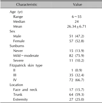
Table 1. Demographic data of patients (n=108)
Characteristic Value
Age (yr)
Range 6∼55
Median 24
Mean 26.34±6.71
Sex
Male 51 (47.2)
Female 57 (52.8)
Sunburns
Never 15 (13.9)
Mild∼moderate 82 (75.9)
Severe 11 (10.2)
Fitzpatrick skin type
II 1 (0.9)
III 35 (32.4)
IV 72 (66.7)
Location
Face and neck 17 (15.7)
Trunk 64 (59.3)
Extremity 27 (25.0)
Values are presented as number only, mean±standard deviation, or number (%).
MATERIALS AND METHODS
A total of 108 patients attending the Department of Der- matology at Bezmialem Vakif University Hospital in Istanbul, a 2-year period were recruited for the study.
Essential inclusion criteria were the presence of at least one nevus and the ability of the participants to state with certainty whether each nevus was present at birth or ap- peared in the first 2 years of life. Lesions which were lo- cated on mucosal, subungual, and acral sites were ex- cluded from our study.
A total of 108 melanocytic nevi in 108 consecutive Caucasian patients were evaluated. After verbal consent was obtained from each patient, all study participants re- ceived clinical and dermoscopic examinations. Clinical data were obtained for each patient and included the fol- lowing: sex, age, skin phototype, topography, diameter, color, dermoscopic pattern, symmetry, borders, atypical pigment network, hypertrichosis, perifollicular hypo- pigmentation, milia-like cysts, radial streaks, regression, atypical dots/globules, structureless areas, and blue-whit- ish veil.
Dermoscopic images of all the lesions were acquired at a 30× magnification and were stored in a digital imaging system (Fotofinder, Digital Dermoscopy; Foto Finder Systems GmbH, Bad Birnbach, Germany). All digital im- ages were examined by the same dermatologist and were evaluated for global and local features. The size of the ne- vi was calculated by using specific software.
Dermoscopic patterns were classified as reticular, glob- ular, cobblestone, reticuloglobular, homogeneous, periph- eral reticular with a central homogeneous area, peripheral globular with a central homogeneous area, and reticular patchy.
CMN were divided into 3 groups, according to the diame- ter of the lesion: Small-sized congenital nevus is defined as having a diameter less than 1.5 cm; medium-sized con- genital nevus is defined as having a diameter more than 1.5 cm but less than 20 cm; large congenital nevus is de- fined as having a diameter more than 20 cm.
Statistical analysis
Descriptive analysis of the sample was performed, includ- ing percentages for categorical variables, and mean and standard deviations for continuous variables. Comparisons between categorical variables were performed with chi-square tests and Fisher corrections when required.
p<0.05 was considered significant.
RESULTS
Clinical and dermoscopic examinations were performed on 108 participants.
Descriptive results
Sixty-two participants (57.4%) were aged less than 16 years, and 46 participants (42.6%) were aged 16 and more. Table 1 provides all patient demographic data.
There were 52 small-sized nevi (48.1%), 49 medium-sized nevi (45.4%), and 7 large-sized nevi (6.5%). Thirteen small-sized nevi (25.0%) were located on the extremities, 31 (59.6%) on the trunk, and 8 (15.4%) on the face and neck area. Twelve medium-sized nevi (24.5%) were lo- cated on the extremities, 29 (59.2%) on the trunk, and 8 (16.3%) on the face and neck area. Two large-sized nevi (28.6%) were located on the extremities, 4 (57.1%) on the trunk, and 1 (14.3%) on the face and neck area. No differ- ences were observed between the location of nevi and size (p=0.999).
Twenty patients with Fitzpatrick skin type III (38.5%) and 32 patients (61.5%) with Fitzpatrick skin type IV had small-sized congenital nevi. One patient (2.0%) with Fitzpatrick skin type II, 13 (26.5%) with Fitzpatrick skin type III, and 35 (71.4%) with Fitzpatrick skin type IV had medium-sized congenital nevi. Two patients with Fitzpatrick skin type III (28.6%) and 5 (71.4%) with Fitzpatrick skin
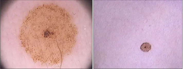
Fig. 2. Peripheral reticular pattern with central homogen area.
Fig. 1. Globular pattern with central dark globules.
type IV had large-sized congenital nevi.
Dermoscopic patterns
1) General dermoscopic structures in congenital nevi On dermoscopic examination, we found the presence of these patterns: reticular pattern, 24.1% (n=26); globular pattern, 32.4% (n=35); reticular-globular pattern, 12%
(n=13); homogeneous pattern, 14.8% (n=16); reticular- homogeneous pattern, 5.6% (n=6); globular-homoge- neous pattern, 1.9% (n=2); cobblestone pattern, 6.5%
(n=7); reticular patchy pattern, 2.8% (n=3) (Fig. 1∼4, Table 2). The globular pattern as the predominant dermo- scopic pattern was more frequent in children younger than 16 years old (32.4%); the reticular pattern as the pre-
dominant pattern was more frequent in adults older than 16 years old (28.3%).
Perifollicular hypopigmentation was present in 32 con- genital nevi (29.6%). An atypical pigment network was observed in 30 congenital nevi (27.8%). Milia-like cysts were present in 23 congenital nevi (21.3%). Hypertrichosis was observed in 17 congenital nevi (15.7%). Radial streaks were observed in 15 congenital nevi (13.9%). Focal hypo- pigmentation was present in 70 congenital nevi (64.8%).
Atypical dots/globules were observed in 72 congenital ne- vi (66.7%). Regression was present in 6 congenital nevi (5.6%). Blue-whitish veil was present in 3 congenital nevi (2.8%). Vascular structures were always absent.
Fig. 3. Globular pattern with hypertrichosis.
Fig. 4. Patchy reticular pattern.
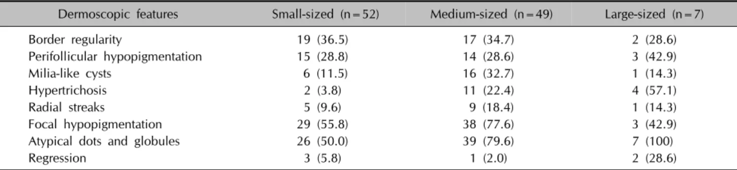
Table 2. Dermoscopic features according to the size of the nevus
Dermoscopic features Small-sized (n=52) Medium-sized (n=49) Large-sized (n=7)
Border regularity 19 (36.5) 17 (34.7) 2 (28.6)
Perifollicular hypopigmentation 15 (28.8) 14 (28.6) 3 (42.9)
Milia-like cysts 6 (11.5) 16 (32.7) 1 (14.3)
Hypertrichosis 2 (3.8) 11 (22.4) 4 (57.1)
Radial streaks 5 (9.6) 9 (18.4) 1 (14.3)
Focal hypopigmentation 29 (55.8) 38 (77.6) 3 (42.9)
Atypical dots and globules 26 (50.0) 39 (79.6) 7 (100)
Regression 3 (5.8) 1 (2.0) 2 (28.6)
Values are presented as number (%).
2) Relation between the size and dermoscopic structures in congenital nevi
On dermoscopic examination, we found the presence of these patterns: reticular pattern, 34.6% (n=18); globular pattern, 36.5% (n=19); homogeneous pattern, 19.2%
(n=10); reticular-homogeneous pattern, 5.8% (n=3); and cobblestone pattern, 3.8% (n=2) in small-sized congenital nevi; reticular pattern, 14.3% (n=7); globular pattern, 30.6% (n=15); reticular-globular pattern, 22.4% (n=11);
homogeneous pattern, 12.2% (n=6); reticular-homoge- neous pattern, 6.1% (n=3); globular-homogeneous pat-
tern, 4.1% (n=2); cobblestone pattern, 6.1% (n=3); retic- ular patchy pattern, 4.1% (n=2) in medium-sized con- genital nevi; reticular pattern, 14.3% (n=1); globular pat- tern, 14.3% (n=1); reticular-globular pattern, 28.6% (n=2);
cobblestone pattern, 28.6% (n=2); reticular patchy pat- tern, 14.3% (n=1) in large congenital nevi.
No association was found between the border regularity (p=0.911), perifollicular hypopigmentation (p=0.744), ra- dial streaks (p=0.442), regression (p=0.072), blue-whitish veil (p=0.543) and size. Medium-sized congenital nevi had a statistically higher ratio of milia-like cysts (p=0.03) and focal hypopigmentation (p=0.031) than the others.
An atypical pigment network was observed in all of the large-sized congenital nevi (100%), 18 (36.7%) me- dium-sized congenital nevi, and 5 (9.6%) small-sized con- genital nevi. There was a statistically significant difference related to the atypical pigment network between groups (p=0.025). Atypical pigmentation is a common dermo- scopic feature in large-sized and medium-sized congenital nevi.
Hypertrichosis was observed in 2 (3.8%) small-sized nevi, 11 (22.4%) medium-sized congenital nevi, and 4 (57.1%) large-sized congenital nevi. There was an association and a highly positive correlation between hypertrichosis (r=
0.485, p=0.000), the presence of atypical dots/globules (r=0.783, p=0.001), asymmetry (r=0.552, p=0.006) and the size of the lesion.
No association was found between asymmetry (p=0.975) border regularity (p=0.353), perifollicular hypopigmentation (p=0.084), radial streaks (p=0.579), regression (p=0.407), focal hypopigmentation (p=0.243) and the location of nevi.
DISCUSSION
Congenital melanocytic nevi (CMN) are described as neu- ral crest-derived hamartomas, which appear at, or shortly after birth as pigmented tumors, by some authors8. The in- cidence of CMN in neonates ranges from 0.2% to 2.1%
regardless of nevus size13. It is known that the large-sized CMN have a higher risk of malignant melanoma (MM) de- velopment than small- and medium-sized nevi. MM aris- ing in CMN usually develops at younger ages and is lo- cated deeper within large CMN and superficially within small- or medium-sized CMN. Two important meta-analy- ses have been conducted to determine the significance of MM in CMN14,15. In one of these reviews, 10 of the 432 patients developed melanoma within their giant con- genital nevi. Krengel et al.15 analyzed 14 studies with a to- tal of 6,571 patients CMN with who were followed for a mean of 3.4∼23.7 years. Forty-six (0.7%) developed a to-
tal of 49 melanomas (mean age at diagnosis: 15.5 years, median age: 7 years). The authors found a markedly in- creased risk of developing melanoma during childhood and adolescence15. Large CMN are sometimes associated with neurocutaneous melanocytosis (NCM). NCM is a melanocytic proliferation including the cranial nervous system; this can increase intracranial pressure. In our study, none of the patients with large CMN had the symp- toms of intracranial pressure. Brain MRI examinations were normal.
It has been estimated that the lifetime risk of developing melanoma is 1 in 100 for patients with small and medium CMN16,17.
Congenital melanocytic nevi should be managed con- servatively if there are no abnormalities at clinical and der- moscopic examination and the patient has no cosmetic disturbance18. Annual clinical and digital dermoscopic ex- amination is indicated in these patients. Therefore, the physician should know the dermoscopic features of CMN to avoid unnecessary excisions, and to recognize melano- ma when it begins to develop. In a nationwide study, mel- anoma developed in 3 of 131 (2.3%) patients with giant CMN18. Diagnosis of large CMNs is simple, regarding their size and appearance since birth19. Therefore, we did not include so many large CMNs in our study. We aimed to identify dermoscopic features of small- and me- dium-sized CMN, especially.
In most cases, dermoscopic examination reveals a glob- ular or cobblestone pattern in large CMN. In our study, re- ticular-globular and cobblestone patterns were the most common patterns of large CMN in line with findings in the literature. The predominant dermoscopic patterns in small-sized and medium-sized nevi were globular, present in 36% of the small-sized group, and in 30% of the me- dium-sized group. Our results are consistent with those of previous studies20,21. Additionally, the predominant der- moscopic pattern was globular pattern in patients younger than 16 years old, whereas it was reticular pattern in pa- tients older than 16 years. Some authors suggest that a predominantly reticular pattern is present particularly in individuals 12 years or older22,23. Our results support this finding.
In general, atypical dots/globules, focal hypopigmentation, and perifollicular hypopigmentation are the most common dermoscopic features of CMN found in our study. Our re- sults are consistent with the results of Stinco et al.24. Atypical dots/globules, focal hypopigmentation, asymme- try, and border regularity were common in both of the children and adults.
Milia-like cysts, hypertrichosis, radial streaks were un- common than atypical dots and globules, focal hypopig-
mentation and perifollicular hypopigmentation, in general.
The rarest dermoscopic feature was regression, according to our study.
The presence of atypical dots/globules, asymmetry, and hypertrichosis correlate with the size of CMN. It is known that these features are typical for giant and large CMN, and our results are consistent with findings in the litera- ture25. Focal hypopigmentation and milia-like cysts are statistically more common in medium-sized nevi than small-sized and large-sized nevi. There is no data about the rates of focal hypopigmentation and milia-like cysts, according to the size of nevi reported previously in the literature. Regression and radial streaks are the rarest der- moscopic features in all sizes of nevi. Perifollicular hypo- pigmentation and border regularity are other common fea- tures in all sizes of nevi without a statistical difference.
Small-sized nevi are primarily located on the trunk (59.6%), and the most frequent dermoscopic features in small-sized nevi are focal hypopigmentation (55.8%) and atypical dots/globules (50.0%). Medium-sized nevi are al- so frequently located on the trunk (59.2%). Atypical dots/globules (79.6%) and focal hypopigmentation (77.6%) are the most prevalent dermoscopic features in me- dium-sized nevi. Large-sized nevi are typically located on the trunk (57.1%); atypical dots/globules (100%) and asymmetry (85.7%) are the most common features in large-sized nevi. All sizes of nevi mostly occur in people with Fitzpatrick skin type 4 without any statistical difference.
We designed our study to define distinct dermoscopic fea- tures based on size to be helpful in distinguishing CMN from dysplastic nevi and melanoma. The most important conclusions of our study are that in children from our pop- ulation: The globular pattern as the predominant dermo- scopic pattern is more frequent in children younger than 16 years old, and the reticular pattern as the predominant pattern is more frequent in adults older than 16 years old;
Atypical dots/globules, focal hypopigmentation, and peri- follicular hypopigmentation are the most common dermo- scopic features of CMN; The rarest dermoscopic feature is the blue-whitish veil; The presence of atypical dots/glob- ules, asymmetry, and hypertrichosis correlates with the size of CMN; Focal hypopigmentation and milia-like cysts are significantly more common in medium-sized nevi than small-sized and large-sized nevi.
Atypical dots/globules, focal hypopigmentation, perifollicular hypopigmentation, and atypical pigment network have been associated with dysplastic nevi up until now. In re- cent years, it has been shown that there was a mistake about the terms dysplastic nevus and congenital nevus12. Most of the small-sized and medium-sized congenital nevi
may have the same dermoscopic patterns and features with dysplastic nevi.
Our findings may provide information for larger studies designed to accurately define the dermoscopic features of CMN.
ACKNOWLEDGMENT
We thank our patients, essential to this work, for con- tributing to continuous and prospective advance in this research.
REFERENCES
1. Marghoob AA. Congenital melanocytic nevi. Evaluation and management. Dermatol Clin 2002;20:607-616.
2. Makkar HS, Frieden IJ. Congenital melanocytic nevi: an update for the pediatrician. Curr Opin Pediatr 2002;14:
397-403.
3. Castilla EE, da Graça Dutra M, Orioli-Parreiras IM.
Epidemiology of congenital pigmented naevi: I. incidence rates and relative frequencies. Br J Dermatol 1981;104:
307-315.
4. Ingordo V, Gentile C, Iannazzone SS, Cusano F, Naldi L.
Congenital melanocytic nevus: an epidemiologic study in Italy. Dermatology 2007;214:227-230.
5. Tannous ZS, Mihm MC Jr, Sober AJ, Duncan LM.
Congenital melanocytic nevi: clinical and histopathologic features, risk of melanoma, and clinical management. J Am Acad Dermatol 2005;52:197-203.
6. Clemmensen OJ, Kroon S. The histology of "congenital features" in early acquired melanocytic nevi. J Am Acad Dermatol 1988;19:742-746.
7. Mizushima J, Nogita T, Higaki Y, Horikoshi T, Kawashima M. Dormant melanocytes in the dermis: do dermal melanocytes of acquired dermal melanocytosis exist from birth? Br J Dermatol 1998;139:349-350.
8. Alikhan A, Ibrahimi OA, Eisen DB. Congenital melanocytic nevi: where are we now? Part I. clinical presentation, epidemiology, pathogenesis, histology, malignant transfor- mation, and neurocutaneous melanosis. J Am Acad Der- matol 2012;67:495.e1-e17; quiz 512-514.
9. Viana AC, Gontijo B, Bittencourt FV. Giant congenital melanocytic nevus. An Bras Dermatol 2013;88:863-878.
10. Bolognia J, Jorizzo JL, Schaffer JV. Dermatology. 3rd ed.
Philadelphia, London: Elsevier Saunders, 2012:1871-1876.
11. Kovalyshyn I, Braun R, Marghoob A. Congenital mela- nocytic naevi. Australas J Dermatol 2009;50:231-240; quiz 241-242.
12. Kittler H, Rosendahl C, Cameron A, Tschandl P. Der- matoscopy. An algorithmic method based on pattern ana- lysis. Vienna: Facultas.wuv, 2011:29-35.
13. Lin J, Koga H, Takata M, Saida T. Dermoscopy of pigmented lesions on mucocutaneous junction and mucous membrane. Br J Dermatol 2009;161:1255-1261.
14. Watt AJ, Kotsis SV, Chung KC. Risk of melanoma arising in
large congenital melanocytic nevi: a systematic review.
Plast Reconstr Surg 2004;113:1968-1974.
15. Krengel S, Hauschild A, Schäfer T. Melanoma risk in congenital melanocytic naevi: a systematic review. Br J Dermatol 2006;155:1-8.
16. Sahin S, Levin L, Kopf AW, Rao BK, Triola M, Koenig K, et al. Risk of melanoma in medium-sized congenital mela- nocytic nevi: a follow-up study. J Am Acad Dermatol 1998;39:428-433.
17. Zaal LH, Mooi WJ, Klip H, van der Horst CM. Risk of malignant transformation of congenital melanocytic nevi: a retrospective nationwide study from The Netherlands. Plast Reconstr Surg 2005;116:1902-1909.
18. Yun SJ, Kwon OS, Han JH, Kweon SS, Lee MW, Lee DY, et al. Clinical characteristics and risk of melanoma develop- ment from giant congenital melanocytic naevi in Korea: a nationwide retrospective study. Br J Dermatol 2012;
166:115-123.
19. Moscarella E, Zalaudek I, Ferrara G, Manzo M, Savarese I, Argenziano G. Problematic melanocytic lesions in children.
Expert Rev Dermatol 2009;4:249-261.
20. Braun RP, Calza AM, Krischer J, Saurat JH. The use of
digital dermoscopy for the follow-up of congenital nevi: a pilot study. Pediatr Dermatol 2001;18:277-281.
21. Oliveria SA, Geller AC, Dusza SW, Marghoob AA, Sachs D, Weinstock MA, et al. The Framingham school nevus study:
a pilot study. Arch Dermatol 2004;140:545-551.
22. Seidenari S, Pellacani G, Martella A, Giusti F, Argenziano G, Buccini P, et al. Instrument-, age- and site-dependent variations of dermoscopic patterns of congenital melanocytic naevi: a multicentre study. Br J Dermatol 2006;155:56-61.
23. Changchien L, Dusza SW, Agero AL, Korzenko AJ, Braun RP, Sachs D, et al. Age- and site-specific variation in the dermoscopic patterns of congenital melanocytic nevi: an aid to accurate classification and assessment of melanocytic nevi. Arch Dermatol 2007;143:1007-1014.
24. Stinco G, Argenziano G, Favot F, Valent F, Patrone P.
Absence of clinical and dermoscopic differences between congenital and noncongenital melanocytic naevi in a cohort of 2-year-old children. Br J Dermatol 2011;165:1303-1307.
25. Haliasos EC, Kerner M, Jaimes N, Zalaudek I, Malvehy J, Hofmann-Wellenhof R, et al. Dermoscopy for the pediatric dermatologist part III: dermoscopy of melanocytic lesions.
Pediatr Dermatol 2013;30:281-293.