DOI : 10.3341/jkos.2009.50.2.313
= 증례보고 =
투명각막 절개를 통한 백내장 수술 후 발생한 각막상피내생의 치료
강희영⋅신민철
한림대학교 의과대학 춘천성심병원 안과학교실
목적: 투명각막 절개를 통한 백내장 수술 후 발생한 각막상피내생 환자에서 외과적 절제와 5-플루오로우라실(5-fluorouracil, 5-FU)의 전방 내 주입술을 시행하여 좋은 결과를 얻었기에 이를 보고하고자 한다.
증례요약: 70세 여자 환자가 2년 전 투명각막 절개법으로 백내장 수술을 시행 받은 후 내원 3개월 전 발생한 좌안 안구 통증 및 각막 부종으로 진료 의뢰되었다. 내원 시 각막실질부종과 함께 미만성의 종이모양 막이 투명각막 절개창 부위의 각막 후면에서부터 동공 주변 홍채까지 관찰 되었다. 저자들은 증식된 각막상피내생조직의 외과적 절제와 5-플루오로우라실(5-fluorouracil, 5-FU)의 전방 내 주입술로 치료하였고, 그 후 2개월 동안 재발되지 않는 상태로 잘 유지되었다.
<대한안과학회지 2009;50(2):313-317>
■ 접 수 일: 2008년 10월 20일 ■ 심사통과일: 2009년 2월 5일
■ 통 신 저 자: 신 민 철
강원도 춘천시 교동 153 한림대학교 춘천성심병원 안과 Tel: 033‐252‐9970, Fax: 033‐255‐5210 E-mail: [email protected]
각막상피내생은 안구를 관통하는 외상이나, 안구 내 수 술적 처지 후 발생할 수 있는 좋지 않은 시력 예후를 보이 는 합병증 중의 하나 이다.1-20최근에는 수술 기구와 수기, 봉합술 및 현미경의 발달로 인하여 미세수술이 점차 보편 화 되면서 안조직 손상의 감소와 수술 시간의 단축으로 안 구 내 수술 후 각막상피내생 뿐 아니라 다른 합병증의 발생 빈도도 감소 되었다.1,8,9,11,16
백내장 수술 또한 투명각막 절 개창을 통한 무봉합 백내장 초음파 유화 수술법이 도입되 면서 이전의 낭외, 낭내적출 방법에 비하여 그 안과적 합병 증이 줄었으며, 각막상피내생의 발생 빈도 또한 감소하였 다.21 각막상피내생은 그 임상 양상이나 세극등 검사 소견 만으로는 확진 할 수 없고, 조직학적 검사가 필요하며, 항대 사제의 전방 내 주입이나 전방 내로 자란 상피조직과 침범 된 홍체 및 모양체의 절제로 호전된 것이 보고되었으나, 병 변의 상태에 따라 그 치료법을 결정하여야 한다.1,5,8,9,11
국 내에서는 아직 백내장 수술 후 발생한 각막상피내생이 보 고된 바 없으며, 라식 수술과 관통성 외상 후에 발생한 경 우만 보고되어 있다.13-20그리고, 증식된 막의 절제와 동시 에 항대사제의 전방 내 주입을 시행하여 치료한 경우는 보 고되어 있지 않다. 이에 저자들은 투명각막 절개를 통한 초 음파 유화술 후 발생한 각막상피내생에 대한 조직학적 진 단과 각막상피내생 조직의 최대 절제와 동시에 5-플루오 로우라실(5-fluorouracil, 5-FU)의 전방 내 주입을 통한
치료 후 양호한 경과를 보여 문헌 고찰과 함께 보고하는 바 이다.
증례보고
70세 여자 환자로 3개월 전부터 시작된 좌안의 흐리게 보임과 안구 통증을 주소로 내원하였다. 환자는 2년전 양안 에 투명각막 절개창을 이용한 초음파 유화술을 시행 받았 으며 합병증은 없는 상태였다. 기저 질환은 당뇨 이외에는 없었으며, 내원 당시 시력은 우안 0.7, 좌안 0.02였고, 골드 만 안압계로 측정한 안압은 우안 12 mmHg, 좌안 56 mmHg였다. 내원 시 시행한 세극등 검사상 좌안 각막 실질 부종이 관찰되었고, 미만성의 종이모양 막이 투명각막절개 창 부위의 각막 후면에서부터 동공 주변 홍채까지 관찰 되 었다(Fig. 1A). 또한, 동공연의 홍채 외반(Fig. 1B) 및 아 래쪽 홍채의 신생혈관도 볼 수 있었다(Fig. 1A). 고삼투압 제제의 전신 투여와 국소 베타차단제의 점안을 통해 좌안 안압이 12 mmHg 정도까지 조절되었고 각막부종 소견도 호전되었다. 입원 3일째 투명각막 절개창 부위에 전방 천자 후 절개 통로의 증식된 막을 홍채 주걱(iris spatula)과 초 퍼(chopper)를 사용하여 최대한 제거하였으며, 각막 후면에 서 동공연까지 홍채 위에서 증식되어 있는 막은 유리체 절 제기와 초퍼(chopper)로 최대한 절제 및 제거 하였다. 동시 에 항대사제인 5-플루오로우라실(5-fluorouracil, 5-FU) 0.5 mg를 완충용액(Balanced Salt Solution, BSS) 0.1 ml 에 섞은 후 0.1 ml를 전방 내 주사하였다. 수술 중 제거한 미만성 종이모양 막에 대한 조직 검사에서 검은 색의 홍채 실질을 둘러싸고 있는 중증 편평상피세포와 결막 기원의
A B
Figure 1. Anterior segment photograph shows diffuse sheet-like epithelial membrane covers from clear cornea incision site to around the pupil margin. Red star is the old clear corneal incision site and vascularization of the incision site (blue arrow head) (A). Everted iris is shown (red arrow head) and diffuse corneal edema. All red arrows indicate the diffuse sheet-like membrane (B).
A
B
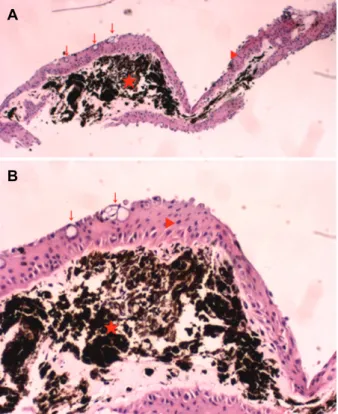
Figure 2. Microscopic examination of the excised tissue shows that black pigmented lesion is a stroma of the iris (red star) is surrounded by stratified epithelium (red arrow head) contains conjunctival origin goblet cells (red arrows). (H&E staining ×40 A, ×100 B)
술잔세포가관찰 되는소견을 보여 각막상피내생으로 확진 할 수 있었다(Fig. 2). 각막상피내생 조직의 최대한 절제 (Fig. 3)와 5-FU의 전방 내 주입 후 2개월 간 재발 의심소
견이 보이지 않고 호전되었다(Fig. 4). 시력은 0.02 (한천 석 3 m 시력 검사용) 정도로 수술 전, 후의 차이는 없었으 나, 각막 부종이 호전되고, 안압 역시 9 mmHg 정도로 잘 유지되고 있다.
고 찰
각막 윤부의 줄기 세포나 결막의 상피세포가 관통성 안 외상이나 백내장, 녹내장 여과 수술 및 각막 이식 수술 등 의 안구 내 수술 후, 또한 상처 부위에서 발생된 샛길이나 불완전한 창상의 치유 및 봉합 부위의 유출 등에 의하여 전 방내로 이동하여, 각막 후면에서 홍채 전방 및 앞쪽 유리체 쪽으로 증식하는 것을 각막상피내생이라고 한다.1,8-17 각막 상피내생이 발생한 후 늦게 진단 되거나 수술적 치료 없이 약물 치료만 시행한 경우에는 안구 적출이 필요한 경우가 있을 만큼 예후가 좋지 않다.11 주로 시력 감소나 충혈, 통 증과 눈물 흘림을 호소하며, 세극등 검사상 각막부종이나 띠각막병증, 각막후면의 증식된 막, 전방각의 폐쇄, 홍채 뒤 집힘 등을 관찰 할 수 있으며, 절개창의 누출 검사를 위한 자이델 검사(Seidel test)와 경면 현미경 검사가 각막상피 내생의 진단에 도움을 줄 수 있다.1-11본 증례의 경우 백내 장 수술 후 시력감소와 안구 통증을 호소하였고, 각막절개 창 부분에서 전방내의 동공주변까지 판상의(sheet like) 증 식된 막이 관찰 되었으며, 각막의 부종 및 홍채 뒤집힘 그 리고 안압의 상승을 보여 각막상피내생을 의심할 수 있었 다. 전방각에서 부분적으로 증식된 막과 얕은 전방에 의해 안압이 상승된 것으로 판단되나, 지속적인 만성섬모체염 또
A B
Figure 3. Anterior segment photographs at one day after removal of the whole membrane in the anterior chamber.
Clear corneal incisioned site was closed with nylon (A). Although there was a corneal edema and everted iris,all the sheet-like membranes were excised successfully (B).
A B
Figure 4. Anterior segment photograph at two months after the total excision of the ingrown epithelium and intracameral 5-FU injection (A). There are no recurrence of epithelial membrane from the clear corneal incision site to the pupil margin on the iris (B).
한 안압상승요인으로 작용하였을 것으로 판단되었다.1고 삼투압제의 정맥주사와 국소 베타차단제의 점안으로 상승 된 안압은 외과적 처치 전 조절되었으며, 외과적 절제 후 증식된 막의 조직학적 검사를 통해 결막 혹은 각막의 비각 질중층편평상피를 확인 할 수 있다. 이처럼 결막 혹은 각막 의 상피세포가 전방내에서 자라는 합병증은 전방내에서의 증식된 양상에 따라 상피진주종(epithelial pearl tumor), 낭성상피(epithelial cyst) 그리고 각막상피내생(epithelial downgrowth, epithelial ingrowth)로 세분되며, 그 분류에 따라 치료법 및 예후도 다르다.9Figure 3에서 결막 기원의 술잔세포가 포함된 중층편평상피 세포가 관찰되는 것으로 본 증례의 환자를 각막상피내생으로 진단 할 수 있었으며, 백내장 수술 시 투명각막 절개창을 만드는 과정에서 결막
상피세포가 직접적으로 파종되었거나, 수술 후 절개창의 불 완전한 치유로 인한 결막상피세포의 이동이 원인일 것으로 생각된다. 저자들은 기존의 백내장 수술 시 만들어진 투명 각막 절개창 부위의 절제와 긁어냄 술 이후 10번 나일론으 로 봉합하였으며, 새로운 절개창을 통해 유리체 절제기와 초퍼(chopper)로 증식되어진 각막상피 조직을 최대한 제거 후 봉합하였다. 그 후, 항대사물질인 5-플루오로우라실 (5-FU) 0.5 mg을 0.1 ml 평형 염액에 희석하여 0.1 ml를 전방내에 주입하였다. 수술 약 2개월 후 각막 부종이 호전 되었으며, 각막상피 내생의 재발 이나 절개 부위의 누출 및 이차성 녹내장에 의한 안압의 상승 등은 보이지 않았다. 그 러나 고안압의 상태가 내원 전 7일 정도 지속되었고, 내원 3개월 전부터 안압 상승에 의한 시신경의 손상으로 시력 호
전은 보이지 않은 것으로 판단 된다. 본 증례의 치료법과는 달리 초음파 유화술로 백내장 수술을 시행 받은 후 전방내 에 발생한 낭성 상피(epithelial cyst)가 발생된 경우, 낭 (cyst)에 대한 내시경적 광응고를 시행함으로써 성공적으 로 치료하였다는 보고도 있고, 본 증례와 동일한 조건인 투 명각막 절개창을 통한 초음파 유화술로 백내장 수술 시행 후 창상 부위 누출에 의해 발생한 각막상피내생에 대하여 증식막에 대한 수술적 제거 없이 여러 차례 5-플루오로우 라실(5-FU)의 전방내 주입 후 실패한 보고도 있다.10,15 이 미 위에서 언급한 바와 같이 본 증례의 경우는 미만성의 판 모양 각막상피내생(diffuse sheet like epithelial ingrowth) 으로 최대한 외과적 절제의 시행과 항대사제 전방내 주입 술을 시행하였고, 2개월까지 재발 없이 잘 유지되어 오고 있다.
투명각막 절개창을 통한 백내장 수술 후 각막상피 내생 의 합병증이 발생 시 임상 양상 및 조직검사를 통한 정확하 고 빠른 진단과 수술적 완전 절제 후 항대사제 사용을 통한 치료가 효과적인 방법이 될 수 있다고 본다.
참고문헌
1) Vargas LG, Vroman DT, Solomon KD, et al. Epithelial Down- growth after Clear Corena Phacoemulsification: report of two cases and review of the literature. Ophthalmology 2002;109:
2331-5.
2) Sullivan GL. Epithelization of the anterior chamber following cataract extraction: a new approach to treatment. Trans Am Acd Ophthalmol 1958;56:606-54.
3) Long JC, Tyner GS. Three cases of epithelial invasion of the anterior chamber treated surgically. Arch Ophthalmol 1957;58:
396-400.
4) Brown SI. Treatment of advanced epithelial downgrowth.
Trans Am Acd Ophthalmol 1973;77:618-22.
5) Friedman AH. Radical anterior segment surgery for epithelial invasion of the anterior chamber: report of three cases. Trans Am Acd Ophthalmol 1977;83:216-23.
6) Stark WJ, Michels RG, Maumenee AE, Cupples H. Surgical management of epithelial ingrowth. Am J Ophthalmol 1978;
85:772-80.
7) Maumenee AE, Paton D, Morse PH, Butner R. Review of 40 histologically proven cases of epithelial downgrowth following
cataract extraction and suggested surgical management. Am J Ophthalmol 1970;69:598-603.
8) Knauf HP, Rowsey JJ, Margo CE. Cystic epithelial down- growth clear-corneal cataract extraction. Arch Ophthalmol 1997;22:330-5.
9) Chen SH, Pineda R 2nd. Epithelial and fibrous downgrowth:
mechanism of disease. Ophthalmol Clin North Am 2008;15:
41-8.
10) Jadav DS, Rhlander NR, Vold SD, et al. Endoscopic photo- coagulation in the management of epithelial downgrowth.
Cornea 2008;27:601-4.
11) Weiner MJ, Trentacoste J, Pon DM, Albert DM. Epithelial downgrowth: a 30-year clinicopathological review. Br J Ophthalmol 1989;73:6-11.
12) Kim SK, Ibarra MS, Syed NA, et al. Development of epithelilal downgrowth several decades after intraocular surgery. Cornea 2005;24:108-9.
13) Cho JS, Ko MK, Shin JC. Epithelial ingrowth of anterior chamber and anterior surface of vitreous. Korean J Ophthal- mol 1998;12:118-21.
14) Lai MM, Haller JA. Resolution of epithelial ingrowth in a patient treated with 5-fluorouracil. Am J Ophthalmol 2002;
133:562-4.
15) Tomlins PJ, Savant V, Quinlan M. Failure of intracameral fluorouracil to resolve an epithelial ingrowth following clear corneal cataract surgery. J Cataract Refract Surg 2007;33:923-4.
16) Lee BL, Gaton DD, Weinreb RN. Epithelial downgrowth following phacoemulsification through a clear cornea. Arch Ophthalmol 1999;117:283.
17) Shikh AA, Damji KF, Mintsioulis G, et al. Bilateral epithelial downgrowth managed in one eye with intraocular 5-fluor- ouracil. Arch Ophthalmol 2008;120:1396-8.
18) Srinivasan S, Jones DH, Jay JL, Roberts F. Epithelial down- growth following clear cornea phacoemulsification in a buphthalmic eye. Br J Ophthalmol 2004;88:152-3.
19) Kim SW, Byun YJ, Kim EK, Kim TI. Treatment of epithelial ingrowth after laser in situ keratomilusis using amniotic membrane patch. J Korean Ophthalmol Soc 2007;48:230-7.
20) Lim TH, Kim MJ, Kim TI, Tchah HW. Lamellar keratoplasty and restoration of traumatic dislocation of LASIK flap using human fibrin adhesive. J Korean Ophthalmol Soc 2005;46:
1741-6.
21) Hunyor AP, Davis Belcher III. BSC. Epithelial and Fibrous Invasion of the Eye. In: Steinert RF, Fine IH, Gimbel HV, et al, eds. Cataract Surgery: Technique, Complications, Management, 2nd ed. Philadelphia: Saunders, 2004; chap. 46.
=ABSTRACT=
The Treatment of Epithelial Ingrowth After Phacoemlusification Through Clear Cornea Incision
Hee Young Kang, MD, Min Chul Shin, MD
Department of Ophthalmology, Chuncheon Sacred Heart Hospital, Hallym University, School of Medicine, Chuncheon, Korea
Purpose: To report a case of epithelial ingrowth treatment by surgical excision of epithelial tissues and intracameral 5-fluorouracil injection.
Case summary: A 70-year-old female patient who underwent phacoemulsification through clear cornea incision in both eyes 2 years before, was referred for her left ocular pain and corneal edema of 3 months' duration. Diffuse sheet-like epithelium grew from the clear cornea incision site to the pupil margin lesion of the iris. The epithelial tissues were excised and 5-fluorouracil was injected intracamerally. There were no recurrences for 2 months.
J Korean Ophthalmol Soc 2009;50(2):313-317
Key Words: Epithelial ingrowth, Excision, Phacoemulsification, 5-Fluorouracil
Address reprint requests to Min Chul Shin, MD
Department of Ophthalmology, College of Medicine, Hallym University
#153 Gyo‐dong, Chunchon‐si, Gangwon‐do 200‐060, Korea Tel: 82‐33‐252‐9970, Fax: 82‐33‐255‐5210, E-mai; [email protected]