Subsequent Hip Fracture in Osteoporotic Hip Fracture Patients
Sang Ho Lee, Tong Joo Lee, Kyu Jung Cho, Sang Hyun Shin, and Kyoung Ho Moon
Department of Orthopedic Surgery, School of Medicine, Inha University, Incheon, Korea.
Received: August 11, 2011 Revised: October 21, 2011 Accepted: October 27, 2011
Corresponding author: Dr. Kyoung Ho Moon, Department of Orthopedic Surgery, School of Medicine, Inha University, 27 Inhang-ro, Jung-gu,
Incheon 400-711, Korea.
Tel: 82-32-890-3663, Fax: 82-32-890-3099 E-mail: [email protected]
∙ The authors have no financial conflicts of interest.
© Copyright:
Yonsei University College of Medicine 2012 This is an Open Access article distributed under the terms of the Creative Commons Attribution Non- Commercial License (http://creativecommons.org/
licenses/by-nc/3.0) which permits unrestricted non- commercial use, distribution, and reproduction in any medium, provided the original work is properly cited.
Purpose: A significant number of patients who have experienced previous surgical treatment for an osteoporotic hip fracture experience a subsequent hip fracture (SHF) on the opposite side. This study aims to analyze the risk factors and the cor- relation between osteoporosis and SHF on the opposite side in order to assess the usefulness of bisphosphonate treatment for the prevention of SHFs. Materials and Methods: We included 517 patients treated from March 1997 to April 2009 in this study. The inclusion criteria included previous unilateral hip fracture, without os- teoporotic treatment, and a T-score less than -3.0 at the time of the fracture. We studied these patients in terms of death, SHF, alcoholism, living alone, dementia, dizziness, health status, osteoporotic treatment after fracture and bone mineral den- sity (BMD). In total, 34 patients experienced a SHF. We selected another 34 pa- tients without a SHF who had similar age, sex, body mass index, BMD, diagnosis, treatment and a follow up period for a matched pair study. We compared these two groups. The average follow up was 8.3 years and 8.1 years, respectively. Results:
The mortality rate of the 517 patients was 138 (27%). The BMD at the time of fracture demonstrated no statistical difference between the two groups (p>0.05).
Nine patients (26%) within the SHF group were prescribed Risedronate and 18 pa- tients (53%) received the same treatment in the non-SHF group. There was a sta- tistical relationship with the treatment of osteoporosis (p=0.026). The average BMD of patients with SHF was -5.13 and -5.02 in patients without SHF was (p>0.05). Conclusion: Although primary surgical treatments are important for an excellent outcome in osteoporotic hip fractures, treatment of osteoporosis itself is just as important for preventing SHFs.
Key Words: Hip fracture, osteoporosis, bone mineral density, subsequent fracture
INTRODUCTION
The incidence of hip fracture is increasing due to an increase in elderly popula- tions. It is expected that 63 million hip fractures will occur globally in 2050, and Melton, et al.1 reported that 6% of males and 17.5% of females will experience hip fracture.2 Hodsman, et al.3 reported that patients with previous hip fractures will experience subsequent hip fractures in the same region. They also suggested that the mortality is 2.7 times higher in these patients than the group without previous fractures.4 There are numerous reports regarding the seriousness of hip fractures;
rosis treatment.
Ten males and twenty four females had a SHF and their ages at the time of the first fracture was 75.1 (65-86) and 79.3 (68-89) years, respectively, at the time of SHF on av- erage. The interval between the first fracture and the SHF was 4.2 years (8 months-9 years) on average. Among the 34 patients who had a SHF, a femur neck fracture was the first fracture in 17 patients (50%), while 13 patients (38%) experienced an intertrochanteric fracture and 4 patients (12%) experienced a subtrochanteric fracture. Sixteen pa- tients received a total hip replacement arthroplasty (THRA), while 7 patients had dynamic hip screws, 7 patients had in- tramedullary nails and 4 patients underwent axial pinning.
Twenty patients (59%) had a femur neck fracture as a SHF, 10 patients (29%) had an intertrochanteric fracture, and a subtrochanteric fracture was noted in 4 patients (12%). Of these, 18 patients received a THRA, while 6 patients had dynamic hip screws, 6 patients had intramedullary nails and 4 patients underwent axial pinning. A Pearson chi-square test, Mann-Whitney test and a Fisher’s exact test were used as statistical methods.
RESULTS
The mortality rate was 27%, as 138 of the 517 patients had died. The average age was 75.8 (56-96) years for 148 males and 369 females with an average BMI of 20.2 (15.1-31.4).
In total, 34 patients (6.6%) experienced a SHF, and 76 pa- tients (15%) experienced a trauma other than a hip fracture, alcoholism was noted in 7 patients (1%), 94 patients (18%) lived alone, 41 patients (8%) suffered from dementia, and dizziness was noted in 20 patients (4%). Their American society of anesthesiologists (ASA) physical status was 1.9 [1-4 standard deviation (SD) 0.7] on average. The average T-score at the time of initial trauma was -5.09 (-3.0 - -6.8, SD 0.37), while an average T-score of -5.29 (-2.4 - -6.9, SD 0.35) was recorded at the final follow up. A total of 190 pa- tients (37%) received osteoporosis treatment after the initial fracture and their mean T-score was -5.15 (-3.0 - -6.8, SD 0.35) initially and -4.92 (-2.4 - -6.7, SD 0.41) at the final follow up. The 34 patients who experienced a SHF had an initial T-score of -5.13 (-3.2 - -6.6, SD 0.34), and a T-score of -5.48 (-3.5 - -6.8, SD 0.38) at the time of the SHF. The 483 patients without a subsequent fracture had T-scores of -5.03 (-3.0 - -6.8, SD 0.37) initially and a T-score of -5.28 (-2.4 - -6.9, SD 0.36) at the final follow up (Table 1).
however, it is difficult to find information on the treatment of osteoporosis, which is the leading cause of hip fractures.
Bone mineral density (BMD) is the most important factor in predicting the strength of bone, and the risk of fracture is higher when BMD is low and age is higher. Despite this fact, only a few patients receive osteoporotic treatment after evaluation of BMD.5,6 The authors studied patients who were treated for a hip fracture, grouping the patients with subsequent hip fracture (SHF) and those without SHF of the contralateral hip joint. We analyzed the risk factors of SHF and the effect of osteoporosis treatment on the preven- tion of SHF.
MATERIALS AND METHODS
In total, we analyzed 517 patients treated from March 1997 to April 2009. The inclusion criteria included patients who had ipsilateral hip fracture with a T-score of BMD lower than -3.0 and who received no osteoporosis treatment at the time of the incident. Exclusion criteria included patients who had a T-score of BMD more than -3.0, patients who received osteoporosis treatment at the time of the incident, and patients whose cause of hip fracture was not a traffic accident, fall from more than height, or pathologic fracture.
The average follow up period was 8.1 years (2-14 years).
Patient survival, presence of SHF, alcohol history, marriage status, dementia, dizziness, osteoporosis treatment after fracture, and BMD were collected via the patients’ medical records and telephone interviews. BMD tests were per- formed on all patients 2 weeks after trauma with Dual-ener- gy X-ray absorptiometry (DEXA, Lunar, GE, Milwaukee, WI, USA). An annual follow up was done in 190 patients who underwent osteoporosis treatment. We included 34 pa- tients (6.6%) with a previous history of surgery due to ipsi- lateral hip fracture and who had SHF on the contralateral side in the SHF group. In order to limit statistical bias using a matched pair study, we also included 34 patients without a contralateral hip fracture in the non-SHF group consider- ing their age, sex, body mass index (BMI), BMD, diagno- sis, surgical method, and follow up period. t-tests were per- formed for the two groups for age, BMI, BMD and follow up period. No statistical difference was found (p>0.05) after a chi-square test on age, diagnosis and surgical method. We also analyzed factors known to cause SHF such as addition- al fractures, alcohol consumption, solitude habitation, de- mentia, dizziness, medical condition, and history of osteopo-
to the ASA physical status, with scores as low as 1.6 on av- erage in cases of patients with the first fracture. Secondly, we, as a university medical institution, encountered patients mostly with internal complications, who are difficult to treat. Although it is correct to say that the decrease in BMD near the hip joint has a strong correlation with bone strength and fracture status of bones, the following compo- nents are also known to be important factors: age, gender, Among the 34 patients with a SHF, 5 patients (15%) ex-
perienced a distal radius fracture. There were no reports of alcoholism in cases with a SHF; however, for those that did have a SHF, 5 (15%) were living alone, 3 (9%) had demen- tia, and 3 (9%) reported dizziness. Their ASA physical sta- tus score was 1.8 (1-4, SD 0.7) on average. The group with- out a SHF comprised 4 cases (12%) of distal radius fractures, 2 cases (6%) of lumbar compression fractures, 6 cases (18%) lived alone, 2 cases (6%) had dementia, 3 cases (9%) reported dizziness and there were no alcoholism cas- es. Their ASA physical status score was 1.6 (1-4, SD 0.5) on average. The two groups demonstrated no statistical dif- ferences for other area fractures, alcoholism, living alone, dementia, dizziness and presence of a medical condition.
The initial T-score in the group with SHF was -5.13 (-3.2 - -6.6, SD 0.34), while the non SHF group recorded a T-score of -5.02 (-3.2 - -6.8), which had no statistical difference (p>0.05) (Table 2). Nine patients in the SHF group were prescribed Risedronate, which belongs to the bisphospho- nate group, after initial trauma, while 18 patients in the non SHF group received the same medication. This was statisti- cally different between the two groups (p=0.026).
DISCUSSION
The occurrence of fracture near the hip joint is increasing as the average life span increases due to medical improve- ment. However, mortality rates and complication rates are very high regardless of technological advances.7-9 Boston10 reported that, in the cases of a first femoral neck fracture, the death rate rose to 13% and 30% in cases of a second fracture. Our own research too demonstrated a high mortal- ity rate ratio of 27%. The mortality rate was high due, first,
Table 1. Demographics of 517 Patients Who Had Hip Fractures
Death 138 (27%)
Age 75.8 (56-95)
Sex (M/F) 148/369
BMI 20.2 (15.1-31.4)
Other site fracture 76 (15%)
Alcoholism 7 (1%)
Living alone 94 (18%)
Dementia 41 (8%)
Dizziness 20 (4%)
Health status (ASA) 1.9 (1-4)
Subsequent hip fracture 34 (6.6%)
Follow up period (yrs) 8.1 (2-14)
T-score
1st visit (n=517) -5.09 (-3.0 - -6.8) Last f/u (n=465) -5.29 (-2.4 - -6.9) Osteoporosis Tx. group
1st visit (n=190) -5.15 (-3.0 - -6.8) Last f/u (n=171) -4.92 (-2.4 - -6.7) Subsequent Fx. group
1st visit (n=34) -5.13 (-3.2 - -6.6) 2nd visit (n=34) -5.48 (-3.5 - -6.8) No subsequent Fx. group
1st visit (n=34) -5.03 (-3.0 - -6.8)
Last f/u (n=34) -5.28 (-2.4 - -6.9)
Osteoporosis treatment (Risedronate) 190 (37%) BMI, body mass index; ASA, American society of anesthesiologists; Tx, treatment; Fx, fracture.
Table 2. Comparison of the Subsequent Fx. Group with No Subsequent Fx. Group
Subsequent Fx. (n=34) No subsequent Fx. (n=34) p value
Age 75.1 (68-89) 76 (60-90) 0.825
Sex (M/F) 10/24 12/22 0.604
BMI 20.7 (16.3-27.2) 20.5 (16.7-29.1) 0.963
BMD (T-score) -5.13 (-3.2 - -6.6) -5.02 (-3.2 - -6.8) 0.847
Alcoholism 0 0 1
Living alone 5 (15%) 6 (18%) 1
Dementia 3 (9%) 2 (6%) 1
Dizziness 3 (9%) 3 (9%) 1
Health status (ASA) 1.8 (1-4) 1.6 (1-4) 0.415
Other site fracture 5 (15%) 6 (18%) 1
Osteoporosis treatment (Risedronate) 9 (26%) 18 (53%) 0.026
BMD, bone mineral density; BMI, body mass index; ASA, American society of anesthesiologists; Fx, fracture.
a resultant long-term stay at the hospital, rehabilitation, and an increase in the mortality rate resulting from localized or systemic complications are of concern.8-10 McClung, et al.18 reported the effectiveness of Risedronate in preventing frac- tures near the hip joint in a case of an aged woman who was diagnosed with osteoporosis. Bilezikian19 also reported a decrease in fractures near the hip joint in osteoporosis pa- tients administered Risedronate. Our research found a sta- tistically significant result in recurrence prevention of frac- tures near the hip joint with the use of Risedronate after first-time fractures near the hip joint. Although Kanis, et al.20 and his associates claimed that there is a correlation be- tween a low BMD and recurrence of fractures in all age groups, as age increases, the ratio of morbidity accompa- nied by other diseases also increases, and this results in less importance of a low BMD that is contributed from many other risk factors of fracture, especially in an old popula- tion. In our research, as both groups demonstrated little dif- ference in low BMD and the ratio of morbidity accompa- nied by other disease and the correlation with other risk factors were relatively low, we were able to verify the ef- fectiveness of treatment with Risedronate in the bisphos- phonate group in preventing SHF. There were a few limita- tions to the current study. First, the source of our data was limited to only one medical institution, so that the number of patients was quite small. We would expect more detailed data and results if we are able to co-work with other medi- alcohol addiction, history of fracture(s), solitude habitation,
dementia and injuries from falls.11-14 Our data did not find any statistical differences for these factors, which are known to affect the recurrence of fractures, between the two groups studied. This is probably due to the low number of the target pool in each group. Jung, et al.15 stated that, while revealing no epidemiological and physical differences in an osteoporo- sis group with a history of fractures and an osteoporosis group without such history, patients within the range of os- teoporosis had a high risk of fracture, even in those without any risk factors, regardless of the existence of the fracture.
In 1994, the WHO defined a T-score of equal to or less than -2.5 as osteoporosis and the same T-score with a history of fractures as severe osteoporosis. Currently, domestic medi- cal insurance states that a T-score equal to or less than -3.0 falls within insurance coverage range and this deserves crit- ical review and modification if the characteristics of osteo- porosis, of which prevention is more important, are consid- ered. Jang, et al.16 and his associates claimed that the risk of fracture near the hip joint increases if the T-score drops be- low -1.5, which was at an intersection for each group.17 Our data suggested that the treatment for osteoporosis has not properly been performed, as we encountered 517 hip frac- ture patients who had never had any medical care or treat- ment before, even though their T-scores were as low as -5.09 on average at the time of trauma. Problems arise not only with the hip fracture itself due to osteoporosis, but also
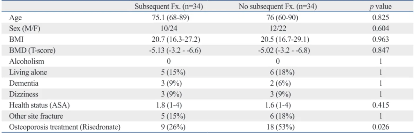
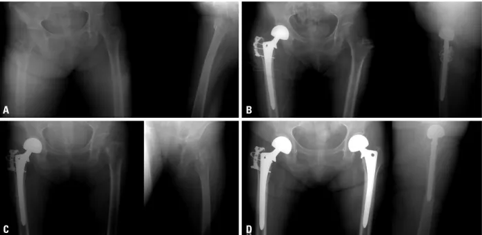
Fig. 1. A 69-year old woman had a subsequent fracture on the left side after a right side intertrochanteric fracture (initial T-score -5.3). (A) Initial radiographs of right hip side intertrochanteric fracture. (B) Postoperative radiograph after a bipolar hemiarthroplasty. (C) Radiographs of subsequent fracture in the left femur neck (T-score -5.7 with patient who did not have treatment for osteoporosis). (D) Postoperative radiograph after a bipolar hemiarthroplasty. The pa- tient started medication for osteoporosis.
A
C
B
D
gy of osteoporosis and osteoporotic fractures. Epidemiol Rev 1985;7:178-208.
8. Eastwood EA, Magaziner J, Wang J, Silberzweig SB, Hannan EL, Strauss E, et al. Patients with hip fracture: subgroups and their outcomes. J Am Geriatr Soc 2002;50:1240-9.
9. Melton LJ, Riggs BL Jr. Further characterization of the heteroge- neity of the osteoporotic syndromes. In: Kleerekoper M, Krane SM, editors. Proceedings of the International Symposium on Clin- ical Disorders of Bone and Mineral Metabolism. New York: Mary Ann Liebert, Inc.; 1989. p.145-52.
10. Boston DA. Bilateral fractures of the femoral neck. Injury 1982;14:207-10.
11. Bell GH, Dunbar O, Beck JS, Gibb A. Variations in strength of vertebrae with age and their relation to osteoporosis. Calcif Tissue Res 1967;1:75-86.
12. Carter DR, Hayes WC. Bone compressive strength: the influence of density and strain rate. Science 1976;194:1174-6.
13. Ryg J, Rejnmark L, Overgaard S, Brixen K, Vestergaard P. Hip fracture patients at risk of second hip fracture: a nationwide popu- lation-based cohort study of 169,145 cases during 1977-2001. J Bone Miner Res 2009;24:1299-307.
14. Cook PJ, Exton-Smith AN, Brocklehurst JC, Lempert-Barber SM.
Fractured femurs, falls and bone disorders. J R Coll Physicians Lond 1982;16:45-9.
15. Jung ES, Lee YK, Baek SI. Diffrences of bone mineral density between osteoporotic group with or without compression fracture of the spine. J Korean Soc Fract 1998;11:629-33.
16. Jang J, Kim WL, Kang SB, Lee JH, Yoon KS. The relationship of osteoporosis and hip fractures in elderly patients. J Korean Hip Soc 2008;20:299-304.
17. Assessment of fracture risk and its application to screening for postmenopausal osteoporosis. Report of a WHO Study Group.
World Health Organ Tech Rep Ser 1994;843:1-129.
18. McClung MR, Geusens P, Miller PD, Zippel H, Bensen WG, Roux C, et al. Effect of risedronate on the risk of hip fracture in el- derly women. Hip Intervention Program Study Group. N Engl J Med 2001;344:333-40.
19. Bilezikian JP. Efficacy of bisphosphonates in reducing fracture risk in postmenopausal osteoporosis. Am J Med 2009;122(2 Suppl):S14-21.
20. Kanis JA, Johnell O, De Laet C, Johansson H, Oden A, Delmas P, et al. A meta-analysis of previous fracture and subsequent fracture risk. Bone 2004;35:375-82.
cal institutions regarding this subject. Second, we did not identify other causes of fragility fracture in terms of calci- um profile, hormonal status, and vitamin D deficiency etc.
In conclusion, there are numerous factors related to de- crease of SHF due to osteoporosis. In this study, the effect of treatment with bisphosphonates or Risedronate was superior to others. In the patients with hip fractures, the authors sug- gest that the treatment of osteoporosis to prevent SHF is as important as primary surgical intervention (Fig. 1).
ACKNOWLEDGEMENTS
This study was supported by a Inha University Grant.
REFERENCES
1. Melton LJ 3rd, Chrischilles EA, Cooper C, Lane AW, Riggs BL.
Perspective. How many women have osteoporosis? J Bone Miner Res 1992;7:1005-10.
2. Cooper C, Campion G, Melton LJ 3rd. Hip fractures in the elder- ly: a world-wide projection. Osteoporos Int 1992;2:285-9.
3. Hodsman AB, Leslie WD, Tsang JF, Gamble GD. 10-year proba- bility of recurrent fractures following wrist and other osteoporotic fractures in a large clinical cohort: an analysis from the Manitoba Bone Density Program. Arch Intern Med 2008;168:2261-7.
4. Lee SR, Kim SR, Chung KH, Ko DO, Cho SH, Ha YC, et al.
Mortality and activity after hip fracture: a prospective study. J Ko- rean Orthop Assoc 2005;40:423-7.
5. Gardner MJ, Brophy RH, Demetrakopoulos D, Koob J, Hong R, Rana A, et al. Interventions to improve osteoporosis treatment fol- lowing hip fracture. A prospective, randomized trial. J Bone Joint Surg Am 2005;87:3-7.
6. Juby AG, De Geus-Wenceslau CM. Evaluation of osteoporosis treatment in seniors after hip fracture. Osteoporos Int 2002;13:
205-10.
7. Cummings SR, Kelsey JL, Nevitt MC, O’Dowd KJ. Epidemiolo-