Introduction
Percutaneous vertebroplasty (PVP) is considered an effec- tive treatment method for relieving the pain of osteoporot- ic vertebral compression fractures (VCFs). Although verte- broplasty is relatively safe and has a low complication rate, however, subsequent fracture after vertebroplasty is not un- common. A possible increase in risk of new VCF after aug- mentation is of concern, especially in osteoporotic patients.
Furthermore, there is no standard parameter for variables such as optimal cement volume, proper cement injection site, and optimal pattern of cement distribution. Factors that may induce subsequent fracture are also uncertain.
The purpose of this study was to evaluate the incidence of and risk factors for subsequent fracture after vertebro- plasty.
Materials and Methods
Patient selection
Between January 1, 2007, and December 31, 2009, a total of 296 patients underwent vertebroplasty at our department.
All VCFs were confirmed by magnetic resonance (MR) im- aging. Patients followed up for a minimum of 6 months were included for analysis. We selected 112 patients with prima- ry fracture (first-time fracture) at a single level who received vertebroplasty at that level (Table 1). We also excluded patho- logic fractures (metastases and multiple myeloma). Symp- tomatic subsequent VCFs were diagnosed using plain ra- diographs and MRI.
Procedure
All vertebroplasty procedures were performed in an op- erating room under C-arm guidance. In most cases, an 11-gauge needle was inserted through the routine unipe- dicular approach. A 13-gauge needle was used in the mid-thoracic level. Bone cement was injected under fluo- roscopic control. The procedure was terminated if cement leakage occurred or if the cement reached the posterior quar-
Risk Factors for Subsequent Fracture after Osteoporotic Vertebral Compression Fracture
Sung Gon Kim, MD, Joo Chul Yang, MD, Tae Wan Kim, MD and Kwan Ho Park, MD
Department of Neurosurgery, VHS Medical Center, Seoul, Korea
Objective: Percutaneous vertebroplasty is an effective treatment that relieves pain caused by vertebral compression frac- ture. However, vertebroplasty may increase the risk for subsequent vertebral compression fracture. The purpose of this study is to evaluate the incidence of and risk factors for subsequent fracture after vertebroplasty.
Methods: A retrospective analysis was performed for 112 patients who were diagnosed with a first osteoporotic compres- sion fracture at a single level and underwent vertebroplasty at that level. Possible risk factors for subsequent fracture, such as age, sex, bone mineral density (BMD), location of treated vertebrae, pattern of cement distribution, cement volume, presence of intradiscal cement leakage, and direction of cement leakage, were analyzed.
Results: During the follow-up period, 18 new subsequent vertebral fractures developed (16.1%). Subsequent fractures were more common in osteoporotic patients (T-score ≤-2.5)(p=0.034, r=0.208). Intravertebral cement volume ≥3.5 cc were also associated with a significantly higher risk of fracture (p=0.012, r=0.238).
Conclusion: Low BMD and volume of intravertebral cement were the factors most strongly associated with subsequent fracture after percutaneous vertebroplasty. (Korean J Neurotrauma 2013;9:120-124) KEY WORDS: Osteoporotic compression fracture ㆍBone mineral density ㆍCement volume.
Received: August 16, 2013 / Revised: October 7, 2013 Accepted: October 7, 2013
Address for correspondence: Tae Wan Kim, MD
Department of Neurosurgery, VHS Medical Center, 53 Jinhwang- do-ro 61-gil, Gangdong-gu, Seoul 134-791, Korea
Tel: +82-2-2225-1363, Fax: +82-2-2225-4152 E-mail: [email protected]
Korean J Neurotrauma 2013;9:120-124 http://dx.doi.org/10.13004/kjnt.2013.9.2.120
ter of the vertebral body. If the injected cement did not cross the midline, the same procedure was repeated through the contralateral pedicle. Patients were encouraged to ambulate 4 hours after the vertebroplasty.
Comparison parameters
Age, sex, bone mineral density (BMD), location of treat- ed vertebrae, pattern of cement distribution, volume of in- travertebral cement, presence of intradiscal cement leakage, and direction of cement leakage were assessed in relation to subsequent vertebral fracture. Averaged T-scores from L1 to L4 were used for BMD assessment. Vertebral levels from T12 to L2 were classified as the thoracolumbar junc- tion. The optimal volume of cement was is not clear. There- fore, in our study, we divided patients according to the vol- ume of cement used: greater than or less than 3.5 cc. In cases in which a bipedicular approach was used, the volume of ce- ment was calculated as the sum of both sides. According to the pattern of cement distribution, cases were divided into two groups as follows: cases in which the cement was evenly distributed in the vertebral body (Figure 1) and cas- es with a partially augmented vertebral body (Figure 2).
TABLE 1. The demographics of the patients Number of cases (n)
Sex
Male 060
Female 052
Mean ages (years) 76.0 (range 61-92) Level of initial compression fracture
Thoracolumbar (T12-L2) 084
Above T12 012
Below L2 016
Total 112
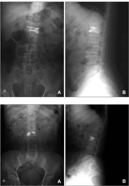
FIGURE 1. Postoperative anteroposterior (A) and lateral (B) plain X-ray show evenly distrib- uted cement in vertebral body.
FIGURE 2. Postoperative anteroposterior (A) and lateral (B) plain X-ray show partially aug- mented cement in vertebral body.
A B
A B
Incidental fractures were designated as either adjacent or non-adjacent to the vertebral level being treated with ver- tebroplasty. Subsequent fractures were also classified as ad- jacent or non-adjacent. Refracture, additional compression at vertebroplasty level, was not assessed in our study due to ambiguity between continued decrease in vertebral height and an additional VCF.
Statistical analysis
All statistical analyses were performed using SPSS ver.
10.0 (SPSS Inc., Chicago, IL). Correlation analysis (Pearson) and Fisher’s exact test were used for statistical analysis. A p-value of less than 0.05 was considered statistically sig- nificant.
Results
The mean age of patients was 76.0 years (range, 61-92 years). There were 60 men and 52 women. No major com- plications occurred during the procedure. The mean fol- low-up period was 15.4 months (range, 6-48 months). Eigh- teen subsequent fractures occurred during the follow-up period. Among them, 6 were adjacent VCFs and 12 were non-adjacent VCFs. The mean interval between the first fracture and an adjacent fracture was 10.8 months (range,
6-24 months), whereas the mean interval between a first fracture and a non-adjacent fracture was 16.5 months (range, 1-26 months). Thus, adjacent fractures tended to occur soon- er than non-adjacent fractures, but non-adjacent fractures were more common. The relationship between subsequent VCFs and these factors is shown in Table 2.
Sex
Our patients consisted of 60 men and 52 women. Subse- quent fractures occurred in 7 men (11.7%) and 11 women (21.2%). Although subsequent fractures were more com- mon in women, the difference was not statistically signifi- cant (p=0.135, 0.173, r=-0.133).
Age
We classified patients according to age; 106 patients were aged 65 years and over, the other 6 less than 65 years. Sev- enteen subsequent fractures occurred in patients aged 65 years and over (16.0%) and 1 in a patient less than 65 years (16.7%). The difference was not statistically significant (p
=0.660, 0.484, r=0.067).
Bone mineral density
Eighty-five patients had T-scores below -2.5, and 27 had scores above -2.5. Seventeen subsequent fractures occurred TABLE 2. The relation of factors and subsequent fractures after percutaneous vertebroplasty
Subsequent
fracture (n=18) Non-subsequent
fracture (n=94) p-value
(Fisher’s exact test) p-value
(Pearson) Correlation coefficient (r-value)
Sex 0.135 0.173 -0.133
Men 07 53
Women 11 41
Age 0.660 0.484 -0.067
More than 65 17 89
Less than 65 01 5
BMD (mean T-score) 0.034 0.028 -0.208
≤-2.5 17 68
>-2.5 01 26
Level of treated vertebrae 0.269 0.377 -0.084
Thoracolumbar (T12-L2) 12 72
Above T12 or below L2 03 25
Pattern of cement distribution 0.476 0.701 -0.037
Evenly distribution 14 69
Partially augmentation 04 25
Cement volume 0.012 0.014 -0.238
More than 3.5 cc 15 49
Less than 3.5 cc 03 45
Intradiscal cement leakage 0.331 0.478 -0.068
Presence 05 24
Absence 13 88
BMD: bone mineral density
in osteoporotic patients (20.0%) and 1 occurred in a non-os- teoporotic patient (3.7%). Low BMD was significantly re- lated to subsequent fracture after vertebroplasty (p=0.034, 0.028, r=0.208).
Location of initial VCF
Initial VCFs occurred at the thoracolumbar region (T12- L2) in 84 patients, and 12 subsequent fractures were in this region (14.3%). Subsequent fractures occurred more frequently in lower lumbar vertebrae (21.4%), but the dif- ference was not statistically significant (p=0.269, 0.377, r=-0.084).
Pattern of cement distribution
Cement was distributed evenly in 83 patients, and 14 sub- sequent fractures occurred in these patients (16.9%). The ce- ment was partially augmented in the vertebral bodies of 29 patients, and 4 subsequent fractures developed in these pa- tients (13.8%). The occurrence of subsequent fracture was not statistically different in these groups (p=0.476, 0.701, r=-0.037).
Cement volume
Of 48 patients in whom the cement volume was less than 3.5 cc, 3 (6.3%) developed subsequent fractures. Of 64 pa- tients in whom the cement volume was greater than 3.5 cc, 15 (23.4%) developed subsequent fractures, a statistically significant difference (p=0.012, 0.014, r=0.238).
Cement leakage
Intradiscal cement leakage occurred in 24 patients (21.4%).
Among them, 18 patients had cement leakage into the upper disc space, and 4 patients had leakage into the lower disc space. Subsequent fractures occurred in 5 patients in whom cement leakage occurred into the disc space. Subsequent fractures occurred more commonly in cases with intradiscal cement leakage (20.8% vs. 14.8%), but the difference was not statistically significant (p=0.331, 0.478, r=0.068).
Discussion
Cement leakage after vertebroplasty is not uncommon, but most of the leakage is not clinically significant. Subse- quent VCFs mean unexpected following new VCF after VCF, commonly occur after percutaneous vertebroplasty.
Subsequent VCFs are classified as adjacent fracture, non- adjacent fracture, and refracture. The incidence of subse- quent VCF is about 12% to 52%.1,3,12) Subsequent VCFs may lead to continued back pain and severe kyphotic deformi-
ty. The relationship between vertebroplasty and subsequent fractures is still uncertain. Risk of new VCFs is greater in patients who undergo vertebroplasty/kyphoplasty than in patients with prior VCFs who do not undergo the proce- dure.11) However, it is difficult to draw strong conclusions about the causal relationship between vertebroplasty and subsequent fractures based on a review of the literature.9,18)
Significant restoration of anterior vertebral height in- creases the risk of subsequent fracture in cemented verte- brae.5) Subsequent VCFs occur more frequently and sooner in adjacent vertebrae than in nonadjacent vertebrae.6,17,18) Our study also showed that adjacent fractures occurred sooner than non-adjacent fractures. In another study, adja- cent fractures were more common than non-adjacent frac- tures,17) however, this was not the case in our study.
The mechanisms of subsequent fractures varied. A direct pillar effect (a difference in strength) may provoke an adja- cent fracture, and a dynamic hammer effect (a difference in segmental mobility) may lead to a non-adjacent fracture.1) Ma et al.l9) performed an extensive review of the literature and stated that there are three strong risk factors for new VCFs: lower BMD, intradiscal cement leakage, and verte- bral height restoration. Yoo et al.21) confirmed that these three factors were related to new VCFs after vertebroplasty.
Although another study did not find a correlation between recurrent fracture and BMD,19) our study did find a signif- icant correlation between lower BMD and subsequent fracture.
A thoracolumbar location of the initial VCF has been found to correlate with adjacent vertebral fracture.3,7) How- ever, in our study, a thoracolumbar location of the initial VCF was less commonly associated with subsequent frac- tures than was a thoracic or lumbar location.
Evenly distributed cement augmentation restores the bio- mechanical properties of vertebrae.15) The distribution of cement augmentation, whether either even or focal, was not related to subsequent fracture in our study.
Cranial vertebrae are most likely to fracture at the adjacent level, whereas thoracic vertebrae are least likely to fracture at the adjacent level.7) About one-fifth of osteoporotic wom- en with VCF treated with vertebroplasty have a subsequent fracture within 1 year.16) In our study, only one patient with a VCF at T11 developed an adjacent fracture (1/12, 8.33%).
Large fill volumes may not be the most biochemically optimal configuration and only a small amount of bone ce- ment (3.5 cc or 14% of vertebral volume fraction) was nec- essary to restore stiffness to predamage levels.4) Another study showed that approximately 3.5 cc of bone cement largely restored normal stress distribution in fractured and
adjacent vertebral bodies and that 7 cc of bone cement was required to restore motion segment stiffness and load-shear- ing between the vertebral bodies and neural arch.8) The oth- er study showed that stiffness and strength are weakly cor- related with the percentage fill volume of cement injected during vertebroplasty.10) Therefore, the standard intraverte- bral cement volume was determined to be 3.5 cc. Intraver- tebral cement volume of more than 3.5 cc of was signifi- cantly associated with subsequent fractures in our study.
Additional vertebroplasties for new fractures exert anal- gesic effects similar to those of the initial procedure. How- ever, the second treatment for new adjacent fractures fre- quently causes more subsequent new fractures than those second treatment for non-adjacent fractures.20)
In cases involving adjacent fractures, lower body mass index and intradiscal cement leakage were significant pre- dictive factors for fracture.1,22) The most important risk fac- tors affecting new VCFs were osteoporosis and interverte- bral discal cement leakage.12-14) In particular, large volumes (≥1 cc) of intradiscal leakage should be avoided.13) How- ever, the importance of intradiscal leakage in subsequent VCF is debatable.2,3) Our study showed no correlation be- tween subsequent VCF and intradiscal leakage. No subse- quent fractures occurred in cases where the cement leaked into the lower disc space, likely because a relatively small- er volume of cement leaked into the lower disc space than into the upper disc space.
The limitations of our study are that the follow-up periods were relatively short and that refractures were excluded.
Conclusion
Low BMD and more than 3.5 cc of intravertebral cement volume were the factors most strongly associated with sub- sequent fractures after PVP. Therefore, patients with lower BMD and those in whom intravertebral cement volume ex- ceeds 3.5 cc may need more careful follow-up after PVP.
■ The authors have no financial conflicts of interest.
REFERENCES
1) Ahn Y, Lee JH, Lee HY, Lee SH, Keem SH. Predictive factors for subsequent vertebral fracture after percutaneous vertebroplasty.
J Neurosurg Spine 9:129-136, 2008
2) Al-Ali F, Barrow T, Luke K. Vertebroplasty: what is important and what is not. AJNR Am J Neuroradiol 30:1835-1839, 2009 3) Lee KA, Hong SJ, Lee S, Cha IH, Kim BH, Kang EY. Analysis of
adjacent fracture after percutaneous vertebroplasty: does intra- discal cement leakage really increase the risk of adjacent vertebral fracture? Skeletal Radiol 40:1537-1542, 2011
4) Liebschner MA, Rosenberg WS, Keaveny TM. Effects of bone ce- ment volume and distribution on vertebral stiffness after vertebro-
plasty. Spine (Phila Pa 1976) 26:1547-1554, 2001
5) Lin WC, Lee YC, Lee CH, Kuo YL, Cheng YF, Lui CC, et al. Re- fractures in cemented vertebrae after percutaneous vertebroplas- ty: a retrospective analysis. Eur Spine J 17:592-599, 2008 6) Liu WG, He SC, Deng G, Guo JH, Fang W, Zhu GY, et al. Risk
factors for new vertebral fractures after percutaneous vertebro- plasty in patients with osteoporosis: a prospective study. J Vasc Interv Radiol 23:1143-1149, 2012
7) Lo YP, Chen WJ, Chen LH, Lai PL. New vertebral fracture after vertebroplasty. J Trauma 65:1439-1445, 2008
8) Luo J, Daines L, Charalambous A, Adams MA, Annesley-Williams DJ, Dolan P. Vertebroplasty: only small cement volumes are re- quired to normalize stress distributions on the vertebral bodies.
Spine (Phila Pa 1976) 34:2865-2873, 2009
Ma X, Xing D, Ma J, Wang J, Chen Y, Xu W, et al. Risk factors for new vertebral compression fractures after percutaneous vertebro- plasty. Spine 38:E713-E722, 2013
9) Molloy S, Mathis JM, Belkoff SM. The effect of vertebral body percentage fill on mechanical behavior during percutaneous verte- broplasty. Spine (Phila Pa 1976) 28:1549-1554, 2003
10) Mudano AS, Bian J, Cope JU, Curtis JR, Gross TP, Allison JJ, et al.
Vertebroplasty and kyphoplasty are associated with an increased risk of secondary vertebral compression fractures: a population- based cohort study. Osteoporos Int 20:819-826, 2009
11) Nam JR, Park SB, Ha SI. Subsequent vertebral fracture after per- cutaneous vertebral augmentation: adjacent and non-adjacent vertebral fractures. Korean J Spine 6:17-21, 2009
12) Nieuwenhuijse MJ, Putter H, van Erkel AR, Dijkstra PD. New vertebral fractures after percutaneous vertebroplasty for painful osteoporotic vertebral compression fractures: a clustered analysis and the relevance of intradiskal cement leakage. Radiology 266:
862-870, 2013
13) Rho YJ, Choe WJ, Chun YI. Risk factors predicting the new symp- tomatic vertebral compression fractures after percutaneous ver- tebroplasty or kyphoplasty. Eur Spine J 21:905-911, 2012 14) Steens J, Verdonschot N, Aalsma AM, Hosman AJ. The influence
of endplate-to-endplate cement augmentation on vertebral strength and stiffness in vertebroplasty. Spine (Phila Pa 1976) 32:E419- E422, 2007
15) Syed MI, Patel NA, Jan S, Harron MS, Morar K, Shaikh A. New symptomatic vertebral compression fractures within a year fol- lowing vertebroplasty in osteoporotic women. AJNR Am J Neu- roradiol 26:1601-1604, 2005
16) Trout AT, Kallmes DF. Does vertebroplasty cause incident verte- bral fractures? A review of available data. AJNR Am J Neurora- diol 27:1397-1403, 2006
17) Trout AT, Kallmes DF, Kaufmann TJ. New fractures after verte- broplasty: adjacent fractures occur significantly sooner. AJNR Am J Neuroradiol 27:217-223, 2006
18) Ye HH, Cha JH, Cho CW, Kim YS, Kim DJ. The incidence of re- current vertebral fracture after kyphoplasty or vertebroplasty. J Korean Neurotraumatol Soc 4:84-88, 2008
19) Yokoyama K, Kawanishi M, Yamada M, Tanaka H, Ito Y, Hirano M, et al. Safety and therapeutic efficacy of the second treatment for new fractures developed after initial vertebroplasty performed for painful vertebral compression fractures. Neurol Res 35:608-613, 20) Yoo CM, Park KB, Hwang SH, Kang DH, Jung JM, Park IS. The 2013 analysis of patterns and risk factors of newly developed vertebral compression fractures after percutaneous vertebroplasty. J Korean Neurosurg Soc 52:339-345, 2012
21) Zhang Z, Fan J, Ding Q, Wu M, Yin G. Risk factors for new osteo- porotic vertebral compression fractures after vertebroplasty: a systematic review and meta-analysis. J Spinal Disord Tech 26:
E150-E157, 2013