pISSN 2383-5702 eISSN 2383-5710
ⓒCopyright 2018 by the Korean Society for Legal Medicine
This is an Open Access article distributed under the terms of the Creative Commons Attribution Non-Commercial License (http://creativecommons.org/licenses/
by-nc/3.0) which permits unrestricted non-commercial use, distribution, and reproduction in any medium, provided the original work is properly cited.
98
Introduction
Methamphetamine is a strong central nervous system stimulant that is mainly used as a recreational drug and is a potent hypertensive agent, and surges in blood pressure may be responsible for the occurrence of spontaneous intracerebral hemorrhage [1]. Furthermore, long-term drug use can gradually induce harmful effects in tissues and organs and can exacerbate some pre-existing diseases. Chronic methamphetamine use
can induce various cardiovascular pathologies such as arteriosclerosis and morphological alterations of the heart, resulting finally in death. The incidence of methamphetamine abuse is growing, especially in younger age groups, and intracerebral hemorrhage in young people secondary to methamphetamine use has been increasingly reported [2]. Blood concentrations are commonly used to diagnose acute toxicity of a drug but are limited for determining the history of drug exposure or chronic toxicity. However, drugs in hair are Korean J Leg Med 2018;42:98-101
https://doi.org/10.7580/kjlm.2018.42.3.98
A Fatal Intracerebral Hemorrhage due to Chronic Methamphetamine Use
Joo-Young Na
1,2, Jung-Joon Kim
3, Jong-Tae Park
41
Biomedical Research Institute,
2
Emergency Medical Center, Chonnam National University Hospital, Gwangju, Korea,
3
Toxicological Division, National Forensic Service Gwangju Institute, Jangseong, Korea,
4Department of Forensic Medicine, Chonnam National University Medical School, Gwangju, Korea
The authors report a case of an otherwise healthy 33-year-old man who presented with intracerebral hemorrhage in the right frontal lobe following chronic methamphetamine use. An autopsy was performed within 2 days after death. The postmortem examination revealed cerebral edema and intracerebral and intraventricular hemorrhage. Microscopic examination revealed endovasculitis in the systemic vessels including the aorta and carotid and coronary arteries, but no aneurysm or arterio-venous malformation. Acute toxicity and chronic methamphetamine use was verified using blood and segmental hair analysis, respectively. Cerebrovascular accidents including stroke and intracerebral and subarachnoid hemorrhage are rare in young persons, but methamphetamine use is a risk factor for cerebrovascular accidents in young adults. Therefore, forensic pathologists should be aware of the acute and chronic harmful effects of methamphetamine. Detailed history taking and toxic screening tests for illicit drug use, especially methamphetamine, as well as a meticulous postmortem examination should be conducted in young patients who died due to cerebrovascular accident.
Key Words: Methamphetamine; Cerebral hemorrhage;
Substance-related disorders; Autopsy Received: August 10, 2018
Revised: August 20, 2018 Accepted: August 21, 2018 Correspondence to Jong-Tae Park
Department of Forensic Medicine, Chonnam National University Medical School, 160 Baekseo-ro, Dong-gu, Gwangju 61469, Korea Tel: +82-62-220-4090
Fax: +82-62-223-4290 E-mail: [email protected]
Case Report
https://doi.org/10.7580/kjlm.2018.42.3.98 http://www.kjlm.or.kr Intracerebral Hemorrhage from Methamphetamine Abuse│Joo-Young Na, et al. 99
stable for a long time, and as the growth rate of hair is approximately 1 cm per month, detection of the drug in hair provides information about the history of drug use and the drug’s chronic toxicity.
Case Report
A 33-year-old man was admitted to the emergency room owing to dyspnea and vomiting. Cardiopulmonary resuscitation was performed, but he died approximately 1 hour after admission. He had no medical history. A medicolegal autopsy was performed within 2 days after death with Court’s warrant requested by the public prosecutor. The patient’s height was 178 cm and his weight was 77 kg. No evidence of acute traumatic injury was noted except needle marks during the external and internal examination. Examination of the brain revealed cerebral edema and congestion. The cerebral hemispheres appeared asymmetric with a right to left midline shift. Intracerebral hemorrhage mainly in the right frontal lobe and intraventricular hemorrhage were noted (Fig. 1). Grossly, there was no vascular abnormality such as aneurysm or arterio-venous malformation.
Microscopic examination revealed endovasculitis in the systemic vessels including the aorta, carotid artery, and coronary artery (Fig. 2).
The patient’s blood alcohol concentration was less than 0.010% and clinicochemical analysis of vitreous humor showed no significant forensic results.
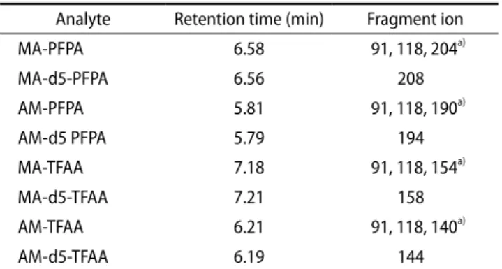
Toxicological analysis was also performed. Confirmatory testing was done using gas chromatography-mass spectrometry (GC-MS). A capillary HP-5MS column was used and the gas flow was set 1 mL/min. The temperature for GC separation was programmed in the range 80-280°C. Mass spectrometry scan mode and selective ion monitoring mode were used for blood and hair samples, respectively. The fragment ions and retention time of analytes are summarized in Table 1.
GC-MS revealed a methamphetamine level of 0.67 mg/L, with an amphetamine (metabolite of methamphetamine) level of 0.08 mg/L in the cardiac blood. Methamphetamine and amphetamine were detected in all hair segments (Fig. 3).
Discussion
Cerebrovascular accident secondary to methamphetamine use, including intracerebral hemorrhage, subarachnoid hemorrhage, and cerebral infarction, have been well- reported [2]. Methamphetamine-related intracerebral hemorrhage occurs most commonly in the supratentorial space [3] and has been reported in all cerebral lobes, with the majority involving the frontal lobe [2]. The etiology of methamphetamine-related intracerebral hemorrhage is unclear. However, methamphetamine is a potent sympathomimetic that results in the release of norepinephrine and dopamine from synaptic nerve endings and causes an elevation of pulse rate and blood pressure [4], which stresses the vessel wall.
Chronic methamphetamine use has additionally been shown to cause long-term systemic harmful effects.
The affected vessels become weak and easy to rupture and hemorrhage, particularly during acute stress, such as following methamphetamine ingestion. In some cases, pathological evidence has been found; for example, a relationship between methamphetamine abuse and necrotizing angitis resulting in intracerebral
Fig. 1. Intracerebral hemorrhage in the right frontal and intraventricular
hemorrhage were noted.
100 Korean Journal of Legal Medicine│2018;42:98-101
http://www.kjlm.or.kr https://doi.org/10.7580/kjlm.2018.42.3.98
hemorrhage has been reported [5]. Cerebral vasculitis is the most commonly described histologic finding in methamphetamine abusers with either hemorrhagic or ischemic stroke [6]. It has been postulated that methamphetamine may cause inflammatory changes within vessel walls [7]. In this report, cerebral vasculitis was unfortunately not proven, but systemic vasculitis involving the aorta, carotid artery, and coronary artery was shown in the histologic examination. Microscopic examination verified the edema and chronic lymphocytic infiltration in the intima of the descending aorta and carotid artery and thickening and infiltration of the lymphocyte in the intima of the coronary arteries.
Hair analysis for drugs is an important tool for
Fig. 2. Microscopic examination verified the edema and chronic lymphocytic infiltration in the intima of the descending aorta (A) and carotid artery (B) (A and B, ×200) and thickening and infiltration of the lymphocyte in the intima of the left descending coronary artery (C) and right coronary artery (D) (C and D, × 100).
A
C
B
D
Table 1. The fragment ions and retention time of analytes Analyte Retention time (min) Fragment ion
MA-PFPA 6.58 91, 118, 204
a)MA-d5-PFPA 6.56 208
AM-PFPA 5.81 91, 118, 190
a)AM-d5 PFPA 5.79 194
MA-TFAA 7.18 91, 118, 154
a)MA-d5-TFAA 7.21 158
AM-TFAA 6.21 91, 118, 140
a)AM-d5-TFAA 6.19 144
MA, methamphetamine; PFPA, pentafluoropropionic anhydride; AM, amphetamine; TFAA, trifluoroacetic anhydride.
a)