MDCT Evaluation of Left Atrium and Pulmonary Vein in the Patients with Atrial Fibrillation:
Comparison with the Non-Atrial Fibrillation Group
1Won Jung Kim, M.D., Eun Jeong Choi, M.D., Ph.D., Hwan Seok Yong, M.D., Ph.D.2, Kyung Sook Yang, Ph.D.3, Soo-Yeon Ham, M.D., Ph.D.,
Yu-Whan Oh, M.D., Ph.D., Young Hoon Kim, M.D., Ph.D.4
1Department of Radiology, Korea University Medical Center
2Department of Radiology, Korea University Guro Hospital
3Department of Biostatistics, Korea University
4Department of Cardiology, Korea University Medical Center Received October 7, 2010 ; Accepted October 28, 2010
Address reprint requests to : Eun Jeong Choi, M.D., Department of Radiology, Anam Hospital, Korea University, Anam 5-ga, Sungbuk-gu, Seoul 136-705, Korea.
Tel. 82-2-920-5931 Fax. 82-2-929-3796 E-mail: [email protected]
Purpose: The anatomy of the left atrium (LA) and the pulmonary veins (PVs) is impor- tant in planning and performing successful electrophysiologic ablation (EPA) for atrial fibrillation (Afib) patients. The authors estimated the findings of LA and PVs of Afib patients by MDCT, and compared these with the findings of LA and PVs of the non- Afib group using coronary CT angiography (CCTA).
Materials and Methods: From September, 2009 to February, 2010, 91 Afib patients un- derwent PVCT (male: female = 72:19, mean age = 55.0-years-old) before EPA. At same time, 90 patients underwent CCTA (male: female = 73:17, mean age = 59.1- years-old). Two radiologists reviewed and analyzed all axial and 3D images of LA and PVs retrospectively with consensus.
Results: The average LA volumes of the Afib group(100.49 mm3) was larger than that of the non-Afib group (78.38 mm3) (p<0.05). The average lengths of the LA right wall in the Afib group (40.25 mm) was longer than that of the non-Afib group (37.3 mm) (p<0.05). The average distances between the PV ostium and first segmental bifurca- tion of the Lt superior PV (LSPV) and the RSPV were shorter in the Afib group (LSPV, 19.38 mm; RSPV, 11.49 mm) than in the non-Afib group (LSPV, 23.23 mm; RSPV, 14.25 mm) (p<0.05). There were higher incidences of anomalous branches such as os- tial, accessory branches, or common ostia in the Afib group versus the non-Afib group (p<0.05).
Conclusion: In Afib group, variable parameters of LA and PVs were obtained and esti- mated by MDCT, and there was statistically significant difference in the parameters of LA and PVs between Afib and non-Afib groups.
Index words :Atrial Fibrillation Heart atria Pulmonary Veins
Tomography, X-Ray Computed
Atrial brillation (Afib), the rapid discharge of multiple electrical impulses from ectopic foci, is one of the most common sustained cardiac arrhythmias with significant morbidity and mortality, The muscular sleeves of the distal pulmonary veins (PVs) are a frequent source of ec- topic foci of electrical activity (1, 2). Electrophysiologic ablation (EPA) of the distal PVs and posterior left atrium (LA) is a helpful treatment for persistent Afib in patients with pharmacologic therapy or cardioversion (1, 2).
The anatomy of the LA and the PVs of an individual Afib patient is complex, and neither fluoroscopy nor echocardiography can adequately depict it (1, 3).
Currently a three-dimensional multi-detector CT (MD- CT) of the LA and distal PVs is an extremely useful modality that can provide the necessary intra-and ex- traatrial anatomic information (1, 2).
Many studies have reported the variability of impor-
tant anatomical findings using CT with contrast en- hancement (1, 2, 4-6). Some of these findings include the number, location, and angulation of PVs and their ostial branches as well as variability of LA volume.
However, there is a paucity of data about the differ- ences of Afib patients with those of control groups by MDCT (7). Therefore, we evaluated and analyzed the anatomical findings of LA and the PVs in Afib patients and normal patients using CT.
Materials and Methods
From September 1, 2009 to February 28, 2010, 91 Afib patients underwent with pulmonary vein CT (PVCT) (male: female = 72:19, mean age = 55.0-years-old) be- fore EPA. Ninety non-Afib patients, at high risk for coro- nary arterial disease or with atypical chest pain under-
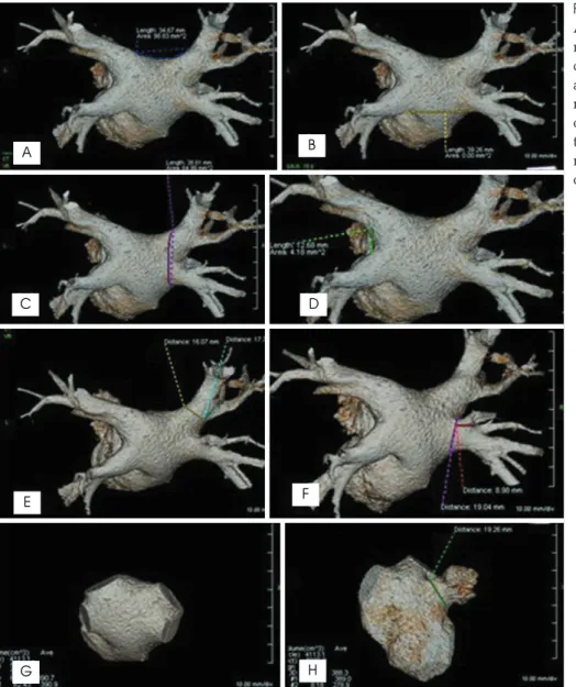
Fig. 1. Measurements of parameters in Afib group. (A) Superior roof, (B) infe- rior roof, (C) right lateral wall, (D) left carina, (E) and (F) ostium of PV, and di- ameter between ostium and first seg- mental bifurcation, (G) LA volume, (H) ostium of LA appendage. Volume of the ostium of the LA appendage was measured as: volume of (H) - volume of (G).
A B
C
E
G H
F D
went coronary CT angiography (CCTA) (male: female = 73:17, mean age = 59.1-years-old). Any patients with a previous history of renal impairment, hypertension, car- diovascular procedure or surgery, pulmonary hyperten- sion (primary or secondary), valve disease, or any kind of cancer in the thorax were excluded from the study.
Results of echocardiography in both groups showed no cardiac functional impairment. All CT studies were per- formed with a 64-multidetector CT (Brilliance 64, Philips Medical System, Cleveland, OH, USA) using this injection protocol: 60 cc~80 cc of iodine contrast media (Ultravist 370, Bayer-Schering Pharma, Berlin, Germany) was injected using 5 cc/sec as an injection rate, followed immediately by 40 cc saline flushing with a 4 cc/sec injection rate. CT protocol for CCTA were as follows: scan range from carina to diaphragm, slice thickness 0.67 mm, slice increment 0.33 mm, 110 Hounsfield unit of scan ROI tracker threshold, 7 sec post injection delay, 7 sec post threshold delay. CT pro- tocol for PVCT were: scan range from aortic arch to di- aphragm, slice thickness 1 mm, slice increment 0.5 mm, 200 Hounsfield unit of scan ROI tracker threshold, 10 sec post injection delay, 7 sec post threshold delay. Two observers individually evaluated all images. PVCT was performed without electrocardiogram (ECG) gating.
Analysis categories for Afib and non-Afib groups were
defined as follows: 1) LA volume, 2) the longest diame- ter of ostium of LA appendage (LAA), 3) the volume of LAA, 4) the length of superior and inferior roofs of LA, 5) the length of right and left walls of LA, 6) the length of bilateral carina of PV, 7) the diameter of each ostium of PVs, and 8) the distance between PV ostium and 1st seg- mental bifurcation (Fig. 1). The wall was from the most superior edge of superior PV to the most inferior edge of inferior PV, and the carina was the shortest diameter be- tween the ostia of superior and inferior PVs at same side. Anomalous branch categories of PVs such as acces- sory branch, common trunk and ostial branch of PV were also evaluated. Evaluation of shortening in the main portion of the PV, an esophageal location behind the LA posterior wall was included. According to the heart rate of non-Afib group, analysis of CCTA was done in mid-diastolic phase (heart rate <65 bpm) or in end-systolic phase (heart rate>65 bpm), to get the best image quality and minimize the image blurring.
Retrospective analysis and review of the 3D LA im- ages was performed with an imaging processing work- station (Aquarius, Terarecon Inc., San Mateo, CA, USA).
Two radiologists were reviewed the images without any information with consensus (kappa coefficient = 0.75).
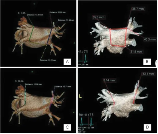
The curvilinear lengths on LA were measured according to the linear ablation sites: bilateral antral ablation line
Fig. 2. Comparison of parameters in Afib group (A and C) with non-Afib group (B and D).
C
A B
D
as PV ostium, roof lines as superior and inferior roofs, left lateral isthmus line as carina, anterolateral and an- teroseptal lines as bilateral walls. Since the LA ap- pendage and the LA have different anatomical charac- teristics, separate analysis of the LA appendage and the LA was performed in reference to the points of inflec- tion on the 3D CT image (7). The absolute and relative volumes of each portion were calculated and compared.
Statistically, the T-test and Mann-Whitney were used to compare each parameter in the two groups.
Anomalous PV branches were evaluated using Fisher’s exact test.
Results
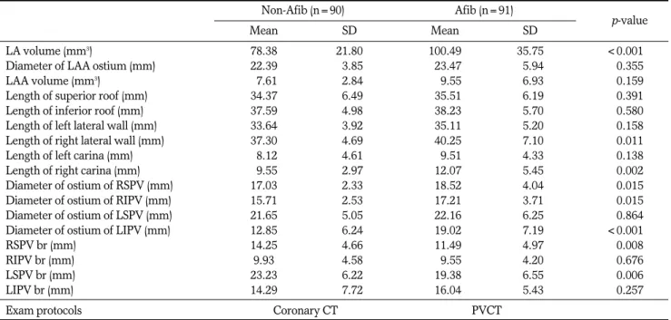
We evaluated 181 CT images and the analyzed data are presented in Table 1. The LA volumes of the Afib group were larger than of the non-Afib group (mean:
100.49 mm3vs. 78.38 mm3, p < 0.05). However, no sig- nificant differences were demonstrated for ostial diame- ter or volume of the LA appendage between the two groups.
The lengths of the superior and the inferior roofs were also similar between the two groups. The length of the LA right wall in the Afib group was longer than in the non-Afib group (mean: 40.25 mm3 vs. 37.30 mm, p <
0.05), but the length of the LA left wall showed no dif-
ference between two groups. On the right side, the length of the carina was greater in the Afib group than the non-Afib group (mean: 12.07 mm3vs. 9.55 mm, p <
0.05), but the lengths of the left side were similar.
In the Afib group, the diameters of the ostia of the RSPV, the RIPV, and the LIPV were significantly larger than those in the non-Afib group (p < 0.05). However, no significant difference was demonstrated for the LSPV ostium. The distances between the PV ostium and the first segmental bifurcation of the LSPV and the RSPV were shorter in the Afib group than in the non-Afib group (p < 0.05).
There was a higher incidence of anomalous branches such as ostial, accessory branches, as well as a greater number of common ostia in the Afib group (57%) versus the non-Afib group (27%) (p < 0.05). The incidence of right, left or both common ostia of the PV was not signif- icantly different between the two groups. There were no significant differences of shortening in the main por- tion of the PV, an esophageal location behind the LA posterior wall. In the non-Afib group, there were 24 pa- tients with significant coronary stenosis, 39 with non- significant coronary stenosis, and 27 with normal coro- nary artery.
Table 1. Characteristics of CT findings
Non-Afib (n=90) Afib (n=91)
Mean SD0 Mean SD0 p-value
LA volume (mm3) 78.38 21.800 100.49 35.750 <0.001<
Diameter of LAA ostium (mm) 22.39 3.85 023.47 5.94 0.355
LAA volume (mm3) 07.61 2.84 009.55 6.93 0.159
Length of superior roof (mm) 34.37 6.49 035.51 6.19 0.391
Length of inferior roof (mm) 37.59 4.98 038.23 5.70 0.580
Length of left lateral wall (mm) 33.64 3.92 035.11 5.20 0.158
Length of right lateral wall (mm) 37.30 4.69 040.25 7.10 0.011
Length of left carina (mm) 08.12 4.61 009.51 4.33 0.138
Length of right carina (mm) 09.55 2.97 012.07 5.45 0.002
Diameter of ostium of RSPV (mm) 17.03 2.33 018.52 4.04 0.015
Diameter of ostium of RIPV (mm) 15.71 2.53 017.21 3.71 0.015
Diameter of ostium of LSPV (mm) 21.65 5.05 022.16 6.25 0.864
Diameter of ostium of LIPV (mm) 12.85 6.24 019.02 7.19 <0.001<
RSPV br (mm) 14.25 4.66 011.49 4.97 0.008
RIPV br (mm) 9.93 4.58 009.55 4.20 0.676
LSPV br (mm) 23.23 6.22 019.38 6.55 0.006
LIPV br (mm) 14.29 7.72 016.04 5.43 0.257
Exam protocols Coronary CT PVCT
Note.─Afib = atrial fibrillation, br = distance between PV ostium and 1st segmental bifurcation, LAA = LA appendage, RSPV = right superior pulmonary vein, RIPV = right inferior pulmonary vein, LSPV = left superior pulmonary vein, LIPV = left inferior pulmonary vein.
Discussion
Atrial enlargement is the most common structural change associated with Afib. The Framingham study es- timated that the hazard ratio for subsequent develop- ment of Afib was 1.39 for every 5-mm incremental in- crease of LA size by echocardiogram (8). Also, other study suggested that Afib could cause LA dilation (9) and reported that the late recurrence of Afib is associat- ed with progressive dilation of LA (3). Our results showed that the LA size was larger in the Afib patients than in the non-Afib group, which was consistent with the previous studies.
Other studies using transesophageal echocardiogra- phy and MDCT reported that the size of the ostium of LAA in Afib patients is larger than in healthy people (10-12). The larger LAA size was related to the higher incidence of LAA thrombi. This finding may indicate a higher risk of thromboembolic events in Afib patients (12). In the present study the diameter of LAA ostium showed no statistical difference between Afib patients and the non-Afib group. However, the LAA diameter demonstrates greater variation in Afib patients than in the the non-Afib group. Wongcharoen et al. (12) report- ed that a longer LA roof and variation in the LA roof morphologies were noted in the Afib patients. In our study, the lengths of the superior and inferior roofs showed no significant difference between the two groups.
Remarkably little has been published concerning nor- mal PV anatomy (13). It is rare to know the PV anatomy of Afib patients before procedures are conducted. Lin et al. (14) were the first investigators to evaluate specifical- ly the PV size in patients with Afib using contrast venog- raphy. They showed that the diameters of the RSPV and LSPV were greater than that of the inferior PVs. In a subsequent article, these authors used MRI to evaluate PV anatomy and reported that the superior PVs were larger than the inferior PVs and that the superior, but not the inferior PVs, were larger in diameter in patients with Afib (15). We thought it is plausible that the stretch and dilation of the superior PVs might change the elec- trophysiologic characteristics of the myocardial sleeve, so that Afib may occur more easily. On the other hand, dilation of the inferior PVs is less significant than in the superior PVs, which may explain in part the lower inci- dence of Afib initiated from ectopic foci in the inferior PVs. Kato et al. (12, 13) reported that the diameters of
the four PVs do not differ, and that there was little varia- tion in the size of the PVs within given patients. In our study, the diameters of the RSPV, RIPV and LIPV in Afib patients were larger than in the non-Afib group.
PVs exhibit a great deal of anatomical variation.
Previous reports described a 23-38% frequency with which variations in PV anatomy were generally ob- served (13, 16). Accessory branches and common trunks were described in these studies. These variations of PV anatomy have not been thoroughly evaluated in Afib pa- tients. In our study, we included accessory branches, common trunks, and ostial branches in the anomalous branch category. A significantly higher incidence of anomalous branching was noted in Afib patients com- pared to the control group. Especially the ostial branch was more common in Afib patient. It is a very interest result.
Other parameters that we assessed, such as the length of LA right and left walls, the length of LA bilateral cari- na and the distance between the PV ostium and the first segmental bifurcation, had never before been evaluated in any previous study. Our results showed that some pa- rameters differed between the two study groups.
Further studies are needed to clarify the clinical signifi- cance of these findings.
Several other anatomical differences between Afib pa- tients and normal patients were previously discussed.
Chaiang et al. (17) reported that the LA isthmus was longer in the Afib patients and that the morphology of the isthmus was variable. They also compared the later- al isthmus to the medial isthmus finding the latter to be longer and having more ridges. They explained the re- gional difference as a result of the orientation and thick- ness of myocardial fibers between the structures of LA.
Also Wongcharoen et al. (12) said the LA roof and the LA septum had variation in Afib patients. These anatomical variations are important to consider in abla- tion therapy, but the pathophysiology and any relation- ship with clinical outcome have not been revealed.
Several limitations of this study should be considered.
First, the PVCT images of Afib group were not always acquired during the period of the cardiac cycle when there was maximal dilation of the LA, due to arrhyth- mia. However, changes of LA morphology and size are probably minimal between the systolic and diastolic phases in Afib patients. Second, the non-Afib group in- cluded patients with normal sinus rhythm who had un- dergone CCTA. Third, the male/female ratios differed between the two groups. Men probably have larger
hearts than women, due to body size.
In contrast to previous studies using MRI or echocar- diography, our study was performed using MDCT. A larger number of Afib patients were examined in our study than in previous studies and we evaluated a wider variety of anatomical differences than previous studies.
In our results, we detected multiple anatomical differ- ences between Afib patients and the control group.
Some of these results were consistent with those of pre- vious studies, but some were not. The incidence of anomalous branches of PV was significantly higher in Afib patients than in the control group. Changes in mul- tiple anatomical parameters that are are presumed to be related to the development, progress and recurrence of Afib. However, there is inadequate information to clear- ly demonstrate any such relationship. Further studies to investigate this question are currently in progress.
References
1. Stanford W, Breen JF. CT evaluation of left atrial pulmonary ve- nous anatomy. Int J Cardiovasc Imaging 2005;21:133-139
2. Lacomis JM, Wigginton W, Fuhrman C, Schwartzman D, Armfield DR, Pealer KM. Multi-detector row CT of the left atrium and pulmonary veins before radio-frequency catheter ablation for atrial fibrillation. Radiographics 2003;23 Spec No:S35-S48
3. Tsao HM, Wu MH, Huang BH, Lee SH, Lee KT, Tai CT, et al.
Morphologic remodeling of pulmonary veins and left atrium after catheter ablation of atrial fibrillation. J Ccardiovasc Electrophysiol 2005;16:7-12
4. Lacomis JM, Goitein O, Deible C, Schwartzman D. CT of the pul- monary veins. J Thorac Imaging 2007;22:63-76
5. Piorkowski C, Hindricks G, Schreiber D, Tanner H, Weise W, Koch A, et al. Electroanatomic reconstruction of the left atrium, pulmonary veins, and esophagus compared with the “true anato- my” on multislice computed tomography in patients undergoing catheter ablation of atrial fibrillation. Heart Rhythm 2006;3:317-327 6. Centonze M, Del Greco M, Nollo G, Ravelli F, Marini M, Della Sala SW, et al. The role of multidetector CT in the evaluation of the left atrium and pulmonary veins anatomy before and after radio-
frequency catheter ablation for atrial fibrillation. Preliminary re- sults and work in progress. Technical note. Radiol Med 2005;110:
52-60
7. Jongbloed MR, Dirksen MS, Bax JJ, Boersma E, Geleijns K, Lamb HJ, et al. Atrial fibrillation: multi-detector row CT of pulmonary vein anatomy prior to radiofrequency catheter ablation--initial ex- perience. Radiology 2005;234:702-709
8. Dittrich HC, Pearce LA, Asinger RW, Mcbride R, Webel R, Zabalgoitia M, et al. Left atrial diameter in nonvalvular atrial fibril- lation: an echocardiographic study. Am Heart J 1999;137:494-499 9. Sanfilippo AJ, Abascal VM, Sheehan M, Oertel LB, Harrigan P,
Hughes RA, et al. Atrial enlargement as a consequence of atrial fib- rillation. A prospective echocardiographic study. Circulation 1990;
82:792-797
10. Li YH, Lai LP, Shyu KG, Hwang JJ, Kuan P, Lien WP. Clinical im- plications of left atrial appendage flow patterns in nonrheumatic atrial fibrillation. Chest 1994;105:748-752
11. Verhorst PM, Kamp O, Visser CA, Verheugt FW. Left atrial ap- pendage flow velocity assessment using transesophageal echocar- diography in nonrheumatic atrial fibrillation and systemic em- bolism. Am J Cardiol 1993;71:192-196
12. Wongcharoen W, Tsao HM, Wu MH, Tai CT, Chang SL, Lin YJ, et al. Morphologic characteristics of the left atrial appendage, roof, and septum: implications for the ablation of atrial fibrillation. J Cardiovasc Electrophysiol 2006;17:951-956
13. Kato R, Lickfett L, Meininger G, Dickfeld T, Wu R, Juang G, et al.
Pulmonary vein anatomy in patients undergoing catheter ablation of atrial fibrillation: lessons learned by use of magnetic resonance imaging. Circulation 2003;107:2004-2010
14. Lin WS, Prakash VS, Tai CT, Hsieh MH, Tsai CF, Yu WC, et al.
Pulmonary vein morphology in patients with paroxysmal atrial fibrillation initiated by ectopic beats originating from the pul- monary veins: implications for catheter ablation. Circulation 2000;
101:1274-1281
15. Tsao HM, Yu WC, Cheng HC, Wu MH, Tai CT, Lin WS, et al.
Pulmonary vein dilation in patients with atrial fibrillation: detec- tion by magnetic resonance imaging. J Cardiovasc Electrophysiol 2003;12:809-813
16. Ho SY, Sanchez-Quintana D, Cabrera JA, Anderson RH. Anatomy of the left atrium: implications for radiofrequency ablation of atrial fibrillation. J Cardiovasc Electrophysiol 1999;10:1525-1533
17. Chiang SJ, Tsao HM, Wu MH, Tai CT, Chang SL, Wongcharoen W, et al. Anatomic characteristics of the left atrial isthmus in pa- tients with atrial fibrillation: lessons from computed tomographic images. J Cardiovasc Electrophysiol 2006;17:1274-1278
대한영상의학회지 2011;64:123-129
심방세동환자의 좌심방 및 폐정맥에 대한 다검출기 CT 소견:
비심방세동환자와의 비교1
1고대의료원 안암병원 영상의학과
2고대의료원 구로병원 영상의학과
3고려대학교 생물통계학교실
4고대의료원 안암병원 심장내과
김원중∙최은정∙용환석2∙양경숙3∙함수연∙오유환∙김영훈4
목적: 좌심방과 폐정맥의 해부학적 구조는 심방세동 환자에서 전기생리학적 절제술을 시행하는데 중요하다. 저자들 은 MDCT를 이용하여 심방세동 환자의 좌심방 및 폐정맥의 소견을 평가하고 이를 심방세동이 없는 관상동맥질환 의심자의 좌심방 및 폐정맥 CT 소견과 비교하고자 하였다.
대상과 방법: 2009년 9월부터 2010년 2월까지 91명의 심방세동 환자가 전기생리학적 절제술을 시행하기 전에 폐정 맥 CT를 시행하였고(남: 여 = 72:19, 평균연령 55세), 같은 시기에 심방세동이 없는 관상동맥질환 의심자 90명에 서 시행한 관상동맥 CT를 비교하였다(남: 여 = 73:17, 평균연령 59.1세). 각 CT의 3차원 영상을 두 명의 영상의 학과 의사가 후향적으로 비교 분석하였다.
결과: 다음의 해부학적 구조들에서 통계적으로 유의한 차이가 나타났다. 심방세동 환자군의 평균 좌심방 용적 (100.49 mm3)이 비교군(78.38 mm3)보다 크며(p < 0.05), 환자군의 좌심방의(환자군의 좌심방) 평균 우측 벽 길 이(40.25 mm)가 비교군(37.3 mm)보다 길었다(p < 0.05). 또한, 좌상폐정맥과 우상폐정맥에서 폐정맥구와 첫 번 째 분지 사이의 평균 거리가 환자군(좌상폐정맥, 19.38 mm; 우상폐정맥, 11.49 mm)에서 비교군(좌상폐정맥, 23.23 mm; 우상폐정맥, 14.25 mm)에서 보다 짧았다(p < 0.05).
결론: 심방세동 환자에서 MDCT를 이용하여 좌심방과 폐정맥의 다양한 측정치를 구할 수 있었으며 심방세동이 없 는 관상동맥질환 의심자와 비교할 때 유의한 차이를 발견할 수 있었다.