68
Open Access
Effect of Aspiration Thrombectomy on Microvascular Dysfunction in ST-Segment Elevation Myocardial Infarction
With an Elevated Neutrophil Count
Hye Young Lee, MD, Jeong Hoon Kim, MD, Byung Ok Kim, MD, Yoon Jung Kang, MD, Hyo Seung Ahn, MD, Mee Won Hwang, MD, Kyoung Min Park, MD, Young Sup Byun, MD, Choong Won Goh, MD and Kun Joo Rhee, MD Division of Cardiology, Department of Internal Medicine, Sanggye Paik Hospital, Inje University College of Medicine, Seoul, Korea ABSTRACT
Background and Objectives: Aspiration thrombectomy (AT) during primary percutaneous coronary intervention (PCI) is an effective adjunctive therapy for ST-segment elevation myocardial infarction (STEMI). An elevated neutrophil count in STE- MI is associated with microvascular dysfunction and adverse outcomes. We evaluated whether AT can improve microvascu- lar dysfunction in patients with STEMI and an elevated neutrophil count. Subjects and Methods: Seventy patients with STEMI undergoing primary PCI from August 2007 to February 2009 in our institution were classified by tertiles of neutro- phil count on admission (<5,300/mm
3, 5,300-7,600/mm
3, and >7,600/mm
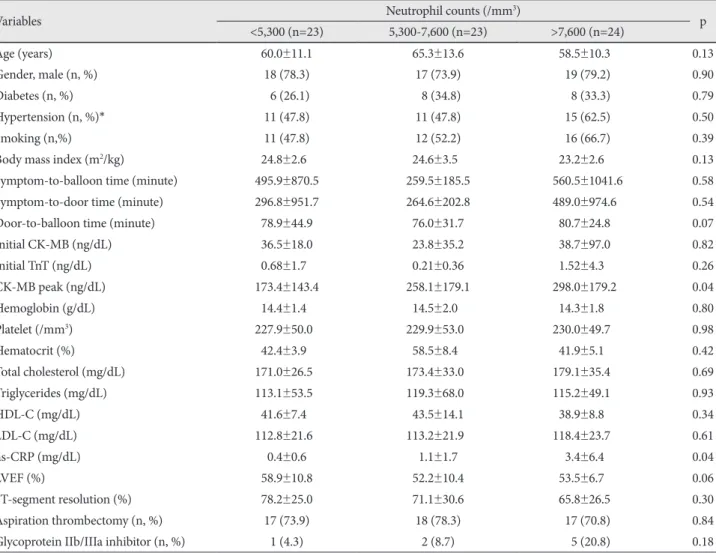
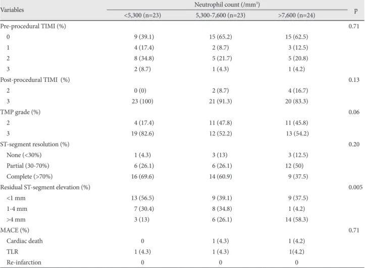
3). The angiographic outcome was post-procedur- al thrombolysis in myocardial infarction (TIMI) flow grade. Microvascular dysfunction was assessed by TIMI myocardial perfusion (TMP) grade and ST-segment resolution on electrocardiography 90 minutes after PCI. The clinical outcome was ma- jor adverse cardiac event (MACE), defined as cardiac death, re-infarction, and target lesion revascularization at 9 months. Re- sults: There were no significant differences in the clinical characteristics and pre- and post-procedural TIMI flow grades be- tween the neutrophil tertiles. As the neutrophil count increased, a lower tendency toward TMP grade 3 (83% vs. 52% vs.
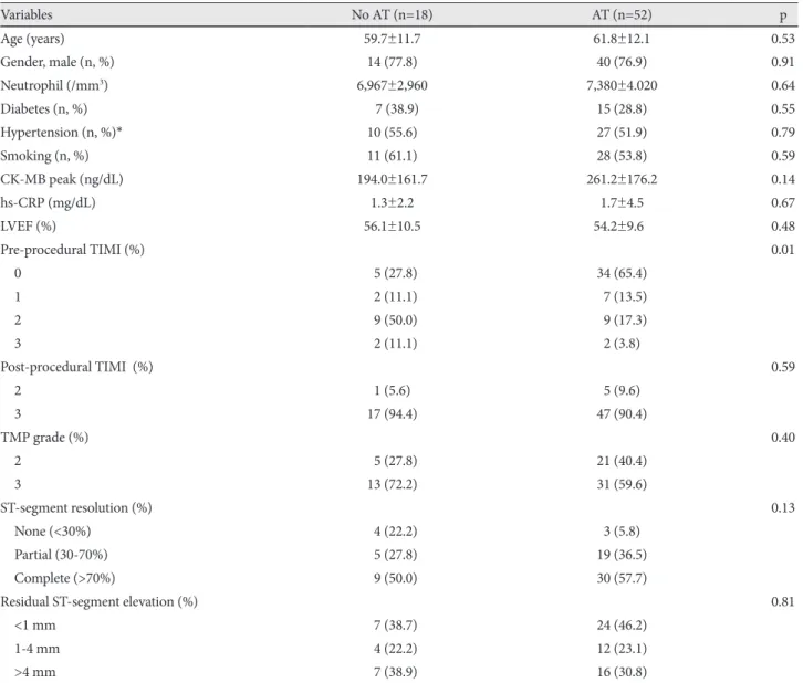
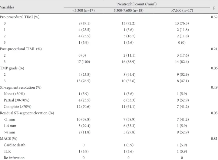
54%, p=0.06) and more persistent residual ST-segment elevation (>4 mm: 13% vs. 26% vs. 58%, p=0.005) was observed. The 9-month MACE rate was similar between the groups. On subgroup analysis of AT patients (n=52) classified by neutrophil tertiles, the same tendency toward less frequent TMP grade 3 (77% vs. 56% vs. 47%, p=0.06) and persistent residual ST-seg- ment elevation (>4 mm: 12% vs. 28% vs. 53%, p=0.05) was observed as neutrophil count increased. Conclusion: A higher neutrophil count at presentation in STEMI is associated with more severe microvascular dysfunction after primary PCI, which is not improved with AT. (Korean Circ J 2011;41:68-75)
KEY WORDS: Myocardial infarction; Neutrophils.
Received: May 27, 2010 Revision Received: June 22, 2010 Accepted: July 19, 2010
Correspondence: Byung Ok Kim, MD, Division of Cardiology, Depart- ment of Internal Medicine, Sanggye Paik Hospital, Inje University College of Medicine, Sanggye-dong, Nowon-gu, Seoul 139-707, Korea Tel: 82-2-950-1266, Fax: 82-2-950-1248
E-mail: [email protected]
• The authors have no financial conflicts of interest.
cc