사람 두경부 편평세포암종 HEp2 세포에서 BCH에 의한 세포성장 억제기전
최봉규1․정규용1․조선호2․김춘성2․김도경2†
1원광대학교 의과대학 약리학교실
2조선대학교 치과대학 구강생리학교실 및 2단계 BK21 프로젝트
Mechanism of Growth Inhibition by BCH in HEp2 Human Head and Neck Squamous Cell Carcinoma
Bong Kyu Choi1, Kyu Yong Jung1, Seon-Ho Cho2, Chun Sung Kim2, and Do Kyung Kim2†
1
Dept. of Pharmacology, Wonkwang University School of Medicine, Jeonbuk 570-749, Korea
2
Dept. of Oral Physiology and The Second Stage of BK21, Chosun University College of Dentistry, Gwangju 501-759, Korea
Abstract
Amino acid transporters are essential for the growth and proliferation in all living cells. Among the amino acid transporters, the system L amino acid transporters are the major nutrient transport system responsible for the Na+-independent transport of neutral amino acids including several essential amino acids. The L-type amino acid transporter 1 (LAT1), an isoform of system L amino acid transporter, is highly expressed in cancer cells to support their continuous growth and proliferation. 2-Aminobicyclo-(2,2,1)-heptane-2-carboxylic acid (BCH) is a model compound for the study of amino acid transporter as a system L selective inhibitor. We have examined the effect and mechanism of BCH on cell growth suppression in HEp2 human head and neck squamous cell carcinoma. The BCH inhibited the L-leucine transport in a concentration-dependent manner with a IC50 value of 51.2±3.8 μM in HEp2 cells. The growth of HEp2 cells was inhibited by BCH in the time- and concentration-dependent manners. The formation of DNA ladder was not observed with BCH treatment in the cells. Furthermore, the proteolytic processing of caspase-3 and caspase-7 in the cells were not detected by BCH treatment. These results suggest that the BCH inhibits the growth of HEp2 human head and neck squamous cell carcinoma through the intracellular depletion of neutral amino acids for cell growth without apoptotic processing.
Key words: amino acid transporter, cancer cells, BCH, growth inhibition, anti-cancer therapy
†Corresponding author. E-mail: [email protected]
†Phone: 82-62-230-6893, Fax: 82-62-232-6896
서 론
아미노산은 단백질 합성의 기질이 되며, 효소, 호르몬 및 신경전달물질들의 합성에 필수적이다. 세포 안에서 필요한 아미노산의 수송은 세포막에 위치한 아미노산 수송체를 통 하여 이루어진다(1). 아미노산 수송계 L은 중성아미노산을 수송하는 세포막 단백질로서 암세포를 포함한 대부분의 세 포에서 중성아미노산의 주경로가 되는 아미노산 수송체로 알려져 있으며(1,2), 상피세포의 기저측 막에 존재하여 소장 상피세포를 통한 중성아미노산의 흡수에 중요한 기능을 한 다(3).
Kanai 등(4)에 의해 아미노산 수송계 L의 첫 번째 아형인 L-type amino acid transporter 1(LAT1)이 동정되었다.
LAT1은 12회 세포막을 관통하는 막 단백질로서 leucine, isoleucine, valine, phenylalanine, tyrosine, tryptophan, me-
thionine 및 histidine 같은 구조가 큰 중성아미노산을 수송하 는 특징을 가지고 있다(4-6). LAT1은 4F2 heavy chain (4F2hc)이라는 1회 세포막을 관통하는 막 단백질과 결합된 heterodimer형 단백질이며, LAT1이 기능을 나타내기 위해 서는 보조인자 4F2hc의 존재가 필수적이다(4,5,7,8). 또한 LAT1은 암세포와 같이 세포 내 대사가 특이적으로 항진되 거나 계속적인 증식과 성장이 필수적인 세포에서 과발현되 어 세포의 계속되는 성장에 중요한 역할을 한다고 알려져 있다(4,5,9,10). LAT1의 분자적 동정 후에 LAT1과 구조적 으로 관련이 있는 아미노산 수송계 L의 두 번째 아형인 L-type amino acid transporter 2(LAT2)가 동정되었다 (3,11-13). LAT1이 암세포에 과발현된다고 알려진 것과는 달리, LAT2는 많은 정상조직에서 발현을 보이며, 구조가 큰 중성아미노산뿐만 아니라 구조가 작은 중성아미노산도 모두 수송한다(12-14).
아미노산 수송체 LAT1과 LAT2의 특성을 살펴보면, 암 세포에서 과발현되는 LAT1의 조절을 통해 암세포 성장억 제의 방법을 제시할 수 있다. 암세포에서 LAT1의 활성을 억제하여 세포 내 중성아미노산의 고갈을 유도한다면 암세 포 성장억제를 유도할 가능성이 있을 것으로 사료된다.
2-Aminobicyclo-(2,2,1)-heptane-2-carboxylic acid(BCH) 는 아미노산 수송체 연구에 주로 이용되는 아미노산 수송계 L의 선택적 억제제이다(4-6,13,15,16). 아미노산 수송계 L이 leucine, isoleucine, valine, phenylalanine, methionine 및 histidine 등의 필수아미노산을 포함한 중성아미노산을 수송 하기 때문에, 만약 세포에서 수송계 L이 BCH 같은 억제제에 의해 차단된다면 세포는 세포성장과 증식에 필수적인 필수 아미노산이 고갈됨으로 큰 손상을 입을 가능성이 있을 것이 다. 그러나 이러한 아미노산 수송억제에 의한 세포성장 억제 기전에 관한 실험적 자료는 거의 없다.
타액선, 구강, 인두 및 후두를 포함하는 두경부 암은 다른 암에 비해 그 발생기전 등 분자생물학적 접근이 가장 되어있 지 않은 암 중의 하나이다. 게다가 정상 두경부 상피세포뿐 만 아니라 두경부 편평세포암종 세포에서 세포 내 필수영양 물질인 아미노산을 수송할 수 있는 아미노산 수송체의 연구 또한 부족한 실정이다. 따라서 본 연구자들은 사람 두경부 편평세포암종 HEp2 세포를 이용하여 아미노산 수송계 L 억 제제인 BCH의 암세포 성장억제에 미치는 효과와 세포성장 억제기전을 밝히고자 하며, 아울러 아미노산 수송체 LAT1 의 억제를 통한 암치료의 효용성을 제시하고자 한다.
재료 및 방법
실험재료
L-[14C]leucine은 Perkin Elmer Life Science Inc.(Boston, MA, USA)로부터 구입하여 사용하였고, BCH와 N-meth- ylthiotetrazole(MTT)는 Sigma(St. Louis, USA)에서 구입 하여 사용하였으며, ECL detection kit는 Amersham Bio- sciences Corp.(Piscataway, NJ, USA)에서 구입하여 사용 하였다. Fetal bovine serum(FBS)과 DMEM 배지는 Invitrogen Co.(Carlsbad, CA, USA)에서 구입하였다. 아미 노산 및 기타시약들은 analytical grade를 구입하여 사용하 였다.
사람 두경부 편평세포암종 HEp2 세포는 American Type Culture Collection(ATCC, Rockville, MD, USA)에서 제공 받아, 10% FBS가 포함된 DMEM 하에서 배양하면서 사용 하였다.
L-[14C]Leucine uptake 측정실험
HEp2 세포에서 중성아미노산 수송에 영향을 미치는 BCH의 특성을 조사하기 위해 Kim 등(15)의 방법을 이용하 여 아미노산 uptake 실험을 시행하였다. HEp2 세포를 37oC 의 성장배지(10% FBS가 포함된 DMEM 배지) 하에서 배양
하였다. 배양접시(10 cm)의 바닥에 세포가 약 90% 정도 채 워져 있을 때, 트립신을 이용하여 세포를 수집하여 24 well plate에 심고(1×10⁵ cells/well), 48시간 후 uptake 실험을 시행하였다. 성장배지를 흡수기를 이용하여 제거한 뒤, 세포 를 Na+-free uptake 용액(125 mM choline-Cl, 4.8 mM KCl, 1.3 mM CaCl2, 1.2 mM MgSO4, 25 mM HEPES, 1.2 mM KH2PO4, 5.6 mM glucose, pH 7.4)을 이용하여 3회 세척한 후 37oC에서 10분간 전배양 하였다. 그 후, 용액을 L- [14C]leucine(3 μCi)이 존재하는 uptake 용액으로 교체하여 1분 배양시켰다. 반응의 정지를 위해 4oC의 같은 용액으로 3회 세척하였다. 세포를 0.1 N NaOH에 녹여 세포 안으로 uptake 된 방사능을 liquid scintillation spectrometry(β- counter)로 측정하였으며, 측정한 방사능을 pmol/mg pro- tein/min으로 산출하였다. 본 논문의 uptake 실험에서 각 실 험군으로 HEp2 세포가 존재하는 4~6개의 well을 이용하였 으며, 각각의 결과를 mean±SEM으로 표시하였다. 각 결과 의 재현성을 확인하기 위하여 3회 이상 반복실험을 수행하 여 결과를 산출하였다. HEp2 세포에서 L-leucine의 수송을 억제하는 BCH의 IC50(최대 억제량의 50%를 유발시키는 농 도)은 L-[14C]leucine의 농도 1 μM(3 μCi) 존재 하에 BCH 농도 0, 1, 3, 10, 30, 100, 300 및 1000 μM에서 uptake(1분)를 수행하여 산출하였다.
MTT 실험
BCH에 의한 세포성장 억제효과를 관찰하기 위해, 24 well plate에 5×10³ cells/well의 HEp2 세포를 심었다. 24시간 배 양한 후, BCH를 다양한 농도와 시간에서 처리하여 37oC에 서 반응시킨 후, 살아있는 세포를 MTT 실험으로 측정하였 다. MTT 실험은 HEp2 세포에 MTT 용액(MTT 최종농도 0.5 μg/μL)을 37oC에서 4시간 처리한 후, MTT 용액을 제거 하고 0.04 N HCl이 함유된 isopropanol로 녹여내어 570 nm 에서 흡광도를 측정하여 시행하였다.
DNA fragmentation 분석
세포사멸의 기전 중 apoptosis의 지표가 되는 DNA frag- mentation 분석을 시행하였다. BCH에 의한 세포 DNA fragmentation 효과를 관찰하기 위해, 10 cm 배양접시 당 5×10⁵개의 HEp2 세포를 심었다. 24시간 배양한 후 BCH 50 mM을 처리하고, 24시간 또는 48시간 동안 37oC에서 배양 한 후, 세포를 수집하여 lysis buffer(0.1 M NaCl, 0.001 M EDTA, 0.3 M Tris-HCl(pH 7.5), 0.2 M Sucrose)를 이용한 통상의 phenol-chloroform extraction법으로 DNA를 추출 하였다(17). 추출한 DNA를 1.5% agarose gel에서 전기영동 (50 volts, 1시간) 하였으며, ethidium bromide로 염색하여 관찰하였다.
Immunoblotting
세포 apoptosis의 지표가 되는 caspase-3와 caspase-7 분 석을 위해 immunoblotting을 시행하였다. 10 cm 배양접시
BCH (μM)
0 100 200 300 400 500 600 700 800 900 1000
[
14C] L- Le u u p ta ke ( % up tak e )
0 20 40 60 80 100 120
Fig. 1. Concentration-dependent inhibition of L-[14C]leu- cine uptake by BCH in HEp2 cells. The L-[14C]leucine uptake (1
μ
M) was measured for 1 min in the presence of various BCH concentrations (0, 1, 3, 10, 30, 100, 300 and 1000μ
M) in the HEp2 cells, and was expressed as a percentage of the control-L-leucine uptake in the absence of BCH. Each data point represents the mean±
SEM for three experiments.당 5×10⁵개의 HEp2 세포를 심고 24시간 배양한 후 BCH 50 mM을 다양한 시간에서 처리하여 37oC에서 반응시킨 후 세포를 수집하였다. 세포를 4oC의 PBS로 2회 세척한 후 4oC 의 lysis buffer(1% Triton X-100, 0.5 mM EDTA, 1 mM phenylmethylsulfonyl fluoride, 5 μg/mL aprotinin 및 5 μg/
mL leupeptin이 포함된 PBS)에서 30분 반응시켰다. 세포 용해물을 12,500×g에서 20분간 원심분리한 후 단백질 시료 를 정량하였다. 단백질 시료를 2배의 SDS sample buffer(60 mM Tris-HCl, pH 6.8, 4% SDS, 25% glycerol, 14.4 mM 2-mercaptoethanol, 0.1% bromophenol blue)에 넣고 100oC 에서 5분간 변성시킨 후, 12% SDS-polyacrylamide gel에 120 volt에서 2시간 전기영동 한 다음, 단백질 transfer를 이 용하여 nitrocellulose membrane(Millipore Co., Billerica, MA, USA)으로 이동시켰다. Membrane을 5% fat-free dry milk-PBST buffer(PBS, 0.2% Tween-20)에서 2시간 동안 blocking하였고, PBST buffer로 15분간 3회 세척하였다. 일 차항체로 anti-caspase-3 antibody와 anti-caspase-7 anti- body 및 anti-β-actin antibody를 Cell Signaling Technol- ogy, Inc.(Danvers, MA, USA)으로부터 구입하여, anti- caspase-3 antibody와 anti-caspase-7 antibody는 1,000배 희석하여 사용하였으며, anti-β-actin antibody는 2,000배 희석하여 사용하였다. 이차항체로 horseradish peroxidase conjugated anti-rabbit IgG를 5,000배 희석하여 사용하였으 며, ECL detection kit를 사용하여 X-ray 필름에 현상한 후 분석하였다.
실험 자료의 통계학적 검정
모든 실험성적은 mean±SEM으로 나타내었고, 각 실험 군 간의 유의성 검정은 ANOVA 후에 Student's t-test를 하였으며, p value가 0.05 미만(p<0.05)의 경우에서 통계적 유의성이 있는 것으로 간주하였다.
결 과
L-leucine수송에 대한 BCH의 효과
HEp2 세포에서 L-[14C]leucine의 수송에 미치는 BCH의 특성을 조사하기 위하여, BCH 존재 하에서 [14C]이 표지된 L-leucine uptake 실험을 시행하였다. L-[14C]Leucine(1 μM) 을 여러 농도의 BCH(0, 1, 3, 10, 30, 100, 300, 1000 μM)와 함께 처리한 결과, BCH는 농도 의존적으로 leucine의 세포 내 수송을 억제하였으며, 그 IC50은 51.2±3.8 μM로 산출되 었다(Fig. 1).
세포성장에 대한 BCH의 효과
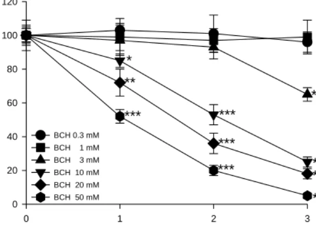
HEp2 세포에서 BCH에 의한 세포성장 억제효과를 조사 하기 위해 MTT 분석을 시행하였다. BCH를 0, 0.3, 1, 3, 10, 20 및 50 mM의 다양한 농도로 3일 동안 HEp2 세포에 투여 한 후 MTT 검사를 시행한 결과, BCH 0.3 mM과 1 mM의
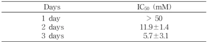
농도에서는 대조군과 비교하였을 때 세포성장 억제의 차이 를 볼 수 없었다(Fig. 2). 그러나 BCH 3, 10, 20 및 50 mM에 서는 대조군과 비교하여 볼 때 뚜렷한 세포성장 억제효과를 볼 수 있었으며, 이 효과는 시간과 농도에 의존적임을 확인 할 수 있었다(Fig. 2). HEp2 세포성장 억제에 대한 BCH의 IC50(BCH를 처리하지 않은 대조군과 비교하여 최대 억제량 의 50%를 유발시키는 농도)은 BCH 처리 1일째는 50 mM 이상이었고, 2일째는 약 11.9 mM이었으며, 3일째는 약 5.7 mM이었다(Table 1).
Time (days)
0 1 2 3
Cell viabi lity ( % of contr o l)
0 20 40 60 80 100 120
BCH 0.3 mM BCH 1 mM BCH 3 mM BCH 10 mM BCH 20 mM BCH 50 mM
***
** ***
***
***
***
***
*** ***
*
Fig. 2. Time-dependent effect of BCH on the cell viability in HEp2 cells. The HEp2 cells were treated with 0, 0.3, 1, 3, 10, 20 and 50 mM BCH for 0~3 days. The cell viabilities were determined by the MTT assays. The percentage of cell viability was calculated as a ratio of A570 nm of BCH treated cells and untreated control cells. Each data point represents the mean
±
SEM for three experiments. *p<0.05 vs. control, **p<0.01 vs. con- trol and ***p<0.001 vs. control (the control cells measured in the absence of BCH).Table 1. Antiproliferative effect of BCH in HEp2 cells
Days IC50 (mM)
1 day 2 days 3 days
> 50 11.9±1.4 5.7±3.1
DNA fragmentation 분석
BCH에 의한 HEp2 세포의 성장억제 기전을 확인하기 위 하여 DNA fragmentation 분석을 시행하였다. BCH 50 mM 을 24시간 또는 48시간 처리한 HEp2 세포의 DNA를 추출하 여 전기영동으로 확인한 결과, 대조군 및 BCH 처리 실험군 에서 DNA fragmentation 현상을 볼 수 없었다(Fig. 3).
BCH에 의한 caspase-7의 degradation
Caspase-3와 caspase-7이 세포 apoptosis의 지표가 되 므로, procaspase-3와 procaspase-7의 발현분석을 위해 immunoblotting을 시행하였다. BCH 50 mM을 24시간 또 는 48시간 처리한 HEp2 세포의 단백질을 추출하여 확인한 결과, 대조군 및 BCH 처리 실험군에서 procaspase-3와 procaspase-7의 proteolytic cleavage 현상을 볼 수 없었다 (Fig. 4).
Time (days) 0 1 2
Fig. 3. Agarose gel electrophoresis of DNA extracted from HEp2 cells. The cells were treated with BCH of 50 mM for in- dicated time periods and nuclear DNA was subjected to 1.5%
agarose gel electrophoresis.
Caspase-7 (35 kDa) β-actin (43 kDa) 0 1 2
Time (days)
Caspase-3 (32 kDa)
Caspase-7 (35 kDa) Caspase-7 (35 kDa) β-actin (43 kDa) β-actin (43 kDa) 0 1 2
Time (days) 0 1 2 Time (days)
Caspase-3 (32 kDa) Caspase-3 (32 kDa)
Fig. 4. Immunoblotting of caspase-3 and caspase-7 in HEp2 cells. The cells were treated with BCH of 50 mM for indicated time periods. The cell lysate was prepared and analyzed by Western blot analysis. The amount of proteins was 30
μ
g per each lane and the 12% SDS-polyacrylamide gels were used.고 찰
본 연구자들은 이전 연구에서 여러 암세포들을 이용하여 아미노산 수송계 L의 발현과 역할을 조사하였다. 사람 두경 부 편평세포암종 HEp2 세포의 세포막에는 아미노산 수송계 L 중 LAT1과 그 보조인자 4F2hc가 발현하고 있었으나, 아 미노산 수송계 L의 또 다른 아형인 LAT2는 발현하지 않았 다(논문투고 중). 따라서 본 연구에서 HEp2 세포를 이용하 여 아미노산 수송계 L 억제제인 BCH의 암세포 성장억제에 미치는 효과와 세포성장 억제기전을 조사하였다.
중성아미노산 중의 하나인 L-leucine 수송실험에서 BCH 는 L-leucine uptake를 농도 의존적으로 억제하였으며, 그 IC50은 약 51 μM로 산출되었다. 본 실험결과는 비슷한 특성 을 지닌 사람 방광암 세포주 T24 세포에서 LAT1과 4F2hc 발현 및 BCH에 의한 L-leucine 수송억제를 확인한 다른 문 헌들의 결과와 일치하는 것이었다(5,15). 따라서 HEp2 세포 를 이용한 본 실험에서의 결과와 T24 세포를 이용한 본 연구 자들의 이전 문헌에서의 결과(5,15) 및 LAT1이 종양세포에 서 과발현되고 세포의 계속되는 성장에 중요한 역할을 한다 고 보고한 이전의 문헌들(4,5,9,10)의 결과를 같이 고찰하여 볼 때, 사람 두경부 편평세포암종 HEp2 세포에서는 아미노 산 수송계 L 중에서 LAT1이 세포 내 필수적인 중성아미노 산들의 수송에 중요한 기능을 하고 있다고 말할 수 있다.
세포성장 억제효과를 조사하기 위한 MTT 실험에서 BCH는 시간과 농도에 의존적으로 HEp2 세포의 성장을 억 제시켰다. 이는 시간과 농도에 의존적으로 암세포의 성장을 억제시키는 항암효과를 지닌 여러 화합물들([6]-paradol, norcantharidin, baccatin 등)에서의 연구결과(17-19)와 일 치하는 것이었다. HEp2 세포에서는 아미노산 수송계 L 중에 서 LAT1이 존재하여 이를 통해 중성아미노산이 수송되며, 또 BCH는 시간과 농도에 의존적으로 HEp2 세포의 성장을 억제시켰다. 이러한 결과를 종합하여 볼 때, HEp2 세포에서 BCH에 의한 세포성장 억제는 중성아미노산을 수송하는 LAT1의 활성을 BCH가 억제하여 세포성장에 필수적인 필 수아미노산을 다수 포함하는 중성아미노산들의 세포 내 고 갈을 유도함으로써 HEp2 세포성장의 억제를 유도할 가능성 이 있는 것으로 사료된다.
BCH에 의한 HEp2 세포 성장억제의 특성을 확인하기 위 하여 DNA fragmentation 분석을 시행한 결과, BCH를 처리 한 어떤 실험군에서도 DNA fragmentation 현상은 볼 수 없 었다. 이는 항암효과를 지닌 여러 화합물들([6]-paradol, norcantharidin, baccatin 등)이 DNA fragmentation을 유도 하는 apoptosis 과정에 의해 세포를 사멸시킨다는 연구결과 (17-19)와 다른 결과로서, BCH에 의해 유도되는 HEp2 세포 성장억제의 과정에는 apoptosis 과정이 포함되지 않는 것으 로 사료된다.
Caspase라 불리는 세포 내 cysteine protease들의 활성은
apoptosis 과정에 중요한 역할을 하며, 그 중 caspase-3과 caspase-7은 apoptosis에 의한 세포사멸의 effector caspase 로 알려져 있다(20,21). Caspase-3과 caspase-7은 각각 32 kDa과 35 kDa의 불활성 proenzyme으로 합성되며, 다양한 자극에 의해 apoptosis가 일어날 때 proteolytic cleavage 현 상이 일어난다(20-22). 본 연구에서 proenzyme인 procas- pase-3과 procaspase-7의 발현분석을 위해 immunoblot- ting을 시행한 결과, BCH 처리 실험군에서 procaspase-3과 procaspase-7의 proteolytic cleavage 현상을 볼 수 없었으 며, 이 결과는 BCH에 의해 유도되는 HEp2 세포 성장억제의 과정에 apoptosis 과정이 포함되지 않음을 시사한다. 그러나 BCH가 유도하는 암세포 성장억제에 관한 세포 및 분자적 기전연구는 더 추구하여야 할 과제로 생각된다.
결론적으로, 사람 두경부 편평세포암종 HEp2 세포에서 아미노산 수송계 L 억제제 BCH는 LAT1 활성을 억제하여 세포성장에 필수적인 L-leucine 등 중성아미노산의 세포 내 고갈을 유도함으로써 HEp2 세포의 성장을 억제시킬 가능성 이 있는 것으로 사료된다. 또한 본 실험의 결과로, LAT1 억제제를 이용한 암세포 성장억제에 관한 또 하나의 방향을 제시할 수 있을 것으로 생각된다.
요 약
사람 두경부 편평세포암종 HEp2 세포를 이용하여 아미노 산 수송계 L 억제제인 BCH의 암세포 성장억제에 미치는 효과와 세포성장 억제기전을 밝히기 위해 HEp2 세포에서 uptake 실험, MTT 분석, DNA fragmentation 분석 및 im- munoblotting 등을 시행하여 다음과 같은 결과를 얻었다.
아미노산 수송계 L 억제제인 BCH는 L-leucine uptake를 농 도 의존적으로 억제하였으며, 그 IC50은 51.2±3.8 μM로 산 출되었다. BCH는 HEp2 세포의 성장을 시간과 농도에 의존 적으로 억제하였다. BCH를 처리한 실험군에서 DNA frag- mentation 현상은 볼 수 없었다. BCH를 처리한 실험군에서 procaspase-3과 procaspase-7의 proteolytic cleavage 현상 은 볼 수 없었다. 본 연구의 결과로서 사람 두경부 편평세포 암종 HEp2 세포에서 아미노산 수송계 L 억제제 BCH는 LAT1 활성을 억제하여 세포성장에 필수적인 L-leucine 등 중성아미노산의 세포 내 고갈을 유도함으로써 HEp2 세포의 성장억제를 유도할 가능성이 있는 것으로 사료된다.
감사의 글
이 논문은 2006년도 원광대학교의 교비 지원에 의해서 수 행되었습니다.
문 헌
1. Christensen HN. 1990. Role of amino acid transport and
countertransport in nutrition and metabolism. Physiol Rev 70: 43-77.
2. Silbernagl S. 1979. Renal transport of amino acids. Klin
Wochenschr 57: 1009-1019.
3. Kanai Y, Endou H. 2001. Heterodimeric amino acid trans- porters: molecular biology and pathological and pharmaco- logical relevance. Curr Drug Metab 2: 339-354.
4. Kanai Y, Segawa H, Miyamoto K, Uchino H, Takeda E, Endou H. 1998. Expression cloning and characterization of a transporter for large neutral amino acids activated by the heavy chain of 4F2 antigen (CD98). J Biol Chem 273:
23629-23632.
5. Yanagida O, Kanai Y, Chairoungdua A, Kim DK, Segawa H, Nii T, Cha SH, Matsuo H, Fukushima J, Fukasawa Y, Tani Y, Taketani Y, Uchino H, Kim JY, Inatomi J, Okayasu I, Miyamoto K, Takeda E, Goya T, Endou H. 2001. Human L-type amino acid transporter 1 (LAT1): characterization of function and expression in tumor cell lines. Biochim
Biophys Acta 1514: 291-302.
6. Uchino H, Kanai Y, Kim DK, Wempe MF, Chairoungdua A, Morimoto E, Anders MW, Endou H. 2002. Transport of amino acid-related compounds mediated by L-type amino acid transporter 1 (LAT1): insights into the mechanisms of substrate recognition. Mol Pharmacol 61: 729-737.
7. Mastroberardino L, Spindler B, Pfeiffer R, Skelly PJ, Loffing J, Shoemaker CB, Verrey F. 1998. Amino-acid transport by heterodimers of 4F2hc/CD98 and members of a permease family. Nature 395: 288-291.
8. Pfeiffer R, Spindler B, Loffing J, Skelly PJ, Shoemaker CB, Verrey F. 1998. Functional heterodimeric amino acid trans- porters lacking cysteine residues involved in disulfide bond.
FEBS Lett 439: 157-162.
9. Sang J, Lim YP, Panzica M, Finch P, Thompson NL. 1995.
TA1, a highly conserved oncofetal complementary DNA from rat hepatoma, encodes an integral membrane protein associated with liver development, carcinogenesis, and cell activation. Cancer Res 55: 1152-1159.
10. Wolf DA, Wang S, Panzica MA, Bassily NH, Thompson NL. 1996. Expression of a highly conserved oncofetal gene, TA1/E16, in human colon carcinoma and other primary cancers: homology to Schistosoma mansoni amino acid per- mease and Caenorhabditis elegans gene products. Cancer
Res 56: 5012-5022.
11. Verrey F, Meier C, Rossier G, Kuhn LC. 2000. Glycop- rotein-associated amino acid exchangers: broadening the range of transport specificity. Pflugers Arch 440: 503-512.
12. Pineda M, Fernandez E, Torrents D, Estevez R, Lopez C, Camps M, Lloberas J, Zorzano A, Palacin M. 1999. Identifi- cation of a membrane protein, LAT-2, that co-expressed with 4F2 heavy chain, an L-type amino acid transport ac- tivity with broad specificity for small large zwitterionic amino acids. J Biol Chem 274: 19738-19744.
13. Segawa H, Fukasawa Y, Miyamoto K, Takeda E, Endou H, Kanai Y. 1999. Identification and functional character- ization of a Na+-independent neutral amino acid transporter with broad substrate selectivity. J Biol Chem 274: 19745- 19751.
14. Rossier G, Meier C, Bauch C, Summa V, Sordat B, Verrey F, Kuhn LC. 1999. LAT2, a new basolateral 4F2hc/ CD98- associated amino acid transporter of kidney and intestine.
J Biol Chem 274: 34948-34954.
15. Kim DK, Kanai Y, Choi HW, Tangtrongsup S, Chairoungdua A, Babu E, Tachampa K, Anzai N, Iribe Y, Endou H. 2002.
Characterization of the system L amino acid transporter in T24 human bladder carcinoma cells. Biochim Biophys Acta
1565: 112-121.
16. Sloan JL, Mager S. 1999. Cloning and functional expression of a human Na+ and Cl--dependent neutral and cationic amino acid transporter B0+. J Biol Chem 274: 23740-23745.
17. Keum YS, Kim J, Lee KH, Park KK, Surh YJ, Lee JM, Lee SS, Yoon JH, Joo SY, Cha IH, Yook JI. 2002. Induction of apoptosis and caspase-3 activation by chemopreventive [6]-paradol and structurally related compounds in KB cells.
Cancer Lett 177: 41-47.
18. Miller MC 3rd, Johnson KR, Willingham MC, Fan W. 1999.
Apoptotic cell death induced by baccatin III, a precursor of paclitaxel, may occur without G(2)/M arrest. Cancer
Chemother Pharmacol 44: 444-452.
19. Kok SH, Hong CY, Kuo MY, Lee CH, Lee JJ, Lou IU, Lee MS, Hsiao M, Lin SK. 2003. Comparisons of norcantharidin cytotoxic effects on oral cancer cells and normal buccal keratinocytes. Oral Oncol 39: 19-26.
20. Datta R, Kojima H, Yoshida K, Kufe D. 1997. Caspase-3- mediated cleavage of protein kinase C theta in induction of apoptosis. J Biol Chem 272: 20317-20320.
21. Liu X, Zou H, Slaughter C, Wang X. 1997. DFF, a hetero- dimeric protein that functions downstream of caspase-3 to trigger DNA fragmentation during apoptosis. Cell 89: 175- 184.
22. Cohen GM. 1997. Caspases: the executioners of apoptosis.
Biochem J 326: 1-16.
(2008년 2월 22일 접수; 2008년 5월 2일 채택)