• 검색 결과가 없습니다.

Growth inhibition in head and neck cancer cell lines by gefitinib, an epidermal growth factor receptor tyrosine kinase inhibitor

N/A
N/A
Protected

Academic year: 2021

Share "Growth inhibition in head and neck cancer cell lines by gefitinib, an epidermal growth factor receptor tyrosine kinase inhibitor"

Copied!
7
0
0

로드 중.... (전체 텍스트 보기)

전체 글

(1)

Introduction

The survival rate for the patients with squamous cell carcino- ma of the head and neck (SCCHN) remains poor despite of advances in diagnosis and treatment

1)

. Head and neck cancers usually develop in areas of the carcinogen-exposed epithelium and likely result from the accumulation of cellular and genetic alterations, leading to aberrant expression of many proteins involved in cell growth regulation

2-4)

. Blockade or modification of the function of one or several of these proteins may impede or delay the development of cancer.

To enable tumors to grow and progress, tumor cells must have the ability to first increase in numbers and then move

into, and survive in, ectopic locations. Cell survival is the result of a balance between programmed cell death and cellular proliferation. Cell membrane receptors, and their associated signal transducing proteins, control these processes. In addi- tion, a subset of these pathways promotes cell migration; this is required for tumor invasion. Of the numerous receptors and signaling proteins described, protein kinases and phosphotases modulate most signaling pathways. Protein kinases selectively transfer phosphate groups from adenosine triphosphate (ATP) to protein substrates; this regulates their activity and/or interac- tions with other signaling molecules.

At least 60 cell surface receptors with intrinsic tyrosine kinase activity have been described. First recognised in 1980, these receptors can be subdivided into several families, for example epidermal growth factor receptor (EGFR), fibroblast growth factor receptor and platelet-derived growth factor receptor

5)

. Epidermal growth factor receptor is an important receptor involved in signaling pathways implicated in the pro- liferation and survival of cancer cells. EGFR is often highly expressed in human tumors, including oral squamous cell car-

송 승 일

443-721

경기도 수원시 영통구 원천동 산

5

아주대학교 의과대학 치과학교실 Seung-Il Song

Department of Dentistry, School of Medicine, Ajou University San 5, Woncheon-dong, Yeongtong-gu, Suwon 443-721, Korea Tel: 83-31-219-5328 Fax: 82-31-219-5329

E-mail: ssi1219@ajou.ac.kr

Growth inhibition in head and neck cancer cell lines by gefitinib, an epidermal growth factor receptor tyrosine kinase inhibitor

Seung-Il Song

1

, Myung-Jin Kim

2

1

Department of Dentistry, School of Medicine, Ajou University

2

Department of Oral and Maxillofacial Surgery, School of Dentistry, Seoul National University

Cell survival is the result of a balance between programmed cell death and cellular proliferation. Cell membrane receptors and their associated sig- nal transducing proteins control these processes. Of the numerous receptors and signaling proteins, epidermal growth factor receptor (EGFR) is one of the most important receptors involved in signaling pathways implicated in the proliferation and survival of cancer cells. EGFR is often highly expressed in human tumors including oral squamous cell carcinomas, and there is increasing evidence that high expression of EGFR is correlated with poor clinical outcome of common human cancers. Therefore, we examined the antiproliferative activity of gefitinib, epidermal growth factor receptor tyrosine kinase inhibitor (EGFR TKI), in head and neck cancer cell lines.

SCC-9, KB cells were cultured and growth inhibition activity of gefitinib was measured with MTT assay. To study influence of gefitinib in cell cycle, we performed cell cycle analysis with flow cytometry. Western blot was done to elucidate the expression of EGFR in cell lines and phosphory- lation of EGFR and downstream kinase protein, Erk and Akt.

Significant growth inhibition was observed in SCC-9 cells in contrast with KB cells. Also, flow cytometric analysis showed G1 phase arrest only in SCC-9 cells. In Western blot analysis for investigation of EGFR expression and downstream molecule phosphorylation, gefitinib suppressed phospho- rylation of EGFR and downstream protein kinase Erk, Akt in SCC-9. However, in EGFR positive KB cells, weak expression of active form of Erk and Akt and no inhibitory activity of phosphorylation in Erk and Akt was observed. The antiproliferative activity of gefitinib was not correlated with EGFR expression and some possibility of phosphorylation of Erk and Akt as a predictive factor of gefitinib response was emerged. Further investiga- tions on more reliable predictive factor indicating gefitinib response are awaited to be useful gefitinib treatment in head and neck cancer patients.

Key words: Epidermal growth factor receptor, Head and neck cancer, Gefitinib, Epidermal growth factor receptor tyrosine kinase inhibitor [원고접수일 2009. 7. 28 / 1차수정일 2009. 8. 13 / 2차수정일 2009. 8. 20 / 게재확정일 2009. 9. 11]

Abstract (J. Kor. Oral Maxillofac. Surg. 2009;35:287-293)

(2)

cinomas, and high expression of this receptor frequently accompanies development and growth of malignant tumors

6-8)

. There is increasing evidence that high expression of EGFR is correlated with advanced tumor stage and metastasis, and poor clinical outcome of common human cancers such as breast, cervix, lung, and head and neck carcinomas

6-8)

.

These observation has prompted the development of specific pharmacologic inhibitors such as gefitinib (AstraZeneca, Macclesfield, UK), which disrupts EGFR kinase activity by binding to the adenosine triphosphate (ATP) pocket within the catalytic domain

9)

. However, in addition to inhibiting the receptor itself, the molecular inhibition of EGFR activity will consequently silence the downstream targets of this pathway (RAS/RAF/MAPK), as well as the EGFR dependent phos- phatidylinositol 3'-kinase/Akt and Stat 3 pathways

10)

. To this end, decreased activity of MAPK (Erk1/2) has been shown to follow decreased activity of EGFR in a recent clinical trial of SCCHN patients treated with gefitinib

11)

. However, most reports showed the limited efficacy of gefitinib in squamous cell carcinomas and other types of head and neck cancers do not have much data about response of gefitinib treatment.

The aim of this study was to examine the effect and mecha- nism of gefitinib with respect to EGFR inhibition in head and neck cancer cell lines and to support data for clinical applica- tion of gefitinib in head and neck cancer patients.

Materials and Methods

1. Cell culture

Head and neck cancer cell lines were used in this study. Cell line SCC-9 was established from the tongue and obtained from ATCC (Manassas, VA, USA). KB cell line was originated from human oral epidermoid carcinoma and this cell line was obtained form Department of Oral and Maxillofacial Surgery, School of Dentistry, Seoul National University. SCC-9 cell line was grown in Ham/F12: Dulbecco's modified Eagle's medium (DMEM) (1:1) (Gibco, Grand Island, NY, USA) with supplemented with 10% fetal bovine serum (FBS), 0.4 μg/ml hydrocortisone (Sigma, St. Louis, MO, USA) and 100 units/ml penicillin and streptomycin. KB cell line was grown in RPMI1640 media (Gibco, Grand Island, NY, USA) containing 10% FBS, 100 units/ml penicillin and streptomycin. EGFR- selective TKI, gefitinib (ZD1839, Iressa) was provided by AstraZeneca Pharmaceuticals (Macclesfield, UK). This drug can be dissolved in dimethylsulfoxide in appropriate concen- trations and stored at -20℃ until use.

2. Cell proliferation assay

The anti-proliferative effect of gefitinib on the in vitro growth profile of each cell line was examined. All cell lines were plated at a concentration of 1 × 10

5

cells/well into 24- well plates in a triplicate. Eighteen hours later, the drug was added in a range of concentration (0-100 μM) for 48 hours. In the time-dependent experiment, cells were treated with drug with fixed concentration of 1 μM. Cell growth inhibition was measured by determining cell density with MTT assay.

Briefly, the MTT solution (2.5 mg/ml, Sigma, St. Louis, MO, USA ) 100 μl was added for 4 hours at 37℃ incubator to allow MTT metabolization. After the supernatant was removed, the formazan crystals on the bottom were dissolved in 200 μl of 0.4 M HCl in isopropanol. And then, each formazan dye solu- tion was transferred to a microplate. The absorbance of for- mazan formed was measured at 570 nm with 690 nm back- ground subtracting using a microplate reader. The all experi- ments were repeated three times.

3. Flow cytometric analysis

Cells (1×10

6

) treated either with or without 1 μM gefitinib for 48 hours were harvested by trypsinization, washed twice with cold phosphate-buffered saline (PBS), and then fixed in ice-cold 70% ethanol and stored at -20℃ for 18 hours prior to DNA analysis. After the removal of ethanol by centrifugation, the cells were washed with PBS left to room temperature. Cells were stained with 500 μl/tube PI/RNase solution (BD pharmin- gen, San Diego, CA, USA) for 15 min at room temperature.

Data were acquired on a FACS Calibur (BD pharmingen, San Diego, CA, USA) using Cell Quest Pro software(version 5.1.1, BD pharmingen, San Diego, CA, USA).

4. Western blot analysis

Cells (1×10

5

) were seeded into 100 mm dish. After grown about 70% confluence, they were treated either with or without 1 μM gefitinib for 72 hours. Cells were washed with cold PBS and lysed in lysis buffer (20 mM Tris-HCl, 0.5 M NaCl, 0.25%

Triton X-100, 1 mM EDTA, 1 mM EGTA, 10 mM b-glyc-

erophosphate, 10 mM NaF, 1 mM benzamidine, 2 mM PMSF,

300 mM Na

3

VO

4

, 1 mM DTT with freshly added protease

inhibitors) for 20 min at 4℃. The lysate was performed cen-

trifugation for 10 min at 14000 rpm, 4℃, and the supernatant

(protein extracts) was used or stored at -80℃. Protein concen-

tration was determined by the Bradford assay using Biorad

Protein Assay reagent (Biorad, Hercules, CA, USA).

(3)

For western blot, twenty microgram of protein extracts was denatured by boiled with 5X SDS buffer. And then, they were performed electrophoresis on 12% of tris-glycine SDS poly- acrylamide gel. The protein was then transferred to Hybond

TM

- ECL nitrocellulose membrane (Amersham Bioscience, Piscataway, NJ, USA). Non-specific binding sites were blocked using 5% skim milk in Tris-buffered saline (TBS) for 2 hours at room temperature and bindings with rabbit anti- human EGFR, p-EGFR and ERK1/2 antibodies (1:200, Santa Cruz Biotechnology, Santa Cruz, CA, USA), rabbit anti-human Akt, p-Akt and p-ERK1/2 antibodies (1:200, Cell Signaling, Danvers, MA, USA) and mouse anti-b-actin antibodies (1:1000, Sigma, St. Louis, MO, USA) carried out overnight at 4℃ in TBS followed by being washed three times with TBS containing 0.1% Tween 20 and incubation with HPR-conjugat- ed goat anti-rabbit or mouse antibodies (1:2000, Santa Cruz Biotechnology, Santa Cruz, CA, USA) for an hour at room temperature. Finally, ECL western blotting substrate (Pierce, Rockford, IL, USA) was added and the membrane was exposed to Kodak film (Rochester, NY, USA). Equal loading of extracts was confirmed using β-actin expression.

5. Statistical Analysis

Data were expressed as mean±standard deviation. Statistical significance of differences between control and treated sam- ples was calculated by Student's t-test. P<0.05 was considered significant.

Results

1. Growth inhibition by gefitinib

The antiproliferative effect of gefitinib was observed on three head and neck cancer cell lines in different manner.

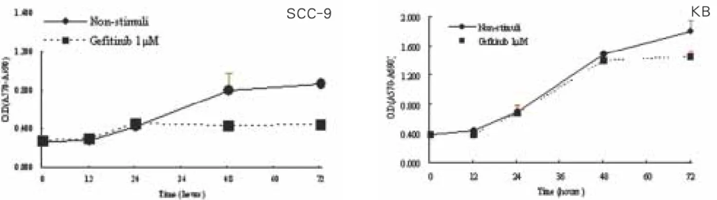
Treatment with gefitinib in several concentrations for 48 hours led to a dose-dependent decrease of cell density in SCC-9 cells, however, in KB cells, no significant growth inhibition by gefitinib was observed (Fig. 1). Similar antiproliferative effect was observed in time-dependent growth curve profile (Fig. 2).

Only in SCC-9 cells, significant growth inhibition was observed.

Fig. 1. Growth inhibition curve of gefitinib in several concentrations. SCC-9 cells were examined significant growth inhibition by gefitinib, while KB cells do not showed significant antiproliferative activity of gefitinib under < 10 μM (p<0.05).

SCC-9 KB

Fig. 2. In 1 μM gefitinib, growth inhibition curve in cell lines. SCC-9 cells were examined significant growth inhibition time- dependently, while KB cells showed no significant antiproliferative activity of gefitinib (p<0.05).

SCC-9 KB

(4)

2. Flow cytometric analysis

To study whether growth inhibition of head and neck cancer cells by gefitinib resulted form cell cycle delay, we examined the distribution of cell cycle by flow cytometry in the presence and absence of gefitinib. Table 1 shows that 1 μM gefitinib treatment for 48 h led to a significant increase in G0-G1 phase and this indicate that gifitinib treatment induced G1 arrest in SCC-9 cells as compared with the control. However, in KB cells, G1 arrest was not observed (Fig. 3).

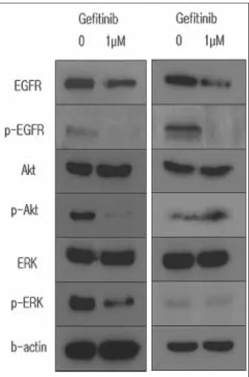
3. Effect on downstream signal pathways of EGFR

To elucidate the molecular mechanism of treatment effect of gefitinib, we evaluated whether EGFR is expressed in cell lines and the EGFR blockade affects the activation of intracel- lular downstream molecules. As shown in Fig. 4, EGFR expression is positive in SCC-9 and KB cells. In SCC-9 cells, gefitinib treatment induced a significant suppression of EGFR autophosphorylation and phosphorylation of Erk, Akt that are involved in downstream signaling of EGFR. However, in KB cell, only EGFR phophorylation was inhibted and phospho- form of Erk and Akt expression was weak and gefitinib do not suppress phosphorylation of Erk, and Akt.

Table 1.

Cell cycle analysis.

SCC-9 KB

Phase control gefitinib control gefitinib

G0-G1 60.0 76.6* 58.4 61.5

S 20.5 11.1* 19.6 21.3

G2-M 11.4 4.7* 15.0 12.5

*means significant difference from control statistically by t-test from three data points (p<0.05).

Fig. 3. Histogram of cell cycle distribution. SCC-9 cells were examined significant G1 arrest with gefitinib in contrast with KB cells.

SCC-9 KB

Fig. 4. Western blot analysis of EGFR expression and phos- phorylation of downstream signaling molecule. EGFR expression is positive in SCC-9 and KB cells. In SCC-9 cells, gefitinib treatment induced a significant suppression of EGFR auto-phosphorylation and phosphorylation of Erk, Akt that are involved in downstream signaling of EGFR.

However, in KB cells, EGFR phophorylation was inhibited

and active form of Erk and Akt expression was weak and

gefitinib do not suppress phosphorylation of Erk and Akt.

(5)

Discussion

The human epidermal growth factor receptor (EGFR) is a 170 KD transmembrane cell surface protein with tyrosine kinase activity which is activated following the binding of sev- eral ligands including EGF, TGF alpha, ampiregulin, HB-EGF, betacellulin, epiregulin and epigen to its external domain

10)

. This receptor is the prototype of the type-I growth factor receptor subfamily, which include erbB2 (HER-2), erbB3 (HER-3) and erbB-4 (HER-4). The EGFR exists as an inactive monomer, however, the binding of ligands to the external domain of the EGFR leads to the formation of homo or het- erodimers with the EGFR and other members of this family and autophosphorylation of several tyrosine residues in its intracellular domain, which ultimately lead to activation of downstream signalling molecules involved in cell proliferation or survival pathways of the ras/raf/Erk and PI-3/Akt respec- tively

12-14)

. It follows that monitoring the phosphorylated (active) status of EGFR, Erk and Akt in cell lines could be potentially used to determine gefitinib efficacy.

In the past 20 years, several groups have investigated the expression of the EGFR in head and neck cancer biopsies and the cell lines established from such tumors

15-26)

. Overall, in the majority of such studies overexpression of the EGFR has been shown to be a characteristic feature of head and neck squa- mous cell carcinoma and this in turn has been correlated with a poor prognosis and resistance to radiotherapy

21,26-29)

.

Gefitinib has been studied in recurrent and metastatic SCCHN as a single agent in single-arm phase II trials

30,31)

, only a minority of patients showed response to this agent like our study. In addition, there was no association between the expression of the EGFR levels and tumor response to gefitinib treatment. In our study, both SCC-9 and KB cell lines showed EGFR expression, but gefitinib response was different. Like other reports, our study showed that EGFR expression can not be a predictive factor of gefitinib response.

Currently, several critical issues about the gefitinib resistance mechanisms are ongoing. Emerging data suggest that EGFR inhibition may not be effective in the presence of independent- ly activated proteins (eg, ERK, AKT, STAT3

32,33)

), tumor cell dedifferentiation (eg, epithelial to mesenchymal transition

34,35)

, or the expression of other cell surface receptors (eg, insulin- like growth factor I receptor

36)

. Erk and Akt are downstream signaling molecules that can be activated by EGFR and other cell surface receptors. If other cell surface receptors such as HER-2, HER-3, HER-4 and insulin-like growth factor receptor are expressed, gefitinib cannot block activation of downstream signaling molecules Erk and Akt. So activated (phosphorylat-

ed) form of Erk and Akt can be a more reliable predictive fac- tor of gefitinib response. In our study, gefitinib sensitive SCC- 9 cells showed strong expression of p-Erk and p-Akt, but in KB cells, weak expression of active form of Erk and Akt was observed and this cell line showed gefitinb resistance. In stud- ies about gefitinib response in NSCLC patients, gefitinib sensi- tivity was correlated with phophorylation of Akt

37,38)

. Cappuzzo et al have reported that patients with phospho-Akt-positive tumors who received gefitinib had a better respose rate in terms of stable disease, disease control rate, and time to dis- ease progression than patients with phospho-Akt-negative tumors

38)

.

Additionally, there is currently studies on the expression pat- tern (i.e. membranous, cytoplasmic, nuclear), and prognostic significance of wild type EGFR, together with phosphorylated EGFR, and EGFRvIII in patients with head and neck cancer.

Such investigation may unravel the relative contribution of each factor to the malignant behavior of the head and neck cancer and response to the EGFR inhibitors.

In common with cytotoxic drugs, the target recognised by the EGFR TKIs is intracellular which may have contributed to the lack of association between the levels of the EGFR expression and response to the EGFR TKIs. In molecular therapy of human cancers using TKIs, a key aim should be the selection of the patients whose tumors are dependent on the phosphory- lation of such antigens for proliferation and metastasis. The potential of several proteins (pEGFR, EGFR, EGFRvIII, HER- 2, HER-3, HER-4, IGF-IR, pErk, pAkt, p27) in determining the biologic behavior of the head and neck cancer and in pre- dicting response to the EGFR inhibitors should be established.

In addition, such investigation should be performed in patients with different types of tumors. The lack of correlation between these factors and response to gefitinib in one type of cancer (eg. NSCLC) dose not indicate that the same could be true in other types of cancer such as head and neck cancer. The results of such investigations may lead to the identification of more reliable predictive factors for the selection of a more specific population of patients who would benefit from gefitinib treat- ment.

Conclusions

We investigate the antiproliferative effect of gefitinib,

EGFR-TKI in head and neck cancer cell lines. SCC-9 cells

were observed significant growth inhibition in contrast with

KB cells. Also, flow cytometric analysis showed G1 phase

arrest only in SCC-9 cell. In Western blot analysis to investi-

gate EGFR expression and downstream molecule phosphoryla-

(6)

tion, gefitinib suppressed phosphorylation of EGFR and down- stream protein kinase Erk, Akt in SCC-9. However, in EGFR positive KB cell, weak expression of active form of Erk and Akt and no inhibitory activity of phosphorylation in Erk, Akt was observed. The antiproliferative activity of gefitinib was not correlated with EGFR expression and some possibility of p-Erk and p-Akt as predictive factor of gefitinib response was emerged. Further investigations about more reliable predictive factor indicating gefitinib response are awaited to be useful gefitinib treatment in head and neck cancer patients.

References

1. Forastiere AA, Koch W, Trotti A, Sidransky D. Head and neck cancer. N Engl J Med 2002;345:1890-1900.

2. Kim ES, Hong WK, Khuri FR. Chemoprevention of aerodiges- tive tract cancer. Annu Rev Med 2003;53:233-43.

3. Shin DM, Hittelman WN, Hong WK. Biomarkers in upper aerodigestive tract tumorigenesis: a review. Cancer Epidemiol Biomark Prev 1994;3:679-709.

4. Jefferies S, Foulkes WD. Genetic mechanisms in squamous cell carcinoma of the head and neck. Oral Oncol 2001;37:115-26.

5. Boyle WJ. Growth factor and tyrosine kinase receptors during development and cancer. Curr Opin Oncol 1992;4:156-62.

6. Hu G, Liu W, Mendelsohn J. Expression of epidermal growth factor receptor and human papillomavirus E6/E7proteins in cer- vical carcinoma cells. J Natl Cancer Inst 1997;89:1271-76.

7. Iihara K, Shiozaki H, Tahara H. Prognostic significance of trans- forming growth factor-alpha in human esophageal carcinoma.

Implication for autocrine proliferation. Cancer 1993;71:2902-9.

8. Grandis JR, Melhem MF, Gooding WE. Levels of TGF-alpha and EGFR protein in head and neck squamous cell carcinoma and patient survival. J Natl Cancer Inst 1998;90:824-32.

9. Janmaat ML, Giaccone G. Small-molecule epidermal growth fac- tor receptor tyrosine kinase inhibitors. Oncologist 2003;8:576-86.

10. Grandis JR, Sol JC. Signaling through the epidermal growth fac- tor receptor during the development of malgnancy. Pharmacol Ther 2004;102:37-46.

11. Albanell J, Codony-Servat J, Rojo F. Activated extracellular sig- nal-regulated kinases: association with epidermal growth factor receptor/ transforming growth factor alpha expression in head and neck squamous carcinoma and inhibition by anti-epidermal growth factor receptor treatments. Cancer Res 2001;61:6500-10.

12. Gullick WJ. The type I growth factoe receptors and their ligands considered as complex system. Endocrine Related Cancer 2001;8:75-82.

13. Yarden Y. The EGFR family and its ligands in human cancer:

Signaling mechanisms and therapeutic apportunities. Eur J Cancer 2002;37:S3-S8.

14. Lui VWY, Grandis JR. EGFR-mediated cell cycle regulation.

Anticancer Res 2002;22:1-12.

15. Cowley GO, Smith JA, Gusterson BA, Hendler F, Ozanne B. The amount of EGF receptor is elevated on squamous cell carcino- mas. Cancer Cells 1984;1:5-10.

16. Partridge M, Gullick WJ, Langdon JD, Sheriff M: Expression of epidermal growth factor receptors on oral squamous cell carci- nomas. Br J Oral Max Surg 1988;26:381-6.

17. Shin DM, Donato NJ, Perez-Soler R, Shin HJC, Wu JY, Zhang P et al. Epidermal growth factor receptor-targeted therapy with C225 and cisplatin in patients with head and neck cancer.

Clinical Cancer Res 2001;7:1204-13.

18. Maurizio M, Almadori G, Ferrandina G, Distefano M, Romanini

ME, Cadoni G et al. Prognostic significance of epidermal growth factor receptor in laryngeal squamous cell carcinoma. Br J Cancer 1996;74:1253-7.

19. Grandis JR, Mellhem MF, Gooding WE, Day R, Holst VA, Wagner MM et al. Levels of TGFα, and EGFR protein in head and neck squamous cell carcinoma and patients survival. J Natl Cancer Inst 1998;90:824-32.

20. Maiorano E, Favia G, Maisonneuve P, Viale G. Prognostic impli- cations of epidermal growth factor receptor immunoreactivity in squamous cell carcinoma of the oral mucosa. J Pathology 2000;185:167-74.

21. Magne N, Pivot X, Bensadoun RJ, Guardiola E, Poissonnet G, Dassonville O et al. The relationship of epidermal growth factor receptor levels to the prognosis of unresectable pharyngeal can- cer patients treated by chemo-radiotherapy. Eur J Cancer 2001;37:2169-77.

22. Ang KK, Berkey BA, Tu X, Zhang HZ, Katz R, Hammond EH et al. Impact of epidermal growth factor receptor expression on sur- vival and pattern of relapse in patients with advanced head and neck carcinoma. Cancer Res 2002;62:7350-6.

23. Hoffmann TK, Ballo H, Braunstein S, Van Lierop A, Wagenmann M, Bier H. Serum level and tissue expression of c- erbB-1 and c-erbB-2 proto-oncogene products in patients with squamous cell carcinoma of the head and neck. Oral Oncol 2001;37:50-6.

24. Khademi B, Shirazi FM, Vasei M, Doroudchi M, Gandomi B, Modjtahedi H et al. The expression of p53, c-erbB-1 and c-erbB- 2 molecules and their correlation with prognostic markers in pa- tients with head and neck tumors. Cancer Letters 2002;184:223- 30.

25. O-charoenrat P, Rhys-Evans PH, Modjtahedi H, Eccles SA. The role of c-erbB receptors and ligands in head and neck squamous cell carcinoma (Review). Oral Oncology 2002;38:627-40.

26. Putti TC, To KF, Hsu HC, Chan AT, Lai GM, Tse G et al.

Expression of epidermal growth factor receptor in head and neck cancers correlates with clinical progression: A multicentre im- munohistochemical study in the Asia-Pacific region.

Histopathology 2002;41:144-51.

27. Haung SM, Harari PM. Modulation of radiation response after epidermal growth factor receptor blockade in squamous cell car- cinoma: Inhibition of damage repair, cell cycle kinetics and tu- mour angiogenesis. Clin Cancer Res 2000;6:2166-74.

28. Liang K, Ang KK, Milas L, Hunter N, Fan Z. The epidermal growth factor receptor mediates radioresistance. Int J Radiation Biol Phys 2003;57:246-54.

29. Ang KK, Andratschke NH, Milas L. Epidermal growth factor re- ceptor and response of head and neck carcinoma to therapy. Int J Radiation Oncology Biol Phys 2004;58:959-65.

30. Cohen EE, Rosen F, Stadler WM. Phase II trial of ZD1839 in re- current or metastatic squamous cell carcinoma of the head and neck. J Clin Oncol 2003;21:1980-7.

31. Cohen EE, Kane MA, List MA. Phase II trial of gefitinib 250 mg daily in patients with recurrent and/or metastatic squamous cell carcinoma of the head and neck. Clin Cancer Res 2005;11:8418- 24.

32. She QB, Solit DB, Ye Q. The BAD protein integrates survival signaling by EGFR/MAPK and PI3K/Akt kinase pathways in PTEN-deficient tumor cells. Cancer Cell 2005;8:287-97.

33. Sriuranpong V, Park JI, Amornphimoltham P. Epidermal growth factor receptor-independent constitutive activation of STAT3 in head and neck squamous cell carcinoma is mediated by the inter- leukin 6/gp130 cytokine system. Cancer Res 2003;63:2948-56.

34. Thompson S, Buck E, Petti F. Epidermal to mesenchymal transi- tion is a determinant of sensitivity of non-small-cell lung carcino- ma cell lines and xenograft to epidermal growth factor receptor inhibition. Cancer Res 2005;65:9455-62.

35. Yauch RL, Januario T, Eberhard DA. Epithelial versus mes-

enchymal phenotype determines in vitro sensitivity and predicts

(7)

clinical activity of erlotinib in lung cancer patients. Clin Cancer Res 2005;11:8686-98.

36. Spector NL, Xia W, Burris H III. Study of the biologic effects of lapatinib, a reversible inhibitor of ErbB1 and ErbB2 tyrosine ki- nases, on tumor growth and survival pathways in patients with advanced malignancies. J Clin Oncol 2005; 23:2502-12.

37. Noro R, Gemma A, Kosaihira S, Kokubo Y, Chen M, Seike M et

al. Gefitinib (IRESSA) sensitive lung cancer cell lines show phosphorylation of Akt without ligand stimulation. BMC Cancer 2006;6:277-98.

38. Cappuzzo F, Magrini E, Ceresoli GL, Bartolini S, Rossi E,

Ludovini V, Gregorc V et al. Akt phosphorylation and gefitinib

efficacy in patients with advanced non-small-cell lung cancer. J

Natl Cancer Inst 2004;96:1133-41.

수치

Fig. 2. In  1  μM  gefitinib,  growth  inhibition  curve  in  cell  lines.  SCC-9  cells  were  examined  significant  growth  inhibition  time- time-dependently, while KB cells showed no significant antiproliferative activity of gefitinib (p&lt;0.05).
Fig. 3. Histogram of cell cycle distribution. SCC-9 cells were examined significant G1 arrest with gefitinib in contrast with KB cells.

참조

관련 문서

PI3K inhibition decreased antioxidants/GD-induced apoptosis in A549 cells, and PI3K inhibitor LY294002 had inhibitory effect on antioxidants/GD-induced caspase-3

The combination treatment of PBA and 6-gingerol induced the cleavage of caspase-3, caspase-4, PARP, GRP78, p-eIF2α, and CHOP in DU145 cells and Western blot analysis for

• Theory can extent to molten polymers and concentrated solutions.. The single-molecule bead spring models.. a)

In this work, processing techniques for producing microcellular silicon carbide with cell densities greater than 10 9 cells/㎤ and cells smaller than 30㎛ have

 The inhibition pattern of microbial growth is analogous to enzyme inhibition..

Results: The human breast cancer cell subline MCF-7/MX5 cells selected in the presence of 5 µg/ml mitoxantrone (MX) were more resistant to MX (15.7... Western blot and

In contrast, cells expressing wild-type form or constitutively active form of RapC (GFP-RapC and GFP-RapC G13V ) showed decreased cell area and cell adhesion, whereas rapC

In this study, platelet-derived growth factors-BB (PDGF-BB), vascular endothelial growth factor (VEGF), and insulin-like growth factor-1 (IGF-1) were quantified in the PRF