Copyrightⓒ 2010, The Korean Academy of Oral Biology
153
Journal of Oral Biology
Growth Inhibition of Human Head and Neck Squamous Cell Carcinomas by Angelica decursiva Extracts
Woo-Cheol Shin, Chun Sung Kim, Heung-Joong Kim, Myoung-Hwa Lee, Hye Ryun Kim, and Do Kyung Kim* Oral Biology Research Institute, Chosun University School of Dentistry, Gwangju, Korea
(received August 25, 2010 ; revised October 29, 2010 ; accepted November 5, 2010)
Angelica decursiva has been used in Korean traditional medicine as an antitussive, an analgesic, an antipyretic and a cough remedy. However, the anti-cancer properties of Angelica decursiva have not yet been well defined. In our current study the cytotoxic activity of ethanol extracts of Angelica decursiva root (EEAD) and the mechanism of cell death exhibited by EEAD were examined in FaDu human head and neck squamous cell carcinoma cells. The cytotoxic effects of EEAD upon the growth of FaDu cells were examined with an MTT assay. In addition, the mechanism of cell death induced by EEAD was evaluated by DNA fragmentation analysis, immunoblotting and caspase activa- tion measurements. EEAD induced apoptotic cell death in FaDu cells in a concentration- and time-dependent manner, as determined by MTT assay and DNA fragmentation analysis.
Furthermore, the proteolytic processing of caspase-3, -7 and - 9 was increased by EEAD treatment of FaDu cells. In addition, the activation of caspase-3 and -7 was detected in living FaDu cells by fluorescence microscopy. These results suggest that EEAD can induce apoptosis and suppress cell growth in cancer cells and may have utility as a future anti- cancer therapy.
Key words: anti-cancer therapy, apoptosis, cell death, EEAD, FaDu human head and neck squamous cell carcinoma
서 론
최근 들어 과일, 야채, 씨앗기름 및 허브 등과 같은 천
연물을 의약품 또는 기능성 식품으로 개발하려는 노력이 활발히 진행되고 있다. 그 예로서 Taxol, Oncovin 또는 Captothecin 같은 약물들이 개발되어졌으며, 이 약물들은 인체 내에서 암의 화학적 예방제로서 또 치료제로서 잠재 적 가능성이 있는 것으로 보고되고 있다(Pezutto, 1997; van Poppel et al., 1997; Christou et al., 2001; Mukherjee et al., 2001).
이러한 생활성 항암물질의 대부분은 세포사의 일종인 apoptosis를 야기하여 암세포의 증식을 억제함으로서 암의 화학적 치료제 역할을 한다(Pezutto, 1997; van Poppel and van den Berg, 1997; Christou et al., 2001; Mukherjee et al., 2001). 따라서 이러한 생활성 항암물질을 사용하여 발 생하는 암세포의 apoptosis는 암으로 인한 인간의 죽음을 제어하고 줄이는 역할을 하는 암 치료법의 결과에 대한 중요한 지표가 되었다(Smets, 1994; Paschka et al., 1998).
세포사를 일으키는 가장 중요한 방법인 apoptosis는 진 핵세포의 항상성과 조직성장의 조절에 중요한 역할을 담 당하고 있다(Green and Reed, 1998; Hengartner, 2000;
Kaufmann and Hengartner, 2001). Apoptosis는 외인성의 death receptor 의존적 경로 또는 내인성의 미토콘드리아 의존적 경로를 따라 일어나는 것으로 보이며, 이는 암의 화학적 치료제에 의해서 일어나기도 한다(Kaufmann and Earnshaw, 2000; Reed, 2001).
우리나라에서 오래전부터 의약품으로 사용되어져 온 전 통 약제 중 하나인 Angelica decursiva는 민간요법으로 진 해제, 진통제, 해열제 또는 기침약 등으로 사용되어져 왔 다. 그러나 Angelica decursiva의 항암효과에 관한 자료는 거의 없다.
구강, 타액선, 인두 및 후두를 포함하는 두경부 암은 다 른 암에 비해 그 발생기전 등 분자생물학적 접근이 가장 되어있지 않은 암 중의 하나이다. 마이크로수술법, 방사선 치료법, 화학적 치료법의 발달에도 불구하고 이러한 종류
*Corresponding author: Do Kyung Kim, Department of Oral Physiology, Chosun University School of Dentistry, 375 Seosuk- dong, Dong-gu, Gwangju 501-759, Korea
Tel : 82-62-230-6893, Fax : 82-62-224-3706 E-mail : [email protected]
의 암으로부터 고통 받는 환자의 임상적 결과에서는 거의 진전을 보이지 않고 있다(Capuani et al., 2008).
따라서 본 연구에서는 사람 두경부 편평세포암종 FaDu 세포를 이용하여 Angelica decursiva의 암세포 성장억제에 미치는 효과와 세포성장 억제기전을 밝히고자 하며, 아울 러 Angelica decursiva에 의한 암 치료의 효용성을 제시 하고자 한다.
재료 및 방법
실험재료
N-methylthiotetrazole (MTT)는 Sigma 사(St. Louis, USA) 에서 구입하여 사용하였고, ECL detection kit는 Amersham Biosciences 사(Piscataway, NJ, USA)에서 구입하여 사용하 였다. Anti-caspase-3, anti-caspase-7 및 anti-caspase-9 항체 는 Cell Signaling Technology 사(Danvers, MA, USA)에 서 구입하여 사용하였고, Cell-permeablefluorogenic sub- strate PhiPhiLux-G1D2는 OncoImmunin 사(Gaithersburg, MD, USA)에서 구입하여 사용하였다. 기타 분석시약들은 analytical grade를 구입하여 사용하였다.
사람 두경부 편평세포암종 FaDu 세포는 American Type Culture Collection (ATCC, Rockville, MD, USA)에서 제 공받아 실험에 이용하였다.
식물재료와 추출(Angelica decursiva 에탄올 추출) 건조된 Angelica decursiva 뿌리를 전남생약조합(전남, 대 한민국)에서 구입하였으며, Angelica decursiva의 동정은 부 산대학교 한의학전문대학원 약물의학부(부산, 대한민국) 조 수인 교수에게 의뢰하였다. Angelica decursiva 뿌리를 1 mm 크기로 분쇄한 후, 95% 에탄올을 이용하여 40oC에서 5시 간 동안 추출하였다. 추출물을 Advantec No. 1 여과지를 이용하여 여과한 후, rotary evaporator (N-1000V-W, Eyela, Japan)를 이용하여 40oC의 진공상태로 증발시켰다. 증발건 조 후, 농축된 추출물을 −40oC에서 3일 동안 동결건조 하 였으며, 2oC에서 보관하며 사용하였다.
세포주와 세포배양
FaDu 세포는 10% fetal bovine serum (FBS, Invitrogen Co., Carlsbad, CA, USA) 및 항생제(100 u/ml penicillin, 100µg/ml streptomysin)가 함유된 37oC의 Dulbecco's Mod- ified Eagles Medium (DMEM, Invitrogen Co., Carlsbad, CA, USA) 성장배지 하에서 배양하면서 실험에 이용하였다.
세포성장 억제실험(MTT 분석)
Angelica decursiva 에탄올 추출물(EEAD)에 의한 세포 성장 억제효과를 관찰하기 위해, 24well plate에 5 × 10³cells/well의 FaDu 세포를 접종하였다. 24시간 배양한
후, EEAD를 다양한 농도와 시간에서 처리하여 37oC에서 반응시킨 후, 세포성장 억제효과를 MTT 분석으로 측정하 였다(Hwang et al., 2007; Kwon et al., 2008). MTT 분 석은 FaDu 세포에 MTT 용액(MTT 최종농도 0.5 µg/µl) 을 37oC에서 4시간 처리한 후, MTT 용액을 제거하고 0.04N HCl이 함유된 isopropanol로 녹여내어 570 nm에서 흡광도를 측정하여 시행하였다.
DNA fragmentation 분석
세포사멸의 기전 중 apoptosis의 지표가 되는 DNA frag- mentation 분석을 시행하였다. EEAD에 의한 세포 DNA fragmentation 효과를 관찰하기 위해, 10cm 배양접시에 5× 10⁵개의 FaDu 세포를 접종하였다. 24시간 배양한 후, EEAD (1µg/ml)를 처리하여 1일 또는 2일 동안 37oC에서 배양한 후, 세포를 수집하여 lysis buffer (0.1M NaCl, 0.001M EDTA, 0.3M Tris-HCl (pH, 7.5), 0.2M sucrose) 를 이용한 통상의 phenol-chloroform extraction법으로 DNA 를 추출하였다. 추출한 DNA를 2% agarose gel에서 전기 영동(50volts, 90분) 하였으며, ethidium bromide로 염색하 여 관찰하였다.
Immunoblotting
세포 apoptosis의 지표가 되는 caspase-3, caspase-7 및 caspase-9 분석을 위해 Hwang 등(2009)의 방법으로 immunoblotting을 시행하였다. 10 cm 배양접시에 5 × 10⁵ 개의 FaDu 세포를 접종하고 24시간 배양한 후, EEAD (1µg/ml)를 처리하여 1일 또는 2일 동안 37oC에서 배양한 후 세포를 수집하였다. 세포를 4oC의 PBS로 2회 세척한 후, 4oC의 lysis buffer (1% Triton X-100, 0.5 mM EDTA, 1 mM phenylmethylsulfonyl fluoride, 5µg/ml aprotinin 및 5µg/ml leupeptin이 포함된 PBS)에서 30분 반응시켰다. 세 포 용해물을 12,500 × g에서 20분간 원심분리한 후, 단백 질 시료를 정량하였다. 단백질 시료를 2배의 SDS sample buffer (60 mM Tris-HCl (pH, 6.8), 4% SDS, 25%
glycerol, 14.4 mM 2-mercaptoethanol, 0.1% bromophenol blue)에 넣고 100oC에서 5분간 변성 시킨 후, 12% SDS- polyacrylamide gel에 120volt에서 2시간 전기영동 한 다 음, 단백질 transfer를 이용하여 nitrocellulose membrane (Millipore Co., Billerica, MA, USA)으로 이동시켰다.
Membrane을 5% fat-free dry milk-PBST buffer (PBS, 0.2% Tween-20)에서 2시간 동안 blocking하였고, PBST buffer로 15분간 3회 세척하였다. 일차항체로 anti-caspase- 3 항체, anti-caspase-7 항체 및 anti-caspase-9 항체를 1,000 배 희석하여 사용하였으며, anti-β-actin 항체는 2,000배 희 석하여 사용하였다. 이차항체로 horseradish peroxidase conjugated anti-rabbit IgG를 5,000배 희석하여 사용하였 으며, ECL detection kit를 사용하여 X-ray 필름에 현상 한 후 분석하였다(Jun 등, 2009).
Caspase-3/-7 활성분석
Caspase-3/-7 활성분석을 위해 세포투과성 형광기질 PhiPhiLux-G1D2 (OncoImmunin, Inc., Gaithersburg, MD, USA)를 이용하였다. FaDu 세포에 EEAD(1 µg/ml)를 처 리하여 1일 동안 37oC에서 배양한 후, 배지를 제거하고 PhiPhiLux-G1D2를 37oC에서 30분간 처리하였으며, caspase- 3/-7의 활성도를 형광현미경(IX71, Olympus, Japan)으로 관찰하였다.
실험자료의 통계학적 검정
모든 실험성적은 mean ± SEM으로 나타내었고, 각 실험 군 간의 유의성 검정은 ANOVA 후에 Student's t-test를 하였으며, p value가 0.05 미만 (p < 0.05)의 경우에서 통 계적 유의성이 있는 것으로 간주하였다.
결 과
세포성장에 영향을 미치는 EEAD의 효과
FaDu 세포에서 EEAD에 의한 세포성장 억제효과를 조 사하기 위해 MTT 분석을 시행하였다. EEAD를 0.01, 0.03, 0.1, 0.3, 1, 3 및 10 µg/ml의 다양한 농도로 1일, 2일 및 3일 동안 FaDu 세포에 투여한 후 MTT 검사를 시행한 결 과, EEAD 처리 1일, 2일 및 3일의 경우에서 EEAD 0.01, 0.03 및 0.1 µg/ml의 농도에서는 대조군과 비교하였을 때 세포성장 억제의 차이를 볼 수 없었다(Fig. 1). 그러나 EEAD 처리 1일, 2일 및 3일의 경우, EEAD 0.3, 1, 3 및 10 µg/
ml에서는 대조군과 비교하여 볼 때 뚜렷한 세포성장 억제
효과를 볼 수 있었으며, 이 효과는 시간과 농도에 의존적 임을 확인할 수 있었다(Fig. 1). FaDu 세포성장 억제에 대 한 EEAD의 IC50(최대 억제량의 50%를 유발시키는 농도) 은 EEAD 처리 1일째는 10 µg/ml 이상이었고, 2일째는 약 0.27µg/ml이었으며, 3일째는 약 0.23 µg/ml이었다(Table 1).
DNA fragmentation 분석
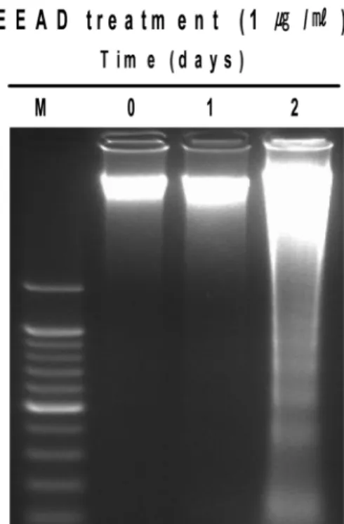
EEAD에 의한 FaDu 세포의 성장억제 기전을 확인하기 위하여 DNA fragmentation 분석을 시행하였다. EEAD 1µg/ml을 0일(대조군), 1일 및 2일 처리한 FaDu 세포의 DNA를 추출하여 전기영동으로 확인한 결과, 대조군과 1 일 처리군에서는 DNA fragmentation 현상을 볼 수 없었 으나, EEAD 1 µg/ml 처리 2일 실험군에서는 DNA frag- mentation 현상을 볼 수 있었다(Fig. 2).
EEAD에 의한 caspase의 활성
Caspase-3, caspase-7 및 caspase-9이 세포 apoptosis의 지 표가 되므로 EEAD를 처리한 FaDu 세포에서 procaspase-
Fig. 1. Cytotoxic effects of EEAD in FaDu cells. The FaDu cells were treated with 0, 0.01, 0.03, 0.1, 0.3, 1, 3 and 10µg/ml EEAD for 1 day (filled circle), 2 days (filled square) and 3 days (filled tri- angle). Cell viability was determined by the MTT assay. The per- centage of cell viability was calculated as a ratio of A570 nm of EEAD treated cells and untreated control cells. Each data point represents the mean ± SEM for three experiments. *P < 0.05 vs.
control, **P < 0.01 vs. control and ***P < 0.001 vs. control (the con- trol cells were measured in the absence of EEAD treatment).
Table 1. Antiproliferative effect of EEAD in FaDu cells.
days IC50 (µg/ml)
1 day > 10
2 days 0.27 ± 0.04
3 days 0.23 ± 0.03
The IC50 values represent the mean ± SEM for three experi- ments.
Fig. 2. Fragmentation of internucleosomal DNA by EEAD in FaDu cells. The cells were treated with 1µg/ml EEAD for 0, 1 and 2 days and nuclear DNA was subjected to agarose gel electro- phoresis.
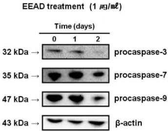
3, procaspase-7 및 procaspase-9 발현분석을 위해 immuno- blotting을 시행하였으며, caspase-3/-7 세포투과성 형광기질 을 처리하여 세포 내에서 발현하는지 확인해 보았다. EEAD 1µg/ml을 처리한 FaDu 세포의 단백질을 추출하여 확인한 결과, 대조군 및 1일 처리군에서는 procaspase-3, procaspase- 7 및 procaspase-9의 단백질 가수분해(proteolytic cleavage) 현상을 볼 수 없었으나, EEAD 2일 처리군에서는 단백질 가수분해 현상을 뚜렷이 볼 수 있었다(Fig. 3). 또한 EEAD 1µg/ml을 1일 처리한 FaDu 세포에 세포투과성 형광기질 을 처리하여 형광현미경으로 확인한 결과, Fig. 4에서와 같 이 caspase-3/-7의 활성을 뚜렷이 확인할 수 있었다.
고 찰
최근, 천연물로부터 항암제를 포함한 새로운 약물들의 개발로 동양의학에 대한 과학계의 관심이 세계적으로 커지 고 있다(Hu et al., 2002; Lee et al., 2002; Cheng et al., 2005; Park et al., 2005; Tan et al., 2005). 그 중 화학요 법제는 암세포에서 다양한 기전을 통해 암세포 독성을 유 발하는 것으로 알려져 있다(Hoshino et al., 1991; Tian et al., 2006). 그 암세포 성장억제 기전들은 세포의 종류와 자 극의 종류에 의존하여 중요한 역할을 담당하며, 최소한의 부작용으로 암세포를 사멸시킬 수 있어야 한다(Hoshino et al., 1991; Tian et al., 2006). Angelica decursiva는 우리 나라에서 전통적으로 사용되어져 온 민간 요법제 중 하나 이다. 그러나 Angelica decursiva의 항암효과에 대해서는 알려진 바가 전혀 없다. 따라서 본 연구에서는 사람 두경 부 편평세포암종 FaDu 세포를 이용하여 Angelica decursiva 의 암세포 성장억제에 미치는 효과와 세포성장 억제기전을 밝히고자 하였다.
세포성장 억제효과를 조사하기 위한 MTT 실험에서, EEAD는 시간과 농도에 의존적으로 FaDu 세포의 성장을 억제시켰다(Fig. 1). 이는 시간과 농도에 의존적으로 암세
포의 성장을 억제시키는 항암효과를 지닌 여러 추출물들 (Echinacea root, Toona sinensis, Willow bark)에서의 연 구결과와 일치하는 것이었다(Yang et al., 2006; Chicca et al., 2007; Hostanska et al., 2007). 더욱이 다른 추출물들 과 비교하여 상대적으로 낮은 농도의 EEAD에서도 충분히 FaDu 세포의 성장을 억제하였다(Yang et al., 2006; Chicca et al., 2007; Hostanska et al., 2007). 이러한 결과는 EEAD가 암세포 성장억제에 대한 특이적인 효과를 가지 고 있다는 것을 시사하며, 또 EEAD의 항암치료제로서의 잠재적인 가치를 시사하고 있다.
Apoptosis는 세포분열과 세포사멸 사이에서 항상성을 유 지시켜주는 중요한 역할을 한다(Green and Reed, 1998;
Hengartner, 2000; Kaufmann and Hengartner, 2001). 암 세포에서 apoptosis의 유도는 천연물들로부터 항암제로의 개발에 가장 유용한 전략이다(Hu and Kavanagh, 2003).
따라서 많은 연구자들이 식물의 추출물을 포함한 많은 천 Fig. 3. Proteolytic cleavage of procaspase-3, -7 and -9 by EEAD treatment in FaDu cells. Expression of procaspase-3, -7 and -9 by EEAD was examined in FaDu cells. The cells were treated with 1µg/ml EEAD for 0, 1 and 2 days. The cell lysate was prepared and analyzed by immunoblotting as described in “MATERIALS AND METHODS”.
Fig. 4. Activation of caspase-3/-7 by EEAD treatment in living FaDu cells. The cells were treated with 1 µg/ml EEAD for 24 hours and added specific cell-permeable substrate Phiphilux-G1D2. Active of caspase-3/-7 was visualized by fluorescence microscopy.
연물들로부터 암세포들의 apoptosis를 유도하는 연구들을 시행하고 있다. 본 연구에서도 EEAD에 의한 FaDu 세포 성장억제 기전에 apoptosis가 포함되는지를 확인하기 위하 여, DNA fragmentation 분석을 시행하였다. EEAD를 0 일, 1일 및 2일 처리한 FaDu 세포의 DNA를 추출하여 전기영동으로 확인한 결과, 대조군과 1일 처리군에서는 DNA fragmentation 현상을 볼 수 없었으나, EEAD 처리 2일 실험군에서는 DNA fragmentation 현상을 볼 수 있 었다(Fig. 2). 이는 암세포의 성장을 억제시키는 항암효과 를 지닌 여러 추출물들이 DNA fragmentation을 유도하 는 apoptosis 과정에 의해 암세포를 사멸시킨다는 연구결 과(Yang et al., 2006; Chicca et al., 2007; Hostanska et al., 2007)와 일치하는 것으로서, EEAD에 의해 유도되는 FaDu 세포 성장억제 과정에는 apoptosis 기전이 포함되는 것으로 사료된다.
Caspase라 불리는 세포 내 cysteine protease들의 활성 은 다양한 자극에 의해 유도된 apoptosis를 개시하고 실 행하는데 중요한 역할을 하며, 포유동물 세포에서 확인된 caspase 중에 caspase-3, caspase-7 및 caspase-9 등이 apoptosis에 의한 세포사멸의 effector caspase로 알려져 있 다(Cohen, 1997; Datta et al., 1997; Liu et al., 1997).
Caspase-3, caspase-7 및 caspase-9은 각각 32kDa, 35kDa 및 47kDa의 불활성 proenzyme으로 합성되며, 다양한 자 극에 의해 apoptosis가 일어날 때 proteolytic cleavage 현 상이 일어난다(Cohen, 1997; Datta et al., 1997; Liu et al., 1997). 본 연구에서 proenzyme들의 발현분석을 위해 immunoblotting을 시행한 결과, EEAD 처리 실험군에서 procaspase-3, procaspase-7 및 procaspase-9의 proteolytic cleavage 현상을 볼 수 있었다(Fig. 3). 또한 EEAD를 처 리한 FaDu 세포에 세포투과성 형광기질을 처리하여 형광 현미경으로 확인한 결과, caspase-3/-7의 활성을 확인할 수 있었다(Fig. 4). 이러한 결과들은 EEAD에 의해 유도되는 FaDu 세포 성장억제 과정에 caspase-3, caspase-7 및 caspase-9에 의존적인 apoptosis가 포함되어 있음을 시사한 다. 그러나 EEAD가 유도하는 암세포 성장억제에 관한 세 포 및 분자적 기전연구는 더 추구하여야 할 과제로 생각된다.
결론적으로, EEAD는 사람 두경부 편평세포암종 FaDu 세포의 apoptosis를 유도하여 암세포 성장을 억제시키는 것 으로 사료되며 본 연구의 결과로, EEAD를 이용한 암세포 성장억제에 관한 또 하나의 방향을 제시할 수 있을 것으 로 생각된다.
감사의 글
이 논문은 2010학년도 조선대학교 학술연구비의 지원 을 받아 연구되었음.
참 고 문 헌
Capuani S, Gili T, Bozzali M, Russo S, Porcari P, Cametti C, D'Amore E, Colasanti M, Venturini G, Maraviglia B, Lazzarino G, Pastore FS. L-DOPA preloading increases the uptake of borophenylalanine in C6 glioma rat model : A new strategy to improve BNCT efficacy. Int J Radiat Oncol.
2008;72:562-7.
Cheng YL, Lee SC, Lin SZ, Chang WL, Chen YL, Tsai NM, Liu YC, Tzao C, Yu DS, Harn HJ. Anti-proliferative activity of Bupleurum scrozonerifolium in A549 human lung cancer cells in vitro and in vivo. Cancer Lett. 2005;222:183-93.
Chicca A, Adinolfi B, Martinotti E, Fogli S, Breschi MC, Pellati F, Benvenuti S, Nieri P. Cytotoxic effects of Echinacea root hexanic extracts on human cancer cell lines. J Ethnophar- macol. 2007;110:148-53.
Christou L, Hatzimichael E, Chaidos A, Tsiara S, Bourantas KL. Treatment of plasma cell leukemia with vincristine, liposomal doxorubicin and dexamethasone. Eur J Hematol.
2001;67:51-3.
Cohen GM. Caspases: the executioners of apoptosis. Biochem J. 1997;326:1-16.
Datta R, Kojima H, Yoshida K, Kufe D. Caspase-3-mediated cleavage of protein kinase C theta in induction of apoptosis.
J Biol Chem. 1997;272:20317-20.
Green DR, Reed JC. Mitochondria and apoptosis. Science.
1998;281:1308-12.
Hengartner MO. The biochemistry of apoptosis. Nature.
2000;407:770-6.
Hoshino T, Hara A, Inoue M, Honda J, Imai Y, Oizumi K, Yokoyama MM. Flowcytometric measurement of NK cell cytotoxicity. J Clin Lab Immunol. 1991;36:39-43.
Hostanska K, Jurgenliemk G, Abel G, Nahrstedt A, Saller R.
Willow bark extract (BNO1455) and its fractions suppress growth and induce apoptosis in human colon and lung cancer cells. Cancer Detect Prev. 2007;31:129-39.
Hu H, Ahn NS, Yang X, Lee YS, Kang KS. Ganoderma lucidum extract induces cell cycle arrest and apoptosis in MCF-7 human breast cancer cell. Int J Cancer. 2002;102:250-3.
Hu W, Kavanagh JJ. Anticancer therapy targeting the apoptotic pathway. Lancet Oncol. 2003;4:721-9.
Hwang IN, Jeong YJ, Jung JY, Lee JH, Kim KM, Kim WJ.
Mechanism underlying NO-induced apoptosis in human gingival fibroblasts. Int J Oral Biol. 2009;34:7-14.
Hwang JH, Kim JY, Cha MR, Park HR. Effect of methanolic extract from silkworm droppings on proliferation and caspase activity in HT-29 human colon cancer cells. J Med Food. 2007;10:467-72.
Jun JH, Ryoo HM, Woo KM, Kim GS, Baek JH. Bone morphogenetic protein 2-induced MAPKs activation is inde- pendent of the Smad1/5 activation. Int J Oral Biol. 2009;
34:115-21.
Kaufmann SH, Earnshaw WC. Induction of apoptosis by cancer chemotherapy. Exp Cell Res. 2000;256:42-9.
Kaufmann SH, Hengartner MO. Programmed cell death : alive and well in the new millennium. Trends Cell Biol.
2001;11:526-34.
Kim JY, Kim HS, Kang HS, Choi JS, Yokozawa T, Chung HY.
Antioxidant potential of dimethyl lithospermate isolated from Salvia miltiorrhiza (red sage) against peroxynitrite. J Med Food. 2008;11:21-8.
Kwon JI, Kim GY, Park KY, Ryu CH, Choi YH. Induction of apoptosis by linoleic acid is associated with the modulation of Bcl-2 family and Fas/FasL system and activation of caspases in AGS human gastric adenocarcinoma cells. J Med Food.
2008;11:1-8.
Lee SM, Li ML, Tse YC, Leung SC, Lee MM, Tsui SK.
Paeoniae Radix, a chinese herbal extract, inhibit hepatoma cells growth by inducing apoptosis in a p53 independent pathway. Life Sci. 2002;71:2267-77.
Liu X, Zou H, Slaughter C, Wang X. DFF, a heterodimeric protein that functions downstream of caspase-3 to trigger DNA fragmentation during apoptosis. Cell. 1997;89:175-84.
Mukherjee AK, Basu S, Sarkar N, Ghosh AC. Advances in cancer therapy with plant based natural products. Curr Med Chem. 2001;8:1467-86.
Park DI, Lee JH, Moon SK, Kim CH, Lee YT, Cheong J, Choi BT, Choi YH. Induction of apoptosis and inhibition of telomerase activity by aqueous extract from Platycodon grandiforum in human lung carcinama cells. Pharmacol Res.
2005;51:437-43.
Paschka AG, Butler R, Young CYF. Induction of apoptosis in prostate cancer cell lines by the green tea component, (−)- epigallocatechin-3-gallate. Cancer Lett. 1998;130:1-7.
Pezutto JM. Plant-derived anticancer agents. Biochem Pharmacol. 1997;53:121-133.
Reed JC. Apoptosis-regulating proteins as targets for drug discovery. Trends Mol Med. 2001;7:314-9.
Smets LA. Programmed cell death (apoptosis) and response to anti-cancer drugs. Anti-Cancer Drug. 1994;5:3-9.
Tan ML, Suaiman SF, Najimuddin N, Smian MR, Tengku Muhammad TS. Methanolic extract of Pereskia bleo (Kunth) DC. (Cactaceae) induces apoptosis in breast carcinama, T47- D cell line. J Ethnopharmacol. 2005;96:287-94.
Tian Z, Chen S, Zhang Y, Huang M, Shi L, Huang F, Fong C, Yang M, Xiao P. The cytotoxicity of naturally occurring styryl lactones. Phytomedicine. 2006;13:181-6.
van Poppel G, van den Berg H. Vitamins and cancer. Cancer Lett. 1997;114:195-202.
Yang HL, Chang WH, Chia YC, Huang CJ, Lu FJ, Hsu HK, Hseu YC. Toona sinensis extracts induces apoptosis via reactive oxygen species in human premyelocytic leukemia cells. Food Chem Toxicol. 2006;44:1978-88.