Synthetic Chenodeoxycholic Acid Derivative
HS-1200-Induced Apoptosis of Human Oral Squamous Carcinoma Cells
In-Ryoung Kim
1, M.S., Hyeon-Jin Sohn
2, D.D.S., Gyoo-Cheon Kim
2, M.S.,Ph.D., Hyun-Ho Kwak
2, M.S.,Ph.D., Bong-Soo Park
2, D.D.S.,M.S.D.,Ph.D.,
Won-Chul Choi
1, M.S.,Ph.D., Myung-Yun Ko
3, D.D.S.,M.S.D.,Ph.D., Yong-Woo Ahn
3, D.D.S.,M.S.D.,Ph.D.
Department of Biology, College of Natural Science, Pusan National University1 Department of Oral Anatomy, College of Dentistry, Pusan National University2 Department of Oral Medicine, College of Dentistry, Pusan National University3
Bile acids and synthetic its derivatives induced apoptosis in various kinds of cancer cells and anticancer effects.
Previous studies have been reported that the synthetic chenodeoxycholic acid (CDCA) derivatives showed apoptosis inducing activity on various cancer cells in vitro. It wasn't discovered those materials have apoptosis induced effects on YD9 human oral squamous carcinoma cells.
The present study was done to examine the synthetic bile acid derivatives(HS-1199, HS-1200) induced apoptosis on YD9 cells and such these apoptosis events. We administered them in culture to YD9 cells. Tested YD9 cells showed several lines of apoptotic manifestation such as activation of caspase-3, degradation of DFF, production of poly (ADP-ribose) polymerase(PARP) cleavage(HS-1200 only), DNA degradation(HS-1200 only), nuclear condensation, inhibition of proteasome activity, reduction of mitochondrial membrane potential(HS-1200 only) and the release of cytochrome c and AIF to cytosol. Between two synthetic CDCA derivatives, HS-1200 showed stronger apoptosis-inducing effect than HS-1199. Therefore HS-1200 was demonstrated to have the most efficient antitumor effect.
Taken collectively, we demonstrated that a synthetic CDCA derivative HS-1200 induced caspases-dependent apoptosis via mitochondrial pathway in human oral sqauamous carcinoma cells in vitro. Our data therefore provide the possibility that HS-1200 could be considered as a novel therapeutic strategy for human orall squamous carcinoma from its poweful apoptosis-inducing activity.
Key words : Apoptosis, Synthetic CDCA derivatives, HS-1200, Human oral squamous cell carcinoma
Correspoding Authur : Yong-Woo Ahn
Department of Oral Medicine, College of Dentistry
Pusan National University, 1-10 Ami-Dong, Seo-Ku, Pusan 602-739, Korea Tel: 82-51-240-7465
Fax: 82-51-247-0955
E-mail: [email protected] Received: 2007-04-30
Accepted: 2007-06-23
*This work was supported by for two years Pusan National University research grant.
I. INTRODUCTION
Carcinoma of the oral cavity, especially oral squamous cell carcinoma(OSCC), are one of the most leading causes of cancer related death and affect nearly 500,000 patients annually world-wide.
And OSCC is one of the most common malignancies that remain incurable with current therapies.
1)Apoptosis, or programmed cell death, is an essential physiological process that is required for the normal development and maintenance of tissue homeostasis. However, apoptosis also is implicated in a wide range of pathological conditions, including immunological diseases, allergy and cancer.
2,3)During apoptosis, cells undergo specific morphological and biochemical changes, including cell shrinkage, chromatin condensation, and internucleosomal cleavage of genomic DNA.
4,5)Bile acids are polar derivatives of cholesterol essential for the absorption of dietary lipids and regulate the transcription of genes that control cholesterol homeostasis. Depending on the nature of chemical structures, each kind of bile acid exhibits distinct biological effects.
6)The natural bile salts were reported to inhibit cell proliferation and induce apoptosis in various cancer cells.
7,8)After synthesis by the liver and excretion into the bile canaliculus and the digestive tract, the primary bile acids, cholic acid (CA) and chenodeoxycholic acid (CDCA), are metabolized by enteric bacteria to produce secondary bile acids; primarily deoxycholic acid, ursodeoxycholic acid (UDCA), and lithocholic acid (LCA). Bile acids are conjugated to glycine, or taurine when the glycine conjugates predominate.
6)Conjugation of bile acids to glycine and taurine is one mechanism by which an organism can decrease the hydrophobicity of a bile acid.
9,10)The conjugation renders the molecules less cytotoxic at physiological concentrations.
11)Numerous studies have shown that elevated concentrations of bile acid within the liver induce hepatocyte apoptosis.
This provides a cellular mechanism for bile acid mediated liver injury.
12)Bile acid hydrophobicity is
correlated with induction of apoptosis and/or growth arrest.
13)To date, there is no report about the apoptotic effect of CDCA derivatives on YD 9 human oral sqauamous carcinoma cell line. Therefore this study was undertaken to examine the molecular mechanism underlying CDCA derivatives-induced apoptosis in human oral sqauamous carcinoma cells
Ⅱ. MATERIALS AND METHODS 1. Reagents
CDCA was obtained from Dae-Woong Pharmaceutical Co.(Seoul, Korea) and Aldrich (Milwaukee, WI, USA). The synthetic bile acid derivatives, HS-1199 and HS-1200 were kindly provided by Professor Young-Hyun Yoo(Depart- ment of Anatomy, Dong-A University College of Medicine, Busan, Korea). The structure and methods of the synthesis of the synthetic bile acid derivatives were described by Im EO et al.
14)HS-1199 is a conjugate form of CDCA with L-phenyl alanine benzyl ester(N-[(3α, 5β, 7α)-3,7-dihydroxyl-24- oxocholan-yl] L-phenyl alanine benzyl ester).
HS-1200 is a conjugate form of CDCA with β -alanine benzyl ester(N-[(3α, 5β, 7α)-3,7-dihydroxyl -24-oxocholan-yl] β-alanine benzyl ester). These bile acids and their derivatives were dissolved in absolute ethanol, and dilutions were made in culture medium. The final concentration of ethanol in the medium was less than 0.1%(vol/vol) in the treatment range(10-100 μM) and showed no influence on cell growth(data not shown). The structures of CDCA and its conjugate forms(HS-1199 and HS-1200) are shown in Fig. 1.
The following reagents were obtained
commercially: Rabbit polyclonal anti-human
caspase-3 and anti-horse cytochrome c, and
anti-human DNA fragmentation factor(DFF), and
goat polyclonal anti-mouse AIF antibodies were
from Santa Cruz Biotechnology, Inc. (Santa Cruz,
CA). Mouse polyclonal anti-human poly(ADP-
ribose) polymerase (PARP) antibody was from
Oncogene (Cambridge, MA); FITC-conjugated goat anti-rabbit and horse anti-mouse IgGs were from Vector (Burlingame, CA); HRP-conjugated donkey anti-rabbit and sheep anti-mouse IgGs were from Amersham Pharmacia Biotech (Piscataway, NJ).
5,5',6,6'-tetrachloro-1,1',3,3'-tetraethylbenzimidazol carbocyanine iodide (JC-1) was from Molecular Probes (Eugene, OR). Dulbecco's modified Eagle's medium (DMEM) and FBS were from Gibco (Gaithersburg, MD). Dimethyl sulfoxide (DMSO), Hoechst 33342, RNase A, proteinase K, aprotinin, leupeptin, PMSF, thiazolyl blue tetrazolium bromide and propidium iodide were from Sigma (St. Louis, MO); SuperSignal West Pico enhanced chemilumin, Gescence Western blotting detection reagent was from Pierce (Rockford, IL).
2. Cell culture
Oral squamous carcinoma cells(YD9 cells) (kindly provided by Professor Jin Kim, Department of Oral Pathology, Yonsei University College of Dentistry, Seoul, Korea) were maintained at 37℃
with 5% CO
2in air atmosphere in minimum essential medium (Eagle) with 2 mM L-glutamine and Earle's BSS adjusted to contain 1.5 ㎍/L sodium bicarbonate, 0.1 mM non-essential amino acids, and 1.0 mM sodium pyruvate, and supplemented with 10% FBS. cells were matained in Dulbecco's modified Eagle's medium with 10%
FBS.
3. MTT assay
Cells were placed in a 96-well plate and incubated 24 h. Then cells treated with 10, 25, 50, 100 μM of CDCA, HS-1199, and HS-1200 for 5 h.
And then cells were treated with 500 μg/ml of thiazolyl blue tetrazolium bromide. Cells were incubated at 37℃ with 5% CO
2for 4h. And then the medium was aspirated and formed formazan crystals were dissolved in the mixture solution of 75 μl DMSO and 75 μl absolute ethanol. Cell viability was measured by a ELISA reader(Sunrise
Remote Control, Tecan, Austria) at 570 nm excitatory emission wavelength.
4. Hoechst staining
Cells were harvested and cell suspension was centrifuged onto a clean, fat-free glass slide with a cytocentrifuge. The samples were stained in 4 ㎍ /ml Hoechst 33342 for 30 min at 37℃ and fixed for 10 min in 4% paraformaldehyde.
5. DNA electrophoresis
2 x 10
6cells were resuspended in 1.5 ml of lysis buffer(10 mM Tris (pH 7.5), 10 mM EDTA (pH 8.0), 10 mM NaCl and 0.5% SDS) into which proteinase K(200 ㎍/ml) was added. After samples were incubated overnight at 48℃, 200 μl of ice cold 5 M NaCl was added and the supernatant containing fragmented DNA was collected after centrifugation.
The DNA was then precipitated overnight at -20℃
in 50% isopropanol and Rnase A-treated for 1 h at 37℃. The DNA from 10
6cells(15 μl) was equally loaded on each lane of 2% agarose gels in Tris-acetic acid/EDTA buffer containing 0.5 ㎍/ml ethidium bromide at 50 mA for 1.5 h.
6. Proteasome activity
After treatment with 50μM of CDCA, HS-1199,
and HS-1200 for 5h, cells were lysed in proteasome
buffer [10 mM Tris-HCl, pH 7.5, 1 mM EDTA, 2
mM ATP, 20% glycerol, and 4 mM dithiothreitol
(DTT)], sonicated, and then centrifuged at 13,000 g
at 4℃ for 10 min. The supernatant (20 ㎍ of
protein) were incubated with proteasome activity
buffer [0.05 M Tris-HCl, pH 8.0, 0.5 mM EDTA, 50
μM Suc-LLVY-AMC] for 1 h 37℃. The intensity
of fluorescence of each solution was measured by a
modular fluorimetric system (Spex Edison, NJ,
USA) at 380 nm excitatory and 460 nm emission
wavelengths. All readings were standardized using
the fluorescence intensity of an equal volume of
free AMC solution (50 μM).
7. Western blot analysis
Cells (2 x 10
6) treated with CDCA, HS-1199 and HS-1200 were washed twice with ice-cold PBS, resuspended in 200 ㎕ ice-cold solubilizing buffer [300 mM NaCl, 50 mM Tris-Cl (pH 7.6), 0.5%
TritonX-100, 2 mM PMSF, 2 ㎕/ml aprotinin and 2
㎕/ml leupeptin] and incubated at 4℃ for 30 min.
The lysates were centrifuged at 14,000 revolutions per min for 15 min at 4℃. Protein concentrations of cell lysates were determined with Bradford protein assay (Bio-Rad, Richmond, CA) and 50 μg of proteins were loaded onto 7.5-15% SDS/PAGE. The gels were transferred to Nitrocellulose membrane (Amersham Pharmacia Biotech, Piscataway, NJ) and reacted with each antibody. Immunostaining with antibodies was performed using SuperSignal West Pico enhanced chemiluminescence substrate and detected with Alpha Imager HP(Alpha Innotech, San Leandro, USA).
8. Immunofluorescent staining
Cells were cytocentrifuged and fixed for 10 min in 4% paraformaldehyde, incubated with each primary antibody for 1 h, washed 3 each for 5 min, and then incubated with FITC-conjugated secondary antibody for 1 hr at room temperature.
Cells were mounted with PBS. Fluorescent images
Fig. 1. Chemical structures of CDCA and its derivatives
were observed and analyzed under Zeiss LSM 510 laser-scanning confocal microscope (Göettingen, Germany).
9. Assay of mitochondrial membrane potential (MMP)
JC-1 was added directly to the cell culture medium (1 μM final concentration) and incubated for 15 min. The medium was then replaced with PBS, and cells were resuspended in 10 ㎍/ml of methanol and incubated at 37℃ for 30 min. Flow cytometry to measure MMP was performed on a Epics XL (Beckman Coulter, FL, USA). Data were acquired and analyzed using EXPO32 ADC XL 4 color software. The analyzer threshold was adjusted on the FSC channel to exclude noise and most of the subcellular debris.
Ⅲ. RESULTS
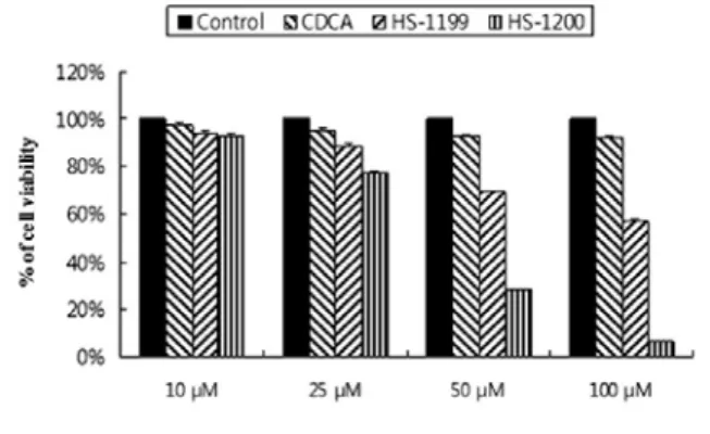
1. Synthetic CDCA derivatives reduced viability in YD9 cells.
As determined by MTT, HS-1200 at 50 μM for 5 h significantly reduced viability of YD9 cells compared to HS-1199. But CDCA at 50 μM did not
Fig. 2. YD9 cells treated with synthetic CDCA derivatives HS-1199 and HS-1200 at 10 μM, 25 μM, 50 μM and 100 μM for 5 h show the reduction of viability in a dose-dependent manner. Result is expressed as percentage of the vehicle-treated control ± SD of three separate experiments.
Fig. 3. YD9 cells treated with synthetic CDCA derivatives HS-1199 and HS-1200 at 50 μM for 5 h show the nuclear condensation or fragmentation compared to the negative control or the CDCA-treated group. (A) Hoechst staining. (B) Quantification of the nuclear condensation determined by Hoechst staining. Result is expressed as percentage of the vehicle-treated control ± SD of three separate experiments.
Fig. 4. OCS9 cells treated for 5 h with synthetic CDCA derivatives HS-1199 and HS-1200 at 50 μM show the reduction of proteasome activity compared to the negative control or the CDCA treated group. Data are presented as the percent of the control.
Fig. 5. YD9 cells treated for 5 h with synthetic CDCA derivatives HS-1199 and HS-1200 at 50 μM produce nuclear events.
Western blot analyses showing degradation of caspase-3, DFF and PARP. HS-1119 induced caspase-3 and DFF degradation. HS-1200 induced caspase-3, DFF and PARP degradtion, and produced 30 kD and 11 kd DFF cleavages and 85 kd PARP cleavage.
Fig. 6. DNA electrophoresis demonstration of YD9 cells treated for 5 h with synthetic CDCA derivatives HS-1199 and HS-1200 at 50 μM. DNA electrophoresis evidently showed DNA ladder in YD9 cells treated with HS-1200.
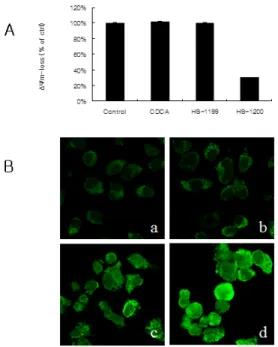
Fig. 7. YD9 cells treated for 5 h with synthetic CDCA derivatives HS-1199 and HS-1200 at 50 μM induce mitochondrial events. (A) Loss of mitochondrial membrane potential (MMP) was significantly showed after treatment with HS-1200 but was not showed after treatment with CDCA and HS- 1119. (B) Confocal microscopy showing the release of cytochrome c from mitochondria into the cytosol after treatment with HS-1119 and HS-1200. a, control cells
; b, CDCA treated cells; c, HS-1119 treated cells; d, HS-1200 treated cells
Both synthetic CDCA derivatives decreased the viability of YD9 cells in a dose-dependent manner(Fig. 2).
2. Synthetic CDCA derivatives induced nuclear condensation and fragmentation in YD9 cells.
Hoechst staining proved that the reduction in viability resulted from apoptosis (Fig. 3A and 3B).
HS-1200 at 50 μM showed stronger cytotoxic effect than HS-1199 at the same dose.
Fig. 8. AIF involves apoptosis in YD9 cells treated with synthetic CDCA derivatives HS-1199 and HS-1200 at 50 μM for 5 h. (A) Expression level of this protein slightly increased compared to the control. (B) Confocal microscopy showing that AIF was released from mitochondria, and that translocation onto nuclei was evident in HS-1119 and HS-1200 treated cells a, control cells ; b, CDCA treated cells; c, HS-1119 treated cells; d, HS-1200 trated cells
3. Synthetic CDCA derivatives inhibited proteasome activity in YD9 cells
Both synthetic CDCA derivatives caused decreases in proteasome activity of YD9 cells.
HS-1200 showed stronger effect than HS-1199 at the same dose(Fig. 4).
4. Nuclear events were demonstrated in YD9 cells after synthetic CDCA derivatives treatment.
Western blot assay showed the activation of
caspases-3, and degradation and cleavages of DFF
and PARP in the treatment of HS-1200. In the
treatment of HS-1199 at same dose, the degradation
of caspase-3 and DFF showed whereas the PARP
degadation and DFF cleavage production did not
(Fig. 5). DNA electrophoresis showed a ladder
pattern of DNA fragments in the treatment of HS-1200 whereas did not show a ladder pattern of DNA fragments in the treatment of CDCA and HS-1199(Fig 6).
5. Synthetic CDCA derivatives induce apoptosis in YD9 cells via mitochondrial pathway.
Mitochondrial membrane potential(MMP) was remarkably reduced after treatment with HS-1200 but was not reduced after treatment with CDCA and HS-1119(Fig. 7A). Immunofluorescent study showed that synthetic CDCA derivatives led to the release of cytochrome c from mitochondria into the cytosol(Fig. 7B). Western blot assay and confocal microscopy were conducted to examine whether another mitochondrial apoptogenic factor AIF is involved or not. Expression level of this protein slightly increased after treatment of HS-1199 and -1200 (Fig. 8A). AIF was shown to release from mitochondria, and tranlocation onto nuclei was evident after treatment of HS-1199 and -1200 (Fig.
8B). These data support that HS-1199 and HS-1200 induce apoptosis via mitochondrial pathway in YD9 cells.
Ⅳ. DISCUSSION
Apoptosis is an evolutionarily conserved, innate process by which cells systemically inactivate, diassemble, and degrade their own structural and functional components to complete their own demise. Cells undergoing apoptosis usually develop characteristic morphological changes, including nuclear condensation and pyknosis, and degradation of DNA into oligonucleosomal fragments.
15,16)It can be activated intracellularly through a genetically defined developmental program or extracellularly by endogenous proteins, cytokines and hormones, as well as drugs, xenobiotic compounds, radiation, oxidative stress, and hypoxia.
15,16)To date several genes involved in regulating apoptotic cell death have been identified. Multiple
lines of evidence indicate that apoptosis can be triggered by the activation of caspase.
17)Among them caspase-3 has been studied the most intensively, which is activated proteolytically when cells are signalled to undergo apoptosis.
18)Several substrates of executive caspases have been demonstrated, including PARP. It is not known yet whether the cleavage of the substrates play a causal role in apoptosis.
A number of studies have been reported the antiproliferative efficacy of synthetic CDCA derivatives in various cancer cells by inducing apoptosis. Those studies demonstrated the decrease of proteasome activity, mitochondrial events, and nuclear condensation in synthetic CDCA derivatives induced apoptosis.
14,19-23)Proteasome is a fundamental non-lysosomal tool that cells use to process or degrade a variety of short-lived proteins. Proteolysis mediated by the ubiquitin-proteasome system has been reported to be implicated in the regulation of apoptosis.
24)The proteasome pathway is mostly known to work upstream of the mitochondrial alterations and caspase activation
25)and can involve in different systems including NF-B, Bax and Bcl-2.
25-28)Proteasome inhibitors, as single or combined with other anticancer agents, are suggested as a new class of potential anticancer agents.
28-34)Also in previous studies a proteasome inhibitor, lactacystin augmented genistein-induced apoptosis of p815 mastocytoma cells.
35)HS-1200 not only produced decrease of proteasome activity, but also induced augmented apoptotic effect in the combination therapy of HS-1200 and lactacystin at low concentration.
20)In this study, synthetic CDCA derivative HS-1200 also produced the reduction of proteasome acitivity.
Mitochondria plays an important role in apoptosis, induction of the mitochondrial permeability transition play a key part in the regulation of apoptosis.
36-38)Permeabilization of the outer mitochondrial membrane (OMM) is modulated by members of the Bcl-2 family of proteins.
Anti-apoptotic members, such as Bcl-2 and Bcl-
XL, inhibit protein release, whereas pro-apoptotic members, such as Bax and Bak, stimulate this release.
39)OMM becomes permeable to intermembrane space proteins such as cytochrome c
34)and AIF (apoptosis inducing factor) during apoptosis. Once released, cytochrome c promotes the activation of pro-caspase-9 directly within the apoptosome complex.
40)Cytochrome c release and disruption of MMP are in fact known features in apoptosis triggered by proteasome inhibition.
41,42)On induction of apoptosis, AIF translocates to the nucleus, resulting in chromatin condensation and large-scale DNA fragmentation.
43)This study also showed that these representative mitochondrial events are involved in synthetic CDCA derivatives induced apoptosis of YD9 cells.
Common final event of apoptosis is nuclear condensation, and this event is controled by caspase, DFF, and PARP. DFF triggers both DNA fragmentation and chromatin condensation during apoptosis.
44)In our study cleavages or degradations of caspase-3, DFF, and PARP were shown in CDCA derivatives-treated YD9 cells.
In this study we analyzed whether a synthetic CDCA derivatives HS-1199 and HS-1200 have apoptotic effects on human oral sqauamous carcinoma cells. Conclusively, we demonstrated a synthetic CDCA derivative HS-1200 induced caspases-dependent apoptosis via mitochondrial pathway in human oral sqauamous carcinoma cells in vitro. But It remains an open question through which exact molecular mechanism synthetic CDCA derivatives exert anticancer activity. Furthermore, identification of the targets of synthetic CDCA derivatives in cancer cell apoptosis is needed.
Future studies may provide important information for understanding the mechanism underlying synthetic CDCA derivatives induced apoptosis and their clinical application.
V. CONCLUSION
Bile acids and synthetic its derivatives induced apoptosis in various kinds of cancer cells and
anticancer effects. Previous studies have been reported that the synthetic chenodeoxycholic acid (CDCA) derivatives showed apoptosis inducing activity on various cancer cells in vitro. It wasn't discovered those materials have apoptosis induced effects on YD9 human oral squamous carcinoma cells.
The present study was done to examine the synthetic bile acid derivatives(HS-1119, HS-1200) induced apoptosis on YD9 cells and such these apoptosis events. We administered them in culture to YD9 cells. Tested YD9 cells showed several lines of apoptotic manifestation such as activation of caspase-3, degradation of DFF, production of poly (ADP-ribose) polymerase(PARP) cleavage(HS -1200 only), DNA degradation(HS-1200 only), nuclear condensation, inhibition of proteasome activity, reduction of mitochondrial membrane potential(HS-1200 only) and the release of cytochrome c and AIF to cytosol. Between two synthetic CDCA derivatives, HS-1200 showed stronger apoptosis-inducing effect than HS-1199.
Therefore HS-1200 was demonstrated to have the most efficient antitumor effect.
Taken collectively, we demonstrated that a synthetic CDCA derivative HS-1200 induced caspases-dependent apoptosis via mitochondrial pathway in human oral sqauamous carcinoma cells in vitro. Our data therefore provide the possibility that HS-1200 could be considered as a novel therapeutic strategy for human oral squamous carcinoma from its poweful apoptosis-inducing activity.
REFERENCES
1. Shen J, Huang C, Jiang L et al.. Enhancement of cisplatin induced apoptosis by suberoylanilide hydroxamic acid in human oral squamous cell carcinoma cell lines. Biochem Pharmacol 2007;73:1901-1909.
2. Zimmermann KC, Bonzon C, Green DR. The machinery of programmed cell death. Pharmacol Ther 2001;92:57-70.
3. Holtzman MJ, Green JM, Jayaraman S, Arch RH.
Regulation of T cell apoptosis. Apoptosis 2000;5:
459-471.
4. Reed JC. Apoptosis-regulating proteins as targets for drug discovery. Trend Mol Med 2001;5:459-471.
5. Kankaanranta H, Giembycz MA, Barnes PJ, el- Haddad B, Saarelainen S, Zhang X, Moilanen E, Lindsay MA. Hydrogen peroxide reverses IL-5 afforded eosinophil survival and promotes constitutive human eosinophil apoptosis. Int Arch Allergy Immunol 2002;127:73-78
6. Hofmann AF. Chemistry and enterohepatic circulation of bile acids. Hepatology 1984;4(5 Suppl):4S-14S.
7. Martinez JD, Stratagoules ED, LaRue JM et al.
Different bile acids exhibit distinct biological effects:
the tumor promoter deoxycholic acid induces apoptosis and the chemopreventive agent ursodeoxycholic acid inhibits cell proliferation. Nutr Cancer 1998;31:111-118.
8. Jones B, Roberts PJ, Faubion WA, Kominami E, Gores GJ. Cystatin A expression reduces bile salt-induced apoptosis in a rat hepatoma cell line. Am J Physiol 1998;275:723-730.
9. Martinez-Diez MC, Serrano MA, Monte MJ, Marin JJ.
Comparison of the effects of bile acids on cell viability and DNA synthesis by rat hepatocytes in primary culture. Biochim Biophys Acta 2000;1500:
153-160.
10. Rust C, Karnitz LM, Paya CV, Moscat J, Simari RD, Gores GJ. The bile acid taurochenodeoxycholate activates a phosphatidylinositol 3-kinase-dependent survival signaling cascade. J Biol Chem 2000;275:
20210-20216.
11. Patel T, Bronk SF, Gores GJ. Increases of intracellular magnesium promote glycodeoxycholate-induced apoptosis in rat hepatocytes. J Clin Invest 1994;94:2183-2192.
12. Patel T, Roberts LR, Jones BA, Gores GJ.
Dysregulation of apoptosis as a mechanism of liver disease: an overview. Semin Liver Dis 1998;18:105-114.
13. Powell AA, LaRue JM, Batta AK, Martinez JD. Bile acid hydrophobicity is correlated with induction of apoptosis and/or growth arrest in HCT116 cells.
Biochem J 2001;356:481-486.
14. Im EO, Choi YH, Paik KJ et al. Novel bile acid derivatives induce apoptosis via a p53-independent pathway in human breast carcinoma cells. Cancer Lett 2001;163:83-93.
15. Wyllie AH, Kerr JFR, Currie AR. Cell death: the
significance of apoptosis. Int Rev Cytol 1980;68:
251-305.
16. Williams GT. Programmed cell death: apoptosis and oncogenesis. Cell 1991;65:1097-1098.
17. Thornberry NA, Rosen A, Nicholson DW. Control of apoptosis by proteases, in Apoptosis (Kaufmann SH eds). Adv Pharmacol 1997;44:155-177.
18. Yuan J. Evolutionary conservation of a genetic pathway of programmed cell death. J Cell Biochem 1996;60:4-11
19. Choi Y H, Im EO, Suh H et al. Apoptotic activity of novel bile acid derivatives in human leukemic T cells through the activation of caspases. Int J Oncol 2001;18:979-984.
20. Seo SY, Jun EJ, Jung SM et al. Synthetic chenodeo- xycholic acid derivative HS-1200-induced apoptosis of p815 mastocytoma cells is augmented by co-treatment with lactacystin. Anticancer Drugs 2003;14:219-225.
21. Jeong JH, Park JS, Moon B et al. Orphan nuclear receptor Nur77 translocates to mitochondria in the early phase of apoptosis induced by synthetic chenodeoxycholic acid derivatives in human stomach cancer cell line SNU-1. Ann N Y Acad Sci 2003;1010:171-177.
22. Choi HJ, Kim HH, Lee HS et al. Lactacystin augments the sulindac-induced apoptosis in HT-29 cells. Apoptosis 2003;8:301-305.
23. Yoon HS, Rho JH, Yoo KW et al. Synthetic bile acid derivatives induce nonapoptotic death of human retinal pigment epithelial cells. Curr Eye Res 2001;22:367-374.
24. Drexler HC, Risau W, Konerding MA. Inhibition of proteasome function induces programmed cell death in proliferating endothelial cells. FASEB J 2000;14:
65-77.
25. Orlowski RZ. The role of the ubiquitin-proteasome pathway in apoptosis. Cell Death Differ 1999;6:
303-313.
26. Grimm LM, Goldberg AL, Poirier GG, Schwartz LM, Osborne BA. Proteasomes play an essential role in thymocyte apoptosis. EMBO J 1996;15:3835-3844.
27. Sadoul R, Fernandez PA, Quiquerez AL et al.
Involvement of the proteasome in the programmed cell death of NGF-deprived sympathetic neurons.
EMBO J 1996;15:3845-3852.
28. Li B, Dou QP. Bax degradation by the ubiquitin/
proteasome-dependent pathway: involvement in tumor survival and progression. Proc Natl Acad Sci
USA 2000;97:3850-3855.
29. Orlowski RZ, Eswara JR, Lafond-Walker A, Grever MR, Orlowski M, Dang CV. Tumor growth inhibition induced in a murine model of human Burkitt's lymphoma by a proteasome inhibitor. Cancer Res 1998;58:4342-4348.
30. Adams J, Palombella VJ, Sausville EA et al.
Proteasome inhibitors: a novel class of potent and effective antitumor agents. Cancer Res 1999;59:2615-2622.
31. Chandra J, Niemer I, Gilbreath J et al. Proteasome inhibitors induce apoptosis in glucocorticoid-resistant chronic lymphocytic leukemic lymphocytes. Blood 1998;92: 4220-4229.
32. Delic J, Masdehors P, Omura S et al. The proteasome inhibitor lactacystin induces apoptosis and sensitizes chemo- and radioresistant human chronic lymphocytic leukaemia lymphocytes to TNF-alpha- initiated apoptosis. Br J Cancer 1998;77:1103-1107.
33. Fanelli M, Minucci S, Gelmetti V et al. Constitutive degradation of PML/RARalpha through the proteasome pathway mediates retinoic acid resistance. Blood 1999;93:1477-1481.
34. Golab J, Stoklosa T, Czajka A et al. Synergistic antitumor effects of a selective proteasome inhibitor and TNF in mice. Anticancer Res 2000;20:1717-1721.
35. Park BS, Baek SJ, Song KH et al. Genistein-induced apoptosis of p815 mastocytoma cells is mediated by Bax and augmented by a proteasome inhibitor, lactacystin. Nutr Cancer 2002;42:248-255.
국문요약
합성 Chenodeoxycholic Acid 유도체 HS-1200이 유도한 사람구강편평상피암종세포 세포자멸사 연구
부산대학교 자연과학대학 생물학과1, 부산대학교 치과대학 구강해부학교실2,
부산대학교 치과대학 구강내과학교실3
김인령
1․손현진
2․곽현호
2․김규천
2․박봉수
2․최원철
1․고명연
3․안용우
3담즙산과 합성담즙산유도체가 여러 종류의 암세포에 세포자멸사(apoptosis)를 유도하고 항암효과가 있다고 알려져 있다. 합성 chenodeoxycholic acid (CDCA) 유도체가 여러 가지 암세포에 유도한 세포자멸사 in vitro
36. Kroemer G, Zamzami N, Susin SA. Mitochondrial control of apoptosis. Immunol Today 1997;18:44-51.
37. Green DR, Reed JC. Mitochondria and apoptosis.
Science 1998;281:1309-1312.
38. Susin SA, Lorenzo HK, Zamzami N et al. Molecular characterization of mitochondrial apoptosis-inducing factor. Nature 1999;397:441-446.
39. Orrenius S. Mitochondrial regulation of apoptotic cell death. Toxicol. Lett 2004;149:19-23.
40. Li P, Nijhawan D, Budihardjo I et al. Cytochrome c and dATP-dependent formation of Apaf-1/caspase-9 complex initiates an apoptotic protease cascade. Cell 1997;91:479-489.
41. Wagenknecht B, Hermisson M, Groscurth P, Liston P, Krammer PH, Weller M. Proteasome inhibitor- induced apoptosis of glioma cells involves the processing of multiple caspases and cytochrome c release. J Neurochem 2000;75:2288-2297.
42. Marshansky V, Wang X, Bertrand R et al. Pro- teasomes modulate balance among proapoptotic and antiapoptotic Bcl-2 family members and compromise functioning of the electron transport chain in leukemic cells. J Immunol 2001;166:3130-3142.
43. Daugas E, Susin SA, Zamzami N et al. Mitochondrio -nuclear translocation of AIF in apoptosis and necrosis. FASEB J 2000;14:729-739.
44. Liu X, Li P, Widlak P et al. The 40-kDa subunit of DNA fragmentation factor induces DNA fragmen- tation and chromatin condensation during apoptosis.
Proc Natl Acad Sci USA 1998;95:8461-8466.