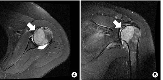
The Shoulder Pain due to Metastatic Breast Cancer -A Case Report-
4
0
0
전체 글
(2)
(3)
(4)
수치
관련 문서
Depending on Tennis Forehand Stroke with elbow pain and without elbow pain Kinematic Analysis of
Based on the above results, the 12-week complex type correction program had a positive effect on the shoulder angle, trunk rotation angle, and body shape
Results after 4 weeks showed that both deep neck bending muscle exercise+general neck strengthening exercise group showed decreased pain and improved functional status
Comparison of pain scale between the conventional and 2-step needle insertion technique according to the injection area ··· 11... The combination of pain scale and
In contrast, Chiropractic has shown better result than sports massage in the range of motion of the shoulder joint, Thoracic Scapular rhythm test, and the
Using this leap motion, a surgeon can perform the virtual shoulder arthroscopic surgery skillfully and reduce mistakes in surgery.. The simulation using leap motion shows
1 John Owen, Justification by Faith Alone, in The Works of John Owen, ed. John Bolt, trans. Scott Clark, "Do This and Live: Christ's Active Obedience as the
The greater tubercle is palpable on the line from the lateral epicondyle of the distal humerus in the direction of the humeral longitudianl axis and just below the acromion
