http://dx.doi.org/10.11620/IJOB.2013.38.3.093
*Correspondence to:Bong-Soo Park, Department of Oral Ana- tomy, School of Dentistry, Pusan National University, Yangsan 626-870, Korea. Tel : 82-51-510-8242, Fax : 82-51-510-8241, E-mail : [email protected]
This is an Open-Access article distributed under the terms of the Creative Commons Attribution Non-Commercial License(http://creati- vecommons.org/licenses/by-nc/3.0) which permits unrestricted non- commercial use, distribution, and reproduction in any medium, pro- vided the original work is properly cited.
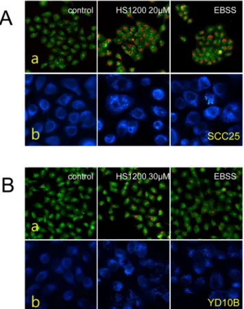
The Role of HS-1200 Induced Autophagy in Oral Cancer Cells
Nam-Mi Jang, Sang-Hun Oh, In-Ryoung Kim, Hae-Ryoun Park
1, and Bong-Soo Park*
Department of Oral Anatomy, School of Dentistry, Pusan National University, Yangsan, Korea
1