Cytotoxic Mechanism of Docosahexaenoic Acid in Human Oral Cancer Cells
Tae-hwa Hong
1†, Hoon Kim
1,4†, Soyeon Shin
1,3†, Kaipeng Jing
1,3, Soyeon Jeong
1,3, Hyun Lim
1, Donghyuk Yun
1, Ki-Eun Jeong
1, Myung-Ryul Lee
1, Jong-Il Park
1, Gi-Ryang Kweon
1, Seung Kiel Park
1, Byung-Doo Hwang
1and Kyu Lim
1,2,3*
1
Department of Biochemisty, College of Medicine,
2Cancer Research Institute and
3Infection Signaling Network Research Center, Chungnam National University, Daejeon 305-764, Korea
4
Department of Dentistry, Eulji University School of Medicine, Daejeon 301-768, Korea Received February 19, 2013 /Revised May 21, 2013 /Accepted May 21, 2013
In the United States, about 40,000 new cases of oral cancer are diagnosed each year and nearly 7,800 patients died from it in 2012. Omega-3 polyunsaturated fatty acids have been found to have anticancer effects in a variety of cancer cell lines and animal models, but their effect in oral cancer remains unclear. This study was designed to examine the effect of docosahexaenoic acid (DHA, a kind of ome- ga-3 fatty acid) on oral cancer cells and the molecular mechanism of its action. We found that ex- posure of squamous cell carcinoma-4 (SCC-4) and squamous cell carcinoma-9 (SCC-9) human oral can- cer cells to DHA induced growth inhibition in a dose- and time-dependent manner. Meanwhile, in addition to the elevated levels of apoptotic markers, such as cleaved PARP, subG1 portion and TUNEL-positive nuclei, DHA led to autophagic vesicle formation and an increase in autophagic flux, indicating the involvement of both apoptosis and autophagy in the inhibitory effects of DHA on oral cancer cells. Further experiments revealed that the apoptosis and autophagy induced by DHA were linked to inhibition of mammalian target of rapamycin (mTOR) signaling by AKT inhibition and AMP-activated protein kinase (AMPK) activation in SCC-9 cells. Together, our results suggest that DHA induces apoptosis- and autophagy-associated cell death through the AMPK/AKT/mTOR signal- ing pathway in oral cancer cells. Thus, utilization of omega-3 fatty acids may represent a promising therapeutic approach for chemoprevention and treatment of human oral cancer.
Key words : AMP-activated protein kinase (AMPK), AKT, autophagy, DHA, mammalian target of rapamycin (mTOR)
†
Tae-hwa Hong, Hoon Kim and Soyeon Shin contributed equally.
*Corresponding author
*Tel:+82-42-580-8223, Fax:+82-42-580-8121
*E-mail : [email protected]
This is an Open-Access article distributed under the terms of the Creative Commons Attribution Non-Commercial License (http://creativecommons.org/licenses/by-nc/3.0) which permits unrestricted non-commercial use, distribution, and reproduction in any medium, provided the original work is properly cited.
Journal of Life Science 2013 Vol. 23. No. 5. 689~697 DOI : http://dx.doi.org/10.5352/JLS.2013.23.5.689
서 론
혀의 squamous cell carcinoma (SCC)는 구강의 일반적인 악성종양[4]으로 미국에서는 2012년에 구강-인두암이 남성에 서는 28,500여명, 여성에서는 11,700명 등 약 40,000여 명의 새 로운 환자가 발생할 것이라 추정되었으며[34], 임파선 전이에 따라 5년 생존율이 25-50%로, 전이가 환자의 생존율에 매우 중요하다고 알려져 있다[38].
식이 지방과 암 발생과의 관련성에 대한 역학 및 실험연구 의 결과가 많이 보고 되어왔다[1, 2, 20, 23, 32]. 특히 alpha-li- nolenic acid (ALA) 등 omega-6 polyunsaturated fatty acids (omega-6 fatty acid, 이하 오메가-6 지방산)은 암의 발생과 밀
접히 관련되어 있으며 eicosapentaenoic acid (EPA) 및 doco- sahexaenoic acid (DHA) 등 omega-3 polyunsaturated fatty acids (이하 오메가-3 지방산)은 암 발생의 위험도를 감소시킨 다고 알려져 있다[1, 2, 6, 20, 23, 32]. 즉 오메가-6 지방산은 암화, 종양성장 및 전이를 자극한다고 하는 반면에 오메가-3 지방산은 이를 억제하는 효과가 있다고 보고 되어 왔다[6, 10, 11, 37]. 이는 오메가-6 지방산 및 오메가-3 지방산의 절대량 보다 오메가-6/오메가-3 지방산 비율이 중요하며 이들 비율의 감소가 암의 예방뿐만 아니라 치료에 중요하다고[6, 9-11, 19, 27, 37]한다. 오메가-3 지방산의 항암 기전에 대해서는 오메가-3 지방산에 의한 전사인자의 조절[24, 29, 30], Cyclooxygenase-2 (Cox-2)를 억제[17, 21, 22], Wnt/β-catenin 신호전달 경로를 차단[21, 22, 35], mirRNA에 의한 조절[7, 8], 혈관신생억제[3, 28, 36] 및 전이억제[25, 33] 등 다양한 작용이 알려져 있다.
세포사멸에는 자가사멸, 괴사 외에도 자가포식이 중요하다
고 알려져 있다[18]. 영양 결핍 시 세포생존을 위해 자가포식이
유도되지만 이는 암세포의 사멸에도 중요한 기전의 하나이다
[5, 13]. 자가포식에는 mammalian target of rapamycin
(mTOR) 신호전달경로가 중요한데 mTOR가 활성화되면 자가
포식을 억제한다. mTOR의 활성은 AMP-activated protein
kinase (AMPK) 및 AKT 등에 의해 조절 될 수 있다. 즉 AMPK 가 활성화 되면 mTOR를 억제함으로 자가포식이 유도된다.
그러나 AKT는 mTOR를 활성화 시킴으로 자가포식을 억제한 다[12, 15, 26, 31]. Jing 등은 자궁경부암세포에서 DHA가 p53 을 억제하여 AMPK를 활성화 시켜 mTOR를 억제함으로 자가 포식이 유도되어 암세포를 사멸 시킨다고 하였다[14]. 그러나 구강암의 세포독성에 대한 오메가-3 지방산의 억제 기전에 대 해서는 명확하게 밝혀져 있지 않았다.
이에 본 연구에서는 먼저 구강암 세포주 SCC-4 및 SCC-9에 서 오메가-3 지방산인 DHA에 의한 세포독성 기전을 규명하여 약간의 지견을 얻었기에 보고하는 바이다.
재료 및 방법
실험재료
Dulbecco's Modified Eagle Medium: Nutrient Mixture F-12 (DMEM/F12), fetal bovine serum (FBS) 등 세포배양에 필요한 시약들은 GIBCO-BRL사(Gaithersburg, MD)에서, PARP, β-actin 등의 항체는 Santa Crut사(California, DA), p-AKT
Thr308, p-AKT
Ser473, p-mTOR, LC3 등의 항체는 Cell Signalig사(Beverly, MA). DHA는 Cayman Chemical사(Ann Arbor, MI)에서, sodium dodecyl sulfate (SDS) 등은 Sigma사 (St. Louis, MO)에서, 구입하였으며 그 외 다른 시약은 특급의 것을 사용하였다.
구강암 세포주의 배양
구강암 세포주 SCC-4 및 SCC-9의 배양은 10% heat-in- activated FBS, penicillin (100 units/ml), streptomycin (100 μ g/ml)을 첨가한 DMEM/F12 (1:1)을 배양액으로 5% CO
2, 37℃ 배양기에서 배양하고, 세포의 밀도가 높아지면 수분간 의 trypsin-EDTA (0.05% trypsin, 0.02% EDTA in Hank’s balanced salt solution without calcium and magnesium)로 처리하고 실험 시 지수기에 있도록 계대배양 하였고, 모든 실험은 FBS가 포함되지 않은 배지로 교환한 다음 24시간 후 시행하였다.
세포 증식능 검색
SCC-4 및 SCC-9세포의 증식능에 대한 DHA 등의 영향은 colorimetric MTT assay를 이용해 측정하였다. 즉 96 well mi- crotiter tissue culture plate에 구강암 세포를 5-10 x 10
3cells/well로 plating하고 하룻밤 배양 후 FBS가 포함되지 않 은 배지로 교환한 다음 24시간 배양하고 DHA 등을 농도별로 처리하였다. 24시간 배양한 다음 세포의 배양액을 제거하고 3-(4,5-dimethylthiazol-2-yl)-2,5-diphenyltetrazolium bromide (MTT) (0.5 mg/ml) 용액으로 37℃에서 2 시간 반응시키고 상 청액을 제거한 후 Dimethyl sulfoxide에 녹여 570 nm에서 au-
tomatic ELISA plate reader로 흡광도를 측정하였다.
Terminal deoxynucleotidyl transferase dUTP nick end labeling (TUNEL) assay
SCC-4 및 SCC-9 세포를 5 x 10
4cells/well로 cover glass가 들어있는 24-well plate에 plating한 후 DHA 처리하여 24시간 동안 배양하였다. 이들 세포를 PBS로 2회 washing한 다음 4%
paraformaldehyde로 4℃에서 20분간 고정시킨 다음 0.2%
TritonX-100으로 처리한 후 DeadEND Fluorometric TUNEL System (Promega, Madison, WI) kit를 사용하여 분해된 DNA 에 anti-digoxigenin-phosphatase를 37℃에서 4시간 동안 반응 시켜 붙였다. 이들 세포는 PBS로 3회 세척한 후 3,3-dia- minobenzidine (DAB)를 이용하여 발색 시킨 후 4’,6- dia- midino-2-phenylindole, dihydrochloride (DAPI)로 핵을 염색 한 것을 mounting medium을 넣고 coverslip으로 밀봉시켜 confocal microscopy (Olympus
TM)로 검경, 촬영하였다.
Western blot
SDS-PAGE 후에 분리된 단백을 전기적 전사법으로 nitro- cellulose membrane에 옮긴 후 전사된 nitrocellulose mem- brane을 5% skim milk로 실온에서 1시간 blocking한 다음 동 일 용액으로 PARP, β-actin 등의 항체(1:1,000)로 4℃에서 하룻 밤 반응시켰다. 이 후 TBS-T로 수회 세척하고 peroxidase con- jugated 된 이차항체를 5% skim milk/TBS-T용액에 1:10,000 으로 희석하여 1시간 반응 시키고 TBS-T로 수회 세척한 후 ECL Western blotting detection system을 이용하여 단백질 band를 확인하였다.
유세포 계측(Flow cytometric analysis)
DHA를 일정시간 동안 처리하여 배양한 세포를 try- sin-EDTA로 처리한 후 세포를 모아 원심분리 하여 pellet를 PBS에 부유시킨 다음 70% ethanol로 20℃에 사용 전까지 보관 하였다. 유세포 계측은 ethanol로 고정된 세포에 RNase A (0.25 μg/ml)을 처리한 후 propidium iodide (50 μg/ml)를 이 용하여 염색하고, spectra/Mesh Nylon로 filter하여 측정하였 다. 세포주기별 분포율은 FACscan (Becton Dickinson)의 FL-2A와 FL-2W linear plot에 근거하여, Cell Quest software 를 이용한 histogram으로 계산하였다.
GFP-LC3 dot 검색
SCC-4 및 SCC-9 세포를 6-well culture plate에 80% con-
fluent하게 plating한 후 0.5 μg/well로 green fluorescence
(GFP)-LC3 plasmid 등을 lipofectamine (Invitrogen,
Camarllo, CA)을 이용하여 transfection 하였다. Transfection
된 세포에 DHA 등의 약물을 일정시간 처리한 후 형광현미경
으로 GFP-LC3 dot를 검경, 촬영하였다.
A B
C D
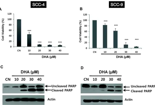
Fig. 1. Effect of DHA on cell growth of human oral cancer cells. A and B: SCC-4 and SCC-9 cells were incubated with different concentrations of DHA for 24 h, and cell growth was determined using the MTT assay as described in Materials and Methods.
C and D: The cells were treated with various concentration of DHA for 24 h. PARP cleavage was analysed by Western blot analysis as described in Materials and Methods. Actin was used as control for the amount of proteins loaded per lane.
CN, control; Columns, mean of four independent experiments; bars, SD; ***, p< 0.005 compared with control.
통계분석
표시된 결과는 3번 이상의 독립적인 실험 결과이며, 실험 결과의 통계 처리는 student's t-test에 준하여 처리하였고, P-value가 최대치 0.05 (p<0.05) 이하인 경우를 유의한 것으로 판정하였다.
결 과
인체 구강암세포 SCC-4 및 SCC-9의 증식에 미치는 DHA의 영향
오메가-3 지방산은 오메가-6 지방산과는 달리 간담도암을 비롯한 수종의 암세포주에 대해 세포독성을 나타낸다[21, 22, 35]고 알려져 있다. 이에 먼저 인체 구강암 세포주 SCC-4 및 SCC-9에 대한 오메가-3 지방산 중 DHA의 세포 독성을 확인 하였다. 이때 96 well tissue plate에 SCC-4 및 SCC-9 세포를 0.5-1 x 10
4cells/well로 배양한 다음 무혈청 배지로 24시간 배양하고 0-40 μM의 DHA를 처리 하여 24시간 후 MTT로 세 포 증식능을 검색하였다. DHA는 SCC-4 및 SCC-9 세포의 증 식을 농도의존적으로 억제하였다(Fig. 1A, 1B). DHA에 의한 SCC-4 및 SCC-9 세포의 증식억제에 자가사멸이 관련되어 있 는지 확인하기 위해 자가사멸 분자 표식자(marcker)의 하나인 PARP cleavage를 검색했을 때 DHA의 처리 후에 cleaved PARP 단백양이 DHA의 농도에 의존적으로 증가하였다(Fig.
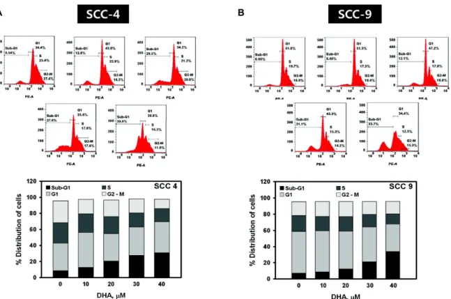
1C, 1D). 세포주기분석에서도 SCC-4 및 SCC-9 세포는 DHA 처리에 의해 자가사멸된 세포가 함유되어 있는 subG1 분획이 증가하였다(Fig. 2). 또한 이를 확인하기 위해 SCC-4 세포에서 DHA 처리 후 TUNEL assay를 시행하였을 때 대조군에 비해 DHA의 농도에 의존적으로 TUNEL 양성세포가 현저히 증가 하였다(Fig. 3). 이는 DHA가 SCC-4 및 SCC-9 세포에 대해 세 포 독성이 있으며, 이는 세포사멸이 밀접히 관련되어 있음을 시사한다.
DHA에 의한 인체 구강암세포 SCC-4 및 SCC-9의 자가 포식 유도
세포사멸에는 자가사멸, 괴사외에도 자가포식이 알려져 있 으며, 최근 Jing 등[14]은 DHA에 의한 자궁경부암세포인 SiHa 의 세포독성기전에 자가 사멸뿐 만 아니라 자가포식도 중요하 다고 하였다. 이에 먼저 구강암세포 SCC-4 및 SCC-9에 대한 DHA의 자가포식을 확인하기 위해 이들 세포에 0-40 μM의 DHA를 처리 후 자가포식의 중요한 분자표식자인 micro- tubule-associated protein1 light chain 3 (LC-3) 단백양을 Western blot으로 검색하였을 때 LC-3II 단백질 양이 처리한 DHA의 농도에 의존적으로 증가하였다(Fig. 4A, 4B). 이는 DHA에 의한 SCC-4 및 SCC-9 세포의 사멸에 자가포식도 관련 되어 있음을 시사한다.
DHA에 의한 LC3-II증가가 자가포식 때문인지 확인하기 위
A B
Fig. 2. DHA increases the cells of subG1 fraction in SCC-4 and SCC-9 cells. The cells were incubated at 37℃ for 24 h with different concentrations of DHA. DNA fluorescence was analyzed by flow cytometry, and the cell cycle distribution was calculated using Cell Quest software as describe in Materials and Methods.
A B
Fig. 3. TUNEL positive cells are induced by DHA in SCC-4 cells. The cells were treated with 30 μM DHA for 24 h and the proportion of apoptotic cells was determined by TUNEL assay as described in Materials and Methods. CN, control; Columns, mean of four independent experiments; bars, SD; ***, p< 0.005 compared with control.
해 autolysosome 형성을 억제하는 약물인 chloroquine (CQ) 처리 후 LC3-II 단백양을 측정하였다. 먼저 CQ 단독 처리 후 LC3-II 단백양이 증가하였으며, DHA에 의해 증가된 LC3-II 단백양은 CQ전처리 후 DHA 단독 처리 보다 더 증가하였다 (Fig. 4C). 이는 DHA에 의해 증가된 LC3-II가 자가포식에 의해 일어남을 강력히 시사하는 것이다.
또한 SCC-4 및 SCC-9 세포에 GFP-LC3를 transfection 한 후 자가포식의 분자표식자인 GFP-LC3 형광 dot를 검색하였
다. 이들 세포에 40 μM의 DHA를 처리 후 GFP-LC3 형광 dot 은 대조군에 비해 현저히 증가 하였다(Fig. 5). 따라서 DHA는 구강암세포를 자가사멸 할 뿐만 아니라 자가포식을 유도하여 세포사멸 시킬 수 있으리라 생각된다.
DHA에 의한 인체 구강암세포 SCC-4 및 SCC-9의 세포 독성에 대한 mTOR 신호전달 경로 억제
최근 Jing 등[14]은 p53 wild type인 자궁경부암세포에서
A B
C D
Fig. 4. Autophagy is induced after DHA treatment in SCC-4 and SCC-9 cells. A and B: Western blot analysis of the expression levels of autophagic protein, LC3 in SCC-4 and SCC-9 cells treated with the indicated dose of DHA for 24 h. C: SCC-4 and SCC-9 cells treated with 30 μM DHA in the presence or absence of 2 μM CQ for 24 h were analyzed by Western blot.
A B
Fig. 5. Representative image of GFP-LC3 dots in GFP-LC3- transfected SCC-4 and SCC-9 cells with or without DHA treatment.
The cells were transfected with GFP-LC3 plasmid for 18 h and then incubated for an additional 3 h with 30 μM DHA.
After that, GFP-LC3 dot was observed using fluorescence microscope (x600). CN, control; Columns, mean of four independent experiments; bars, SD; **, p< 0.01 compared with control.
DHA가 자가포식을 유도하는데 이때 mTOR가 중요하다고 하 였다. mTOR 신호전달 경로에는 AMPK와 AKT에 의한 인산 화가 중요함으로 DHA 처리 후 이들 단백양의 변동을 검색하 였다. 먼저 구강암세포 SCC-9에서 AKT 및 mTOR의 인산화에 대한 DHA의 영향을 검색하였다. SCC-9세포에 DHA를 24시 간 처리 후 농도에 따른 영향을 검색한 바 AKT의 thr308 및 ser473의 인산화가 DHA의 농도에 의존적으로 감소하였으며 또한 mTOR의 인산화도 감소하였다(Fig. 6A). 이는 DHA가 AKT의 인산화를 억제를 통한 mTOR의 활성 억제에 의해 자
가포식이 유도될 수 있음을 시사한다.
AMPK의 인산화에 대한 DHA의 영향을 검색한 바 DHA
처리 후 AMPK의 인산화는 DHA의 농도에 의존적으로 증가
하였다. AMPK의 인산화에 따른 그 효소 활성의 증가를 확인
하기 위하여 AMPK의 내인성 기질의 하나인 acetyl CoA car-
boxylase (ACC)의 인산화를 측정하였을 때 DHA의 농도 의존
적으로 ACC의 인산화가 증가 하였다(Fig. 6B). 이는 DHA에
의한 자가포식의 유도에는 AMPK 인산화의 증가에 따른
mTOR의 억제도 중요함을 시사한다.
A B
Fig. 6. DHA triggers autophagy through mTOR inhibition by activation of AMPK and inhibition of AKT in SCC-9 cells. The cells were treated with 40 μM of DHA in serum free medium for indicated time periods. The cell lysates were obtained for Western blot analysis using antibodies against p-AKTs, and p-mTOR as well as p-AMPK as described in Materials and Methods. Actin was used as control for the amount of proteins loaded per lane.
고 찰
오메가-6 지방산은 암화, 암세포의 성장, 침윤 및 전이 등을 촉진시키지만 오메가-3 지방산은 반대로 억제한다고 알려져 있다[1, 2, 6, 9-11, 19, 20, 23, 27, 32, 37]. 이는 유방암 등 동물실 험에서도 잘 밝혀져 있다. 그러나 구강암세포에 대한 오메가-3 지방산의 세포독성에 대해서는 전혀 알려져 있지 않다.
Lim 등[21, 22]은 오메가-3 지방산이 간암 및 간담도암에서 자가사멸을 유도한다고 하였다. 이에 먼저 구강암세포주인 SCC-4 및 SCC-9에 대한 DHA의 세포독성을 검색했을 때 PARP cleavage 증가, subG1 분획 증가, TUNEL 양성세포의 증가 등을 볼 때 이는 자가사멸을 통한 세포독성을 유발함을 시사하였다.
세포사멸에는 자가사멸 외에도 자가포식이 중요하다고 알 려져 있다[5, 13, 18]. Jing 등[14]은 DHA가 자가포식에 의해 SiHA 세포 등 인체 자궁경부암세포를 사멸할 수 있음을 보고 하였다. 이에 DHA가 구강암세포에서도 자가포식을 유도할 수 있는지 검색하기 위하여 SCC-4 및 SCC-9 세포에 DHA 처 리 후 자가포식의 표식자인 LC3-II 및 GFP-LC3 dot를 측정한 바 DHA에 의해 증가함으로 DHA는 구강암세포 SCC-4 및 SCC-9에서 자가포식을 유도할 수 있음이 확인 되었다. 형성된 autophagosome은 lysosome과 fusion 되어 autolysosome을 형성한 후 포식한 물질들을 파괴함으로 자가포식과정이 마무 리 된다. 그러나 형성된 autophagosome이 자가포식 때문인지 혹은 자가포식과 무관하게 증가된 것인지 확인하기 위해서는 autolysosome의 형성을 억제하는 CQ 등의 약물의 전처리로 autophagic flux assay를 시행해야 한다[16]. 본 실험에서 SCC-4 및 SCC-9 세포에CQ 전처리 후 DHA를 처리했을 때 DHA 단독보다 LC3-II양이 각각 증가하였는데 이는 CQ가 au- tolysosme 형성을 억제함으로 LC3-II의 lysosome에 의한 분해
가 감소되어 나타난 것으로, DHA가 구강암세포를 자가포식 시킴을 강력히 시사하며, 따라서 DHA에 의한 구강암세포 SCC-4 및 SCC-9의 세포사멸에 자가포식도 중요하리라 생각 된다.
자가포식의 조절에는 mTOR를 통한 신호전달 경로가 중요 함으로 따라서 이를 조절함으로 자가포식이 조절될 수 있는 데, 즉 mTOR는 자가포식의 음성조절자로서 알려져 있다.
AMPK는 체내 에너지가 부족할 때 인산화되어 그 활성을 나 타내는 효소로써 AMPK의 활성화는 mTOR의 신호전달을 억 제함으로 자가포식의 양성조절자이다[12, 15, 26, 31]. Jing 등 [14]은 DHA에 의한 p53 wild type인 암세포에서 p53의 증가 가 AMPK를 활성화 시켜 mTOR를 억제한다고 보고하였다.
이에 구강암 세포주에 대한 DHA의 자가포식 기전을 검색한 바 SCC-9에서 DHA 처리 후 p-AMPK 단백양이 증가 하였으
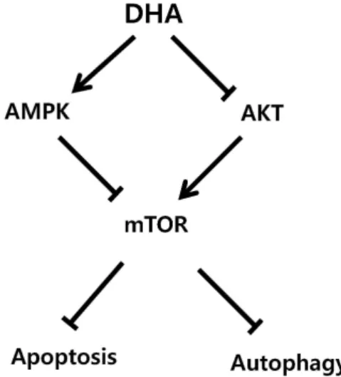
Fig. 7. Proposed mechanism of DHA-induced cell death in oral
cancer cells.
며, AMPK의 내인성 기질인 ACC의 인산화도 증가된 것을 감안할 때 이는 DHA가 SCC-9 세포에서 AMPK 활성화를 증 가 시켜 mTOR를 억제하여 자가포식을 유도할 수 있음을 시사 한다.
한편 AKT는 mTOR의 양성조절자로서 AKT의 활성화는 mTOR를 활성화 시켜 자가포식을 억제한다고 알려져 있다[5, 12, 15, 31]. 이에 구강암 세포주 SCC-9에서 DHA 처리 후 p-AKT의 단백양을 측정했을 때 p-AKT
Thr308및 p-AKT
Ser473단 백양이 감소하였을 뿐만 아니라 p-mTOR도 감소하였는데 이 는 구강암 세포주 SCC-9에서 DHA가 AKT의 활성화를 억제 함으로 mTOR가 억제되어 자가포식이 유도될 수 있음을 시사 한다.
이상의 결과로 DHA는 구강암세포에서 첫째, 자가사멸 및 자가포식을 통해 세포사멸을 유도하며, 둘째, AKT의 활성 억 제에 따른 mTOR 억제, 셋째, AMPK 활성 증가에 의한 mTOR 억제 등을 통해 자가포식을 유도하여 자가사멸과 더불어 세포 사멸을 유도하리라 생각되며(Fig. 7) 따라서 이는 구강암의 치 료 및 예방 목적으로 유용하게 사용할 수 있으리라 시사된다.
감사의 글
본 연구는 2010년도 충남대학교 학술연구비에 의해 지원되 었음.
References